Radiologically Evident Oesophageal Dilation in Paediatric Rumination Syndrome With Recurrent Aspiration Pneumonia: A Rare Case Without Achalasia
Akina Nigi, Keisuke Iwamoto, Hidetoshi Itani, Shigeto Kondou

TL;DR
A 15-year-old autistic boy with rumination syndrome showed rare oesophageal dilation on CT, unrelated to achalasia, highlighting the role of CT in diagnosing such cases.
Contribution
This case presents a rare example of structural oesophageal change caused by a behavioral disorder, not achalasia.
Findings
Progressive oesophageal dilation was observed in a patient with rumination syndrome.
Achalasia was ruled out using imaging techniques.
CT proved valuable for diagnosis when manometry was not feasible.
Abstract
A 15‐year‐old autistic boy with rumination syndrome presented with progressive oesophageal dilation on CT. Achalasia was excluded based on imaging. This case illustrates a rare example of structural change likely due to behavioural disorder and highlights the diagnostic value of CT when manometry is unfeasible. We present a rare example of structural change likely due to behavioural disorder in a 15‐year‐old autistic boy with rumination syndrome who presented with progressive oesophageal dilation on CT. Achalasia was excluded based on imaging.
Genes, proteins, chemicals, diseases, species, mutations and cell lines named across the full text — each resolved to its canonical identifier and authoritative record.
Click any figure to enlarge with its caption.
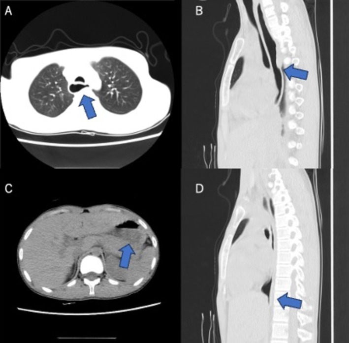 Figure 1
 Figure 2
Peer Reviews
No public reviews on file for this paper yet. If you reviewed it on a platform where reviews are public (OpenReview, ICLR, NeurIPS, ICML), you can paste yours below so the community can read it here.
Videos
No videos yet. Explain this paper in a talk, walkthrough, or lecture? Add one.
Taxonomy
TopicsGastroesophageal reflux and treatments · Restraint-Related Deaths · Dysphagia Assessment and Management
A 15‐year‐old boy with severe intellectual disability and autism had long‐standing rumination behaviour with recurrent vomiting and aspiration pneumonia. Chest CT revealed progressive, air‐filled oesophageal dilation without food stasis, while gastric contents were preserved (Figure 1A–D). Endoscopic or manometric evaluation could not be performed because of his severe autistic condition, but the absence of food residue, fluid level, or distal tapering suggested no overt achalasia or obstruction. Repetitive rumination behaviour, involving abdominal straining and air swallowing, may transiently increase intraesophageal pressure, resulting in functional dilation. While rumination syndrome is classically defined as a behavioural disorder without structural abnormality, secondary changes such as reflux esophagitis or mild dilation have been reported [1, 2]. This case highlights that severe and persistent rumination is likely to have induced functional oesophageal dilation, suggesting that behavioural mechanisms may potentially cause morphological changes even without primary motility disorder. To our knowledge, there have been no previous reports of radiologically evident oesophageal dilation in rumination syndrome without evidence of achalasia or obstruction. Although this may appear theoretically plausible due to chronic aerophagia and straining, such structural consequences of behavioural disorders remain rarely documented.
Consent
The authors declare that written informed consent was obtained for the publication of this manuscript and accompanying images using the consent form provided by the Journal.
Conflicts of Interest
The authors declare no conflicts of interest.
The reference list from the paper itself. Each links out to its DOI / PubMed record.
- 1A. Sawada and Y. Fujiwara , “Belching Disorders and Rumination Syndrome: A Literature Review,” Digestion 105, no. 1 (2024): 18–25.37844547 10.1159/000534092 · doi ↗ · pubmed ↗
- 2S. Pomenti and D. A. Katzka , “Current State of Rumination Syndrome,” Diseases of the Esophagus 37, no. 9 (2024): doae 041.38741462 10.1093/dote/doae 041 · doi ↗ · pubmed ↗