Epidemiological and Clinical Profile of Fixed Pigmented Erythema at the Departmental University Hospital Center Borgou/Alibori (Benin)
Fabrice Akpadjan, Laura Dotsop, Nadège Agbessi, Christiane Koudoukpo

TL;DR
This study describes the occurrence and characteristics of fixed pigmented erythema, a skin reaction to drugs, at a hospital in Benin from 2009 to 2022.
Contribution
The study provides a detailed epidemiological and clinical profile of fixed pigmented erythema in a specific hospital setting in Benin.
Findings
Fixed pigmented erythema had a prevalence of 0.73% with male predominance.
Cotrimoxazole was the most commonly associated drug, followed by paracetamol and quinine.
Over half of the patients self-medicated, suggesting a link to the condition's prevalence.
Abstract
Fixed pigmented erythema (FPE) is a common toxidermia characterized by the appearance of one or more annular, erythematous and hyperpigmented spots, following the systemic administration of a drug. The main aim of this study was to describe the epidemiological and clinical aspects of fixed pigmented erythema at the Departmental University Hospital Center Borgou/Alibori (DUHC‐B/A) from 2009 to 2022. This was a descriptive cross‐sectional study with retrospective data collection, based on the records of patients seen in the Dermatology‐Venerology Unit for FPE. Initially, all files bearing the diagnosis of toxidermia were identified; then, those with the diagnosis of FPE with usable data were retained. Data were entered using EpiData 3.1 and analyzed using EpiData Analysis. Sixty‐four patients were enrolled during the study period. The prevalence of FPE was 0.73%, with a male…
Genes, proteins, chemicals, diseases, species, mutations and cell lines named across the full text — each resolved to its canonical identifier and authoritative record.
Click any figure to enlarge with its caption.
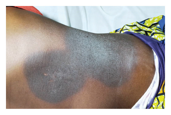 FIGURE 1
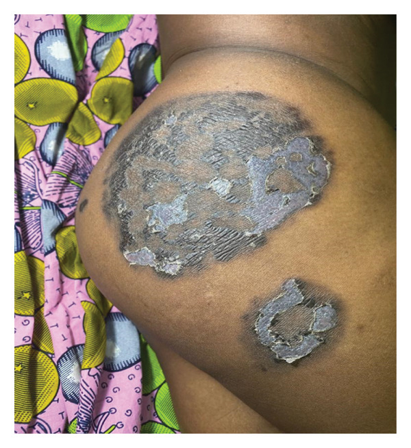 FIGURE 2
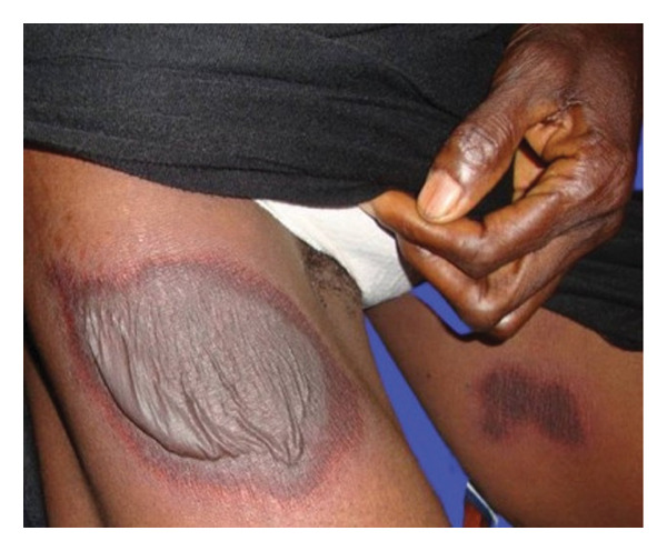 FIGURE 3
|
|
| |
| None | 45 | 70.3 |
| Sinusitis | 6 | 9.3 |
| High blood pressure | 5 | 7.8 |
| Asthma | 4 | 6.2 |
| Diabetes | 1 | 1.6 |
| Physical disability | 1 | 1.6 |
| Allergic rhinitis | 1 | 1.6 |
| Syphilis | 1 | 1.6 |
| Total | 64 | 100 |
|
|
| |
| Antibiotics ( | ||
| Cotrimoxazole | 9 | 14.0 |
| Amoxicillin | 2 | 3.1 |
| Ceftriaxone | 1 | 1.6 |
| Metronidazole | 1 | 1.6 |
| Clarithromycin | 1 | 1.6 |
| Analgesics–NSAIDs ( | ||
| Paracetamol | 6 | 9.3 |
| Metamizole sodium | 1 | 1.6 |
| Diclofenac | 1 | 1.6 |
| Ibuprofen | 1 | 1.6 |
| Anti parasitic drugs ( | ||
| Quinine | 3 | 4.6 |
| Sulfadoxine–pyrimethamine | 2 | 3.1 |
| Artemether–lumefantrine | 1 | 1.6 |
| Artesunate | 1 | 1.6 |
|
|
| |
| Not specified | 49 | 76.6 |
| Upon 7 days | 7 | 10.9 |
| In the first 24 h | 5 | 7.8 |
| In the first 4 weeks | 2 | 3.1 |
| More than a month | 1 | 1.6 |
| Total | 64 | 100.0 |
|
|
| |
| Macules | 60 | 93.8 |
| Scales | 17 | 26.6 |
| Erosions | 12 | 18.8 |
| Crust | 4 | 6.3 |
| Blisters | 3 | 4.7 |
| Papules | 2 | 3.1 |
| Vesicles | 1 | 1.6 |
| Edema | 1 | 1.6 |
|
|
| |
| Thighs | 33 | 51.6 |
| Thorax | 24 | 37.5 |
| Face | 22 | 34.4 |
| Legs | 22 | 34.4 |
| Arms | 21 | 32.8 |
| Fore arms | 21 | 32.8 |
| Back | 20 | 31.3 |
| Hands | 19 | 29.7 |
| Abdomen | 16 | 25.0 |
| Genitalia | 15 | 23.4 |
| Feet | 14 | 21.9 |
| Neck | 10 | 15.6 |
| Bottom | 9 | 14.1 |
| Palms | 7 | 10.9 |
| Eyebrows | 6 | 9.4 |
| Nasal alae | 6 | 9.4 |
| Scalp | 6 | 9.4 |
| Feet soles | 3 | 4.7 |
|
|
| |
| Pruritus | 40 | 62.5 |
| Pain | 10 | 15.6 |
| Fever | 2 | 3.1 |
| Burn sensation | 1 | 1.6 |
Peer Reviews
No public reviews on file for this paper yet. If you reviewed it on a platform where reviews are public (OpenReview, ICLR, NeurIPS, ICML), you can paste yours below so the community can read it here.
Videos
No videos yet. Explain this paper in a talk, walkthrough, or lecture? Add one.
Taxonomy
TopicsDrug-Induced Adverse Reactions · Dermatological and COVID-19 studies · Acne and Rosacea Treatments and Effects
1. Introduction
Fixed pigmented erythema (FPE) is a common toxidermia characterized by the appearance of one or more annular, oval, erythematous and hyperpigmented spots, following the administration of a systemic drug [1, 2]. Upon re‐exposure to the concerned drug(s), lesions may reappear on the same and/or different sites. The lesions leave behind postinflammatory hyperpigmentation [3]. The rash appears a few days to 2 weeks after initial exposure to the causative agent and 30 min to 16 h after re‐exposure [3]. The drugs involved vary according to the common illnesses and prescription habits in each part of the world. The list is not exhaustive [4, 5]. Cases of FPE to plants, dietary supplements, food colorants, preservatives and tinctures have been reported [3–6].
The precise mechanisms by which FPE occurs are unknown, but a cell‐mediated process involving CD8 T lymphocytes has been incriminated [7]. A genetic predisposition has been described, involving the HLA‐B22 and Cw1 antigens [8]. Histopathological examination shows interface dermatosis, vacuolar change, keratinocyte necrosis and pigment incontinence in the dermis. Mild acanthosis, hyperkeratosis, spongiosis, dermal edema and the presence of eosinophils and rare neutrophils may also be present [1, 3].
The aim of this study was to describe the epidemiological and clinical aspects of FPE in the Dermatology‐Venerology Unit of the Departmental University Hospital Center Borgou/Alibori (DUHC‐B/A).
2. Patients and Methods
This was a descriptive cross‐sectional study with retrospective data collection by review of the files of all patients seen in the Dermatology‐Venerology Unit for FPE irrespective of age and gender. The study period was from January 2009 to June 2022. Initially, all files with the diagnosis of toxidermia were identified, followed by those with the diagnosis of FPE with exploitable data. The dependent variable was FPE, and the independent variables were epidemiological and clinical data. Data were recorded and analyzed using EpiData 3.1 and IBM SPSS Statistics 21 software, respectively. Patient data confidentiality was protected.
3. Ethics Statement
This work was conducted in accordance with current ethical standards. The research protocol was approved by the Local Ethics Committee for Biomedical Research of the University of Parakou (LECBR‐UP).
4. Results
4.1. Epidemiological Data
During the study period, 123 cases of toxidermia were recorded in the Dermatology‐Venerology Unit of DUHC‐B/A, out of a total of 8829 patients who consulted during that period. Of the 123 cases, 64 patients suffered from FPE, representing a frequency of 52.03% of all toxidermia, and of 0.73% of all consultations recorded in the Dermatology‐Venerology Unit of DUHC‐BA from January 2009 to June 2022. The mean age of patients was 32.34 ± 18.79 years, with extremes ranging from 1 to 83 years. Patients aged 21–35 were the most represented, with a proportion of 34.4%. The study population comprised 29 women (45.3%), giving a sex ratio (M/F) of 1.20. The most common level of education was university (29.7%), followed by primary education (18.8%), secondary education (15.6%) and the unschooled (10.9%).
Most of the patients were pupils, students and apprentices (34.4%), followed by the unemployed (21.9%) and civil servants (14.1%).
4.2. Clinical Data
Seventy percent of the patients had no previous medical history (Table 1).
The most frequently incriminated drug was cotrimoxazole (14.0%), followed by paracetamol (9.3%) and quinine (4.6%), as shown in Table 2.
Self‐medication was not specified for 30 patients. Of those for whom it was specified, 52.9% were self‐medicating.
The time interval between medication intake and the onset of symptoms was 1–7 days for 10.9% of the patients (Table 3).
In this study, macules were the most common dermatological lesions (93.8%), followed by scales (26.6%) and erosions (18.8%) as shown in Table 4.
The most common sites of lesions were the thighs in 51.6% of the patients, followed by the thorax (37.5%) and face (34.4%) as shown in Table 5.
The most common associated sign was pruritus in 62.5% of the patients (Table 6).
In this study, FPE presented in its typical form (Figure 1) in 52 patients (81.25%) and in its bullous form (Figures 2 and 3) in 12 patients (18.75%).
Fixed pigmented erythema in its typical form on the abdomen (source: Dermatology-Venerology Unit DUHC-B/A).
Bullous fixed pigmented erythema on the right buttock and thigh, scaling phase (source: Dermatology-Venerology Unit DUHC-B/A).
Bullous fixed pigmented erythema on the right thigh of a 65‐year‐old woman (source: Dermatology-Venerology Unit DUHC-B/A).
5. Discussion
The hospital frequency of FPE at DUHC‐B/A was 0.73% for all consultations recorded in the Dermatology‐Venerology Unit. This value is lower than those reported by Atadokpèdé et al. In Cotonou (Benin) in 2011 (1.1%) [9], Saka et al. in Togo in 2012 (1.1%) [10] and Ayanlowo in Nigeria in 2015 (1.8%) [11]. This low frequency could be explained by the fact that FPE is a benign toxidermia that does not worry patients in our setting to the point of motivating a consultation.
In this study, young people were the most affected, with a higher frequency in patients aged between 21 and 50 (34.4%). The mean age of patients was 32.34 ± 18.79 years. This is similar to the mean age reported by Atadokpèdé et al. (33 years), Ayanlowo et al. (32.12 ± 15.5 years) and Saka et al. (31.27 ± 14.01 years) [9–11]. The predominance of younger patients may be linked to an easy tendency to self‐medicate in this age group and to the repeated use of products known to cause toxidermia.
The study population was composed of 29 women (45.3%), giving a sex ratio (M/F) of 1.20. This male predominance was also found in the Togolese series [10]. On the other hand, a slight female predominance was reported in Cotonou (Benin) (M/F = 0.98) [9] and Nigeria [11]. Thus, the sex ratio varies between studies in the subregion. This difference may be linked to each population’s hospital‐attending habits and attitude to FPE.
In our series, 70% of the patients had no previous history of FPE. Among the remaining 30%, the most common antecedents were atopy and hypertension. In the study by Atadokpèdé et al., 50% of the patients had one or more pathological conditions associated with FPE. These were mainly atopy, polymedication, diabetes and HIV infection [9]. The role of pathological history in the occurrence of toxidermia in general, and EPF in particular, may be explained not only by changes in drug pharmacokinetics in certain pathological states, such as renal or hepatic insufficiency, but also by the frequency of polymedication in patients with an existing medical history [12]. As far as we know, there is no direct link between the onset of FPE and a history of atopy.
The most frequently incriminated drug was cotrimoxazole (14.0%), followed by paracetamol (9.3%) and quinine (4.6%). In African series, cotrimoxazole also accounts for a significant proportion of the drugs involved, ranging from 19.0% to 37.4% [9–11]. In European countries, on the other hand, anti‐infective drugs are not always the first responsible for FPE. Brahimi et al. in France [13] and Savin in the United Kingdom [14] found paracetamol to be the first drug responsible for FPEs, followed by nonsteroidal anti‐inflammatory drugs. The most incriminated drugs are also the most accessible in terms of cost and availability in health facilities, pharmacies and markets. These drugs in our countries are not yet subject to strict regulations governing their use.
Of the patients who provided information on the practice of self‐medication, 52.9% resorted to it. High percentages were also found by Atadokpèdé et al. and Saka et al. who reported 57% and 66.9%, respectively. These frequencies reflect the extent of this practice in our country, which can be explained by the poverty of the population, the sometimes limited access to healthcare, the phenomenon of street medicines available without prescription and illiteracy.
The time lapse between drug intake and the onset of FPE symptoms was 1–7 days in 10.9% of the patients. Atadokpèdé et al. and Saka et al. reported an average time lapse of 3 days (1–14 days) and 2.52 ± 3.04 days, respectively. From these results, we can see that FPE most often occurs less than a week after drug intake.
In this study, macules were the most common dermatological lesions (93.8%), followed by scales (26.6%) and erosions (18.8%). This high frequency of macules can be explained by the fact that macules are the most common lesion in FPE and are also the lesion found as a pigmentary sequela.
The most common sites were the thighs in 51.6% of the patients, followed by the thorax (37.5%) and face (34.4%). In the Nigerian and Togolese series, the most common sites were the trunk and lower limbs, the face and genitalia [10, 11].
In this study, FPE presented predominantly in its typical form (81.25%). This predominance of the hyperpigmented macular form was also found by Atadokpèdé et al. (83.87%) and Saka et al. (76.95%) [9, 10], in contrast to the French series by Brahimi et al. where bullous forms accounted for over 50% [13]. These same French authors also reported nonpigmented FPEs in 20.3% of the cases in their series [13].
Fixed drug eruption (FDE) is a T cell–mediated cutaneous adverse drug reaction characterized by the persistence of drug‐specific CD8^+^ resident memory T cells in previously affected skin sites, leading to lesion recurrence upon re‐exposure to the causative drug [15]. Individual susceptibility to FDE appears to be modulated by genetic factors, particularly polymorphisms in Class I human leukocyte antigen (HLA) molecules, which play a central role in antigen presentation to cytotoxic T lymphocytes [16, 17].
Associations between specific HLA alleles, mainly within the HLA‐B locus, and FDE induced by commonly implicated drugs such as antibiotics, nonsteroidal anti‐inflammatory drugs and paracetamol have been reported, although these associations are less consistent and less well established than those described for severe cutaneous adverse reactions, including Stevens–Johnson syndrome and toxic epidermal necrolysis [16, 18]. These genetic associations appear to be highly drug‐specific and population‐dependent, reflecting differences in HLA allele distribution across ancestral backgrounds.
The heterogeneous distribution of HLA alleles among populations may partly account for epidemiological differences in FDE incidence and in the spectrum of causative drugs reported worldwide [17]. Furthermore, immunogenetic variability may influence clinical phenotypes, including lesion extent, generalized or bullous forms and mucosal involvement [19]. In addition, postinflammatory hyperpigmentation, which is more pronounced in darker phototypes, may accentuate perceived phenotypic differences between populations, independently of the underlying immunological mechanisms [19].
At present, the lack of standardized genetic data precludes routine HLA screening in clinical practice for FDE. Multicenter studies involving diverse populations and detailed clinical phenotyping are needed to better define the immunogenetic determinants of this condition [17, 19].
6. Conclusion
This study confirms the relatively low frequency of FPE even though it is the most frequently observed toxidermia at the DUHC‐B/A and also the role of anti‐infective sulfonamides among FPE‐inducing drugs in sub‐Saharan Africa. It underlines the great responsibility of self‐medication in the occurrence of this toxidermia in Parakou. It is important to raise public awareness on the risks of self‐medication and to recognize the serious forms of FPE that can be life‐threatening. It should be noted that these results cannot be generalised to developed countries where practices are not the same.
Funding
No funding was received for this manuscript.
Conflicts of Interest
The authors declare no conflicts of interest.
The reference list from the paper itself. Each links out to its DOI / PubMed record.
- 1Breathnach S. M. , Burns T. , Breathnach S. , Cox N. , and Griffiths , Drug Reactions: Fixed Drug Eruptions, Rooks Textbook of Dermatology, 2004, 4, 7th edition, Blackwell Science Ltd, UK, 20-9–73.
- 2Ozkaya-Bayazit E. , Specific Site Involvement in Fixed Drug Eruption, Journal of the American Academy of Dermatology. (2003) 49, no. 6, 1003–1007, 10.1016/s 0190-9622(03)01588-3, 2-s 2.0-0344430227.14639376 · doi ↗ · pubmed ↗
- 3Butler D. F. , Ilse J. R. , Schwartz R. A. et al., Fixed Drug Eruptions, https://emedicine.medscape.com/article/1336702-overview?form=fpf.
- 4Sehgal V. N. and Srivastava G. , Fixed Drug Eruption (FDE): Changing Scenario of Incriminating Drugs, International Journal of Dermatology. (2006) 45, no. 8, 897–908, 10.1111/j.1365-4632.2006.02853.x, 2-s 2.0-33746652581.16911371 · doi ↗ · pubmed ↗
- 5George A. O. and Ogunbiyi A. O. , Fixed Drug Eruption and Fixed Drug-Like Eruption, International Journal of Dermatology. (2005) 44, no. 4, 349–350, 10.1111/j.1365-4632.2005.02323.x, 2-s 2.0-17444414625.15811099 · doi ↗ · pubmed ↗
- 6Klevansky H. and Kingsley H. J. , Fixed Drug Eruptions Caused by Dyes, South African Medical Journal. (1964) 38.14133047 · pubmed ↗
- 7Shiohara T. , Fixed Drug Eruption: Pathogenesis and Diagnostic Tests, Current Opinion in Allergy and Clinical Immunology. (2009) 9, no. 4, 316–321, 10.1097/aci.0b 013e 32832 cda 4c, 2-s 2.0-68549106084.19474709 · doi ↗ · pubmed ↗
- 8Pellicano R. , Ciavarella G. , Lomuto M. , and Di Giorgio G. , Genetic Susceptibility to Fixed Drug Eruption: Evidence for a Link With HLA-B 22, Journal of the American Academy of Dermatology. (1994) 30, no. 1, 52–54, 10.1016/s 0190-9622(94)70007-9, 2-s 2.0-0028039983.8277031 · doi ↗ · pubmed ↗