Unfolding dermatological spectrum of Still’s disease: a cohort study from the International AIDA Network Still’s Disease Registry
Laura Calabrese, Martina D’Onghia, Alessandra Cartocci, Andrea Hinojosa-Azaola, Jiram Torres-Ruiz, Giuseppe Lopalco, Jessica Sbalchiero, Valeria Caggiano, Henrique A Mayrink Giardini, Ibrahim A Almaghlouth, Piero Ruscitti, Ilenia Di Cola, Petros P Sfikakis, Katerina Laskari

TL;DR
This study explores skin symptoms in Still’s disease patients, finding that symptoms vary with age, ethnicity, and disease severity.
Contribution
The study identifies correlations between skin manifestations and demographic and clinical factors in Still’s disease.
Findings
Salmon-coloured evanescent skin rash was the most common skin manifestation in Still’s disease patients.
Pruritus was more frequent in younger patients and White individuals.
Atypical skin manifestations were more common in Hispanic patients compared to Arabs and Whites.
Abstract
To investigate cutaneous manifestations in Still’s disease patients, evaluating any correlation with ethnic origin, age at disease onset, disease patterns, occurrence of macrophage activation syndrome (MAS) and systemic activity scores. Data were retrospectively drawn from the International AutoInflammatory Disease Alliance (AIDA) Network Registry dedicated to Still’s disease. A total of 518 patients (41.3% males) were enrolled. Salmon-coloured evanescent skin rash (n = 304, 63.9%), macules (n = 40, 7.7%), urticarial eruptions (n = 31, 5.9%), erythema (n = 27, 5.2%) and persistent pruritic papules and plaques (PPPP) (n = 25, 4.8%) accounted for the most frequent skin manifestations observed in Still’s disease. Overall, atypical skin rash were described in 110 (21.2%) patients. Salmon-coloured evanescent skin rash and pruritus were more common among patients aged <16 years compared…
Genes, proteins, chemicals, diseases, species, mutations and cell lines named across the full text — each resolved to its canonical identifier and authoritative record.
Click any figure to enlarge with its caption.
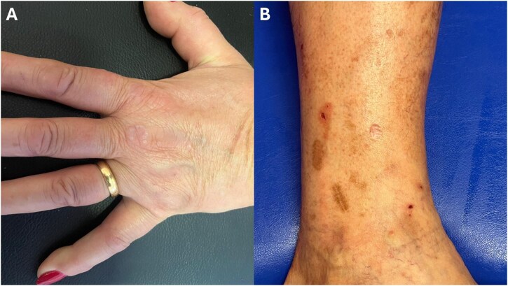 Figure 1
|
| |
|---|---|
| Male sex | 213 (41.3) |
| Ethnic origin | |
| Arab | 50 (9.9) |
| Asian | 6 (1.2) |
| Black | 7 (1.4) |
| Hispanic | 45 (8.9) |
| Jewish | 1 (0.2) |
| Native American | 1 (0.2) |
| White | 390 (77.5) |
| Other | 3 (0.6) |
| Age at disease onset (years), mean (S.D.) | 31.93 (17.39) |
| Type of Still’s disease | |
| Chronic-articular | 101 (21.6) |
| Monocyclic | 162 (34.7) |
| Polycyclic | 137 (29.3) |
| Undefined course | 67 (14.3) |
| Higher body temperature reached during the first episode (°C) (mean (S.D.)) | 39.35 (2.01) |
| Skin manifestations | |
| Salmon-coloured evanescent skin rash | 304 (63.9) |
| Pruritus associated to skin lesions | 104 (29.6) |
| Other skin manifestations | 122 (25.8) |
| Macules | 40 (7.7) |
| Erythema | 27 (5.2) |
| Persistent pruritic papules and/or plaques (PPPP) | 25 (4.8) |
| Urticarial eruptions | 31 (5.9) |
| Pustular lesions on the extremities and the trunk | 4 (0.8) |
| Lichenoid papules | 3 (0.6) |
| Alopecia | 3 (0.6) |
| Pigmented plaques | 3 (0.6) |
| Eczema-like lesions | 2 (0.4) |
| Prominent linear dermographism‐like lesions | 2 (0.4) |
| Vesiculopustular eruptions | 2 (0.4) |
| Acneiform lesions | 2 (0.4) |
| Erythematous maculopapules mimicking Gottron papules | 2 (0.4) |
| Insect bite-like papulonodules | 1 (0.2) |
| Erythema chronicum migrans | 1 (0.2) |
| Erysipelas-like | 1 (0.2) |
| Oedema of the eyelids | 1 (0.2) |
| Flagellate erythema-type manifestations | 1 (0.2) |
| Prurigo pigmentosa-like rash | 1 (0.2) |
| Variables | <16 years | 16–60 years | >60 year |
| Effect size |
|---|---|---|---|---|---|
|
|
|
|
| ||
| Male, | 41 (48.2) | 144 (39.3) | 16 (41.0) | 0.068 | |
| Still subtype, | |||||
| Chronic-articular | 21 (25.3) | 67 (19.5) | 12 (34.3) | 0.089 | 0.102 |
| Monocyclic | 30 (36.1) | 122 (35.6) | 9 (25.7) | 0.491 | 0.055 |
| Polycyclic | 22 (26.5) | 107 (31.2) | 6 (17.1) | 0.182 | 0.086 |
| Cutaneous Still manifestations | |||||
| Salmon-coloured evanescent skin rash, | 65 (80.2) | 212 (60.2) | 23 (62.2) | 0.003 | 0.156 |
| Atypical skin manifestations, | 15 (18.5) | 99 (28.4) | 6 (17.1) | 0.090 | 0.102 |
| Macules, | 5 (5.9) | 34 (9.3) | 1 (2.6) | 0.242 | 0.076 |
| PPPP, | 2 (2.4) | 22 (6.0) | 1 (2.6) | 0.291 | 0.071 |
| Erythema, | 1 (1.2) | 24 (6.6) | 2 (5.1) | 0.146 | 0.089 |
| Pruritus, | 10 (14.7) | 85 (33.2) | 7 (30.4) | 0.012 | 0.160 |
| Variables | Arab | White | Hispanic | Other |
| Effect size |
|---|---|---|---|---|---|---|
|
| 50 | 390 | 45 | 18 | ||
| Male, | 15 (30.0) | 171 (44.0) | 20 (44.4) | 3 (16.7) | 0.038 | 0.129 |
| Age at disease onset, mean (S.D.), years | 28.11 (12.20) | 31.98 (18.58) | 34.55 (10.83) | 32.11 (16.33) | 0.334 | 0.007 |
| Still subtype, | ||||||
| Chronic-articular | 10 (20.8) | 81 (23.4) | 2 (4.7) | 6 (33.3) | 0.025b,c | 0.144 |
| Monocyclic | 25 (52.1) | 113 (32.7) | 15 (34.9) | 4 (22.2) | 0.040 | 0.135 |
| Polycyclic | 3 (6.2) | 102 (29.5) | 23 (53.5) | 5 (27.8) | <0.001 | 0.232 |
| Cutaneous Still manifestations | ||||||
| Salmon-coloured evanescent skin rash, | 37 (77.1) | 224 (63.5) | 22 (50.0) | 10 (58.8) | 0.059 | 0.127 |
| Atypical skin manifestations = yes (%) | 9 (18.4) | 86 (24.4) | 19 (44.2) | 6 (33.3) | 0.020 | 0.146 |
| Macules, | 3 (6.0) | 25 (6.4) | 9 (20.0) | 3 (16.7) | 0.006 | 0.156 |
| PPPP, | 1 (2.0) | 12 (3.1) | 8 (17.8) | 4 (22.2) | <0.001 | 0.248 |
| Erythema, | 2 (4.0) | 22 (5.6) | 3 (6.7) | – | 0.703 | 0.053 |
| Pruritus, | 3 (7.7) | 76 (29.3) | 18 (56.2) | 5 (38.5) | <0.001 | 0.243 |
| Variables | Pouchot < 7 | Pouchot ≥ 7 |
| Effect size |
|---|---|---|---|---|
|
| 340 | 96 | ||
| Male, | 136 (40.0) | 39 (40.6) | 1.000 | 0.005 |
| Age at disease onset, mean (S.D.), years | 32.14 (17.32) | 32.57 (17.94) | 0.831 | 0.001 |
| Still subtype, | ||||
| Chronic-articular | 78 (24.1) | 15 (16.0) | 0.127 | 0.071 |
| Monocyclic | 111 (34.3) | 32 (34.0) | 1.000 | 0.002 |
| Polycyclic | 87 (26.9) | 32 (34.0) | 0.219 | 0.067 |
| Cutaneous Still manifestations | ||||
| Salmon-coloured evanescent skin rash, | 198 (58.8) | 80 (84.2) | <0.001 | 0.220 |
| Atypical skin manifestations, | 87 (26.0) | 23 (24.5) | 0.872 | 0.014 |
| Macules, | 30 (8.8) | 10 (10.4) | 0.782 | 0.023 |
| PPPP, | 18 (5.3) | 6 (6.2) | 0.913 | 0.017 |
| Erythema, | 20 (5.9) | 4 (4.2) | 0.691 | 0.031 |
| Pruritus, | 71 (29.7) | 23 (28.0) | 0.885 | 0.016 |
Peer Reviews
No public reviews on file for this paper yet. If you reviewed it on a platform where reviews are public (OpenReview, ICLR, NeurIPS, ICML), you can paste yours below so the community can read it here.
Videos
No videos yet. Explain this paper in a talk, walkthrough, or lecture? Add one.
Taxonomy
TopicsAutoimmune and Inflammatory Disorders Research · Immune Cell Function and Interaction · Inflammasome and immune disorders
Introduction
Still’s disease is a systemic autoinflammatory disease more frequently affecting children and young adults [1]. Fever, arthralgia, arthritis, lymphadenopathy, splenomegaly, liver involvement and cutaneous eruption account for the commonest signs observed in affected patients. The course of the disease is classically divided into a systemic and a chronic-articular subtype and can be complicated by the onset of macrophage activation syndrome (MAS), characterized by excessive activation of macrophages and T-lymphocytes, with overwhelming systemic inflammation and potentially fatal multi-organ failure [2, 3]. Other potentially life-threatening complications encompass acute respiratory distress syndrome (ARDS), fulminant hepatitis and cardiovascular complications such as myocarditis and pericarditis [4].
The hallmark cutaneous manifestation of Still’s disease is a transient, salmon-coloured maculopapular rash that typically occurs during febrile episodes [5]. The appearance of the rash can be fleeting, frequently resolving within a few hours, which can pose challenges for clinical observation. In addition to the classic salmon-coloured rash, patients with Still’s disease may present with other atypical skin manifestations, the most common being persistent pruritic papules and plaques (PPPP) [6]. The occurrence of atypical skin lesions is well documented in adult-onset Stills disease (AOSD) and has been occasionally described in systemic juvenile idiopathic arthritis (sJIA) [7]. Given the plethora of skin manifestations in Still’s disease, recognizing them is crucial for dermatologists to achieve an accurate diagnosis and effective management, as atypical skin lesions can often lead to misdiagnosis. Indeed, due to the rarity of the disease, large observational studies on a wide number of patients with Still’s disease, particularly focusing on skin manifestations, are lacking. The aim of this study was to thoroughly explore a large international cohort of patients with Still’s disease, with a focus on skin lesions, and to correlate these with patients’ ethnicities, age at onset, disease course and severity, as well as with the onset of specific clinical complications.
Methods
Study design and participants
This study was based on data from the International AutoInflammatory Disease Alliance (AIDA) Network Registry dedicated to Still’s disease [8]. Data were retrospectively collected. The enrolment period started in July 2021; we retrospectively extrapolated information on 518 Still’s disease patients up to January 2024. Still’s disease was classified according to internationally accepted criteria proposed by Yamaguchi et al. and/or Fautrel et al. for adult patients [9, 10]; the International League of Associations for Rheumatology (ILAR) and/or Pediatric Rheumatology International Trials Organization (PRINTO) criteria were employed for patients aged <16 years [11, 12].
Age, sex, ethnic origin, age at disease diagnosis and onset, clinical features, and patterns of the disease, cutaneous manifestations, the occurrence of life-threatening complications such as MAS were collected. Three clinical patterns (monocyclic, polycyclic and chronic) of the disease course were considered. A monocyclic course was defined as a single episode lasting >2 months but <1 year, followed by sustained remission through the entire follow-up period. A polycyclic course was characterized by recurrent systemic flares with remission between inflammatory episodes. A chronic course was defined as a disease pattern characterized by persistent disease activity with prominent joint involvement and less intense systemic inflammatory manifestations [13]. The MAS was defined according to either the 2016 classification criteria proposed by Ravelli et al. [14], and/or the HLH-2004 criteria developed by the Histiocyte Society [15], and/or the HScore, which has been developed and validated for the diagnosis of reactive HLH in both rheumatologic and non-rheumatologic conditions [15].
Cutaneous manifestations of the disease were documented, including the characteristic salmon-coloured skin rash, PPPP as the most frequent atypical presentation of Still’s disease, and a variety of less common atypical skin lesions, ranging from flagellate erythema to vesiculopustular eruptions. Moreover, the presence of pruritus as a symptom was assessed. Additional clinical characteristics recorded included: fever, sore throat, arthralgia or arthritis, myalgia, lymphadenopathy, splenomegaly, hepatomegaly, or liver dysfunction, abdominal or thoracic pain, pleuritis, pericarditis and ocular, renal, neurological and cardiac involvement. Skin lesions were considered as atypical cutaneous manifestations of Still’s disease if occurring during active phases of the disease, characterized by the presence of at least fever, hepatosplenomegaly, lymphadenopathy or severe joint involvement, accompanied by markedly increased ESR, CRP and ferritin levels. Alternatively, skin manifestations were considered atypical if histopathological examination demonstrated features consistent with predominant neutrophilic infiltration. The severity of the disease was assessed using the Pouchot score, with a threshold of 7 [16, 17]. It assigns points to fever, rash, pleuritis, pneumonia, pericarditis, hepatomegaly or abnormal liver function tests, splenomegaly, lymphadenopathy, leukocytes ≥15 000 mm^3^, sore throat, myalgia and abdominal pain as key clinical and laboratory findings, with each item worth 1 point. Conversely, the Rau score, also known as modified Pouchot score, has been proposed and employed to assess disease activity, with a threshold of 4 between active and inactive disease [18]. It focuses on a slightly different range of systemic features and inflammatory parameters, as it incorporates serum ferritin levels ≥3000 µg/l and arthritis, replacing splenomegaly and abdominal pain. The term ‘liver involvement’ was defined as the presence of abnormal liver function and/or hepatomegaly, as identified by US and/or other radiological documentation. Fulminant hepatitis was defined as the rapid onset of acute liver failure characterized by severe hepatic dysfunction, coagulopathy and hepatic encephalopathy in a patient with no pre-existing liver disease [19].
The aim of the study was to investigate cutaneous manifestations in a large international cohort of patients with Still’s disease, evaluating their correlation with ethnic origin, age at disease onset, patterns of the disease, life-threatening complications such MAS, Pouchot score and modified Pouchot score.
Protocol approval
The study was approved by the Ethics Committee of the University Hospital of Siena, Siena, Italy (Protocol Number 14951) as part of the AIDA Program. The study protocol conformed to the tenets of the Helsinki Declaration. Written informed consent to participate in the international AIDA Registry for Still’s disease patients was obtained from all patients and/or their legal guardians.
Statistical analysis
Descriptive statistics included mean and standard deviation (S.D.) for continuous variables, while frequency and percentages were reported for qualitative variables. To determine whether the data distribution was normal or not, the Shapiro–Wilk test was employed. Chi-squared test, Fisher’s exact test and Student’s t test were performed to compare two groups. To evaluate the difference of quantitative variables between age ranges and ethnicities, ANOVA test was used. As post hoc analysis of ANOVA, the Tuckey procedure was assessed. Instead, multiple Fisher exact tests with false discovery rate correction were performed as post hoc of the chi-squared test. The P-values reported for the post hoc analysis have already been adjusted for multiple comparisons.
Effect sizes were calculated to complement statistical significance testing and to provide an estimate of the strength of associations. For categorical variables, Cramer’s V was used, while for continuous variables analysed via ANOVA, Eta-squared was reported. Both statistics range from 0 to 1, with values closer to 1 indicating stronger associations and 0 representing no association. The interpretation of Cramer’s V depends on the size of the contingency table: for 2 × 2 or 3 × 2 contingency tables, values of 0.30 or higher are indicative of a strong association, while larger tables require higher thresholds (≥ 0.50 for 4 × 2 tables) to denote strong associations. Regarding Eta-squared, values ≥0.14 were interpreted as indicative of a strong difference.
A P < 0.05 was considered statistically significant. All data were assessed using software version R 4.1.0 (RStudio Team, 2023; Boston, MA, USA: Posit Software, PBC).
Results
Distribution of Still’s disease manifestations
A total of 518 patients (41.3% males) were included in the present study. Demographic data and manifestations referring to Still’s disease onset, including cutaneous manifestations, are summarized in Table 1. The frequency of extracutaneous signs observed up to the time of the enrolment is listed in Supplementary Table S1.
Overall, 390 (77.5%) patients were White, 50 (9.9%) Arabian, 45 (8.9%) Hispanic and 18 (3.4%) recorded as ‘others’ (including Asians, Black, Jewish, Native Americans). The mean age at disease onset was 31.93 ± 17.39 years. Regarding skin manifestations, 304 (63.9%) patients presented with a salmon-coloured evanescent skin rash. In addition, macules were the most common cutaneous findings in 40 (7.7%) patients, followed by urticarial eruptions (5.9%), erythema (5.2%) and PPPP (4.8%). Less common manifestations were represented by pigmented plaques (0.6%), erythematous maculopapular lesions mimicking Gottron papules (0.4%), prominent linear dermographism‐like lesions (0.4%), flagellate erythema (0.2%) and prurigo pigmentosa-like rash (0.2%). Finally, pruritus was associated with skin lesions in 29.6% of cases. Arthralgia (84.5%) and arthritis (57%), splenomegaly (36.1%) and liver involvement (35%), lymphadenopathy (50.2%) and sore throat (56.4%) represented the most common extracutaneous manifestations at disease onset. Overall, 101 (21.6%) patients presented with a chronic-articular pattern of Still’s disease, 162 (34.7%) showed a monocyclic course and 137 (29.3%) patients had a polycyclic one, while 67 (14.3%) were not classified.
Distribution of skin manifestations among different patient groups
Considering age at disease presentation, we noted that the occurrence of a salmon-coloured evanescent skin rash was more common in the <16 age group (80.2%) compared with the 16–60 (60.2%) and >60 (62.2%) age groups; however, the statistical significance was achieved only comparing the <16 age group with the 16–60 group (P-adjusted = 0.002). With regard to pruritus, it was more frequently observed in the 16–60 (33.2%) and >60 (30.4%) age groups compared with the <16 group (14.7%), with statistical significance being observed in the 16–60 group vs the <16 group (P-adjusted = 0.008) (Table 2).
Regarding ethnicities, Hispanics showed a significantly higher rate of atypical skin manifestations (44.2%) compared with Arabs (18.4%, P-adjusted = 0.036) and White (24.4%, P-adjusted = 0.036). Also, Hispanic patients showed a significantly higher prevalence of macules (20.0%) compared with White (6.4%, P-adjusted = 0.027). Consistently, PPPP were significantly more common in Hispanics (17.8%) and in the ‘other’ ethnicity group (22.2%) vs Arabs (2%) and White (2%) (Hispanic vs Arabs: P-adjusted = 0.023; ‘other’ group vs Arabs: P-adjusted = 0.023; Hispanic vs White: P-adjusted = 0.002; ‘other’ group vs White: P-adjusted = 0.011). Finally, a higher percentage of Hispanic patients (56.2%) reported pruritus associated with skin lesions compared with Arabs (7.7%, P-adjusted < 0.001) and White (29.3%, P-adjusted = 0.008). Moreover, the frequency of pruritus among Arabs was significantly lower compared with White (P-adjusted = 0.008) and the ‘other’ ethnicity group (38.5%, P-adjusted = 0.026) (Table 3). Figure 1 shows two examples of atypical cutaneous manifestations of Still’s disease.
Two examples of atypical cutaneous manifestations of Still’s disease. (A) Slightly erythematous, annular plaque with a moderately firm, rope-like border and central clearing on the dorsal aspect of the third metacarpophalangeal joint of the left hand. Discrete 1–2 mm papules are noted at the periphery of the lesion. The lesion closely resembles the granuloma annulare. (B) Pigmented brown macules distributed on the dorsal aspect of the right leg. The lesions are flat, assume a linear morphology and are not pruritic
When analysing disease patterns, no specific cutaneous manifestation of Still’s disease was significantly different in any disease course. The frequency of atypical cutaneous manifestations was higher in the chronic-articular pattern (32.3%) compared with the monocyclic (23.2%) and polycyclic (26.1%) patterns, but statistical significance was not achieved (P = 0.275). Moreover, pruritus was observed more frequently in the polycyclic group (36.9%) than in the chronic-articular (33.8%) and monocyclic (21.6%) groups, with a statistically significant difference between polycyclic vs monocyclic (P-adjusted = 0.049) groups (Supplementary Table S2).
Demographic, clinical and cutaneous manifestations of Still’s disease contributing to the Pouchot score at baseline are listed in Table 4. A significantly higher percentage of patients with Pouchot score ≥7 during the first febrile episode had a salmon-coloured evanescent skin rash (84.2%) compared with those with Pouchot score <7 (58.8%) (P < 0.001). This finding was still significant when the Rau score was considered (P < 0.05) (Supplementary Table S3).
No statistically significant association between the occurrence of MAS and the presence of salmon-coloured evanescent skin rash or other skin manifestations was observed (P > 0.05) (Supplementary Table S4).
Discussion
Recognizing Still’s disease based on its cutaneous manifestations remains critical among dermatologists, especially due to the wide range of different atypical skin manifestations. This lack of familiarity can result in diagnostic delays, which may contribute to potential complications. Early detection is essential for timely intervention, as it helps prevent the progression of systemic involvement and reduces the risk of severe outcomes. Based on current evidence, AOSD might be characterized by a wide range of dermatological manifestations, including salmon-coloured rash, PPPP, and various other eruptions such as prurigo pigmentosa-like lesions, vesiculopustular lesions or flagellate erythema [20]. In contrast, the skin manifestations in sJIA are generally more uniform, often limited to the characteristic evanescent salmon-coloured rash, and less varied in terms of morphology and distribution [21]. For this reason, only the typical rash of Still’s disease has been attributed to this disorder for a long timeframe, with atypical presentations misleading clinicians when facing with the diagnosis.
To date, some studies in the literature have explored the cutaneous manifestations of Still’s disease and their correlation with other clinical features. However, these studies have generally been conducted on relatively small patient cohorts. In this perspective, Ruscitti et al. analysed a cohort of 100 AOSD patients, correlating various clinical manifestations to disease progression patterns (monocyclic, polycyclic and chronic). The typical skin rash was found to be more frequent in patients with a ‘monocyclic’ and ‘chronic-articular’ course, even though a statistical significance was not achieved [16].
Regarding any association between skin rash and the development of MAS, the results observed in the present study are in line with what observed in a further study on 119 patients by Ruscitti et al. [22]. In particular, no association was found between the typical skin rash and the occurrence of MAS. However, while Ruscitti and colleagues only addressed cutaneous manifestations in terms of ‘skin rash’, referring to the typical one, this current study explores the full spectrum of AOSD skin lesions, confirming a lack of association with MAS.
At current, only a few studies have specifically focused on the various types of cutaneous lesions possibly associated with AOSD. In detail, Sato et al. analysed typical and atypical skin lesions and their frequency in a cohort of AOSD 28 patients [21]. They correlated the various types of lesions to the age at disease onset, distinguishing between patients younger and older than 65 years. As in our study, they found a significantly higher prevalence of typical rash in younger patients compared with older ones. Conversely, among the atypical skin rashes, PPPP was more common in patients over 65 years old. However, this last finding was not confirmed in our study, where none of the atypical skin lesions showed a significant association with the age of AOSD onset.
In the study by Narváez Garcia et al., typical and atypical cutaneous manifestations were analysed in 81 patients deriving from case reports and series, evaluating their impact on prognosis and mortality of AOSD. They observed that the development of atypical rash was associated with a worse prognosis due to a higher mortality rate compared with patients with typical rash, as well as with a Pouchot score ≥7 [23]. Nevertheless, our study did not confirm this finding. Indeed, typical rash was significantly associated with greater disease severity, according to the Pouchot score. Conversely, no statistically significant association was found between atypical cutaneous manifestations and higher or lower severity scores, whether according to Pouchot or Rau scores.
A further study from Eveillard et al. investigated the spectrum of skin involvement of sJIA. These authors found that sJIA patients with atypical skin presentations were more likely to experience long-term, persistent disease, requiring more intensive or prolonged treatments [7].
Interestingly, no studies have explored the cutaneous manifestations of Still’s disease across different ethnic groups so far. This represents a significant gap in our understanding of the disease and investigating these variations could provide valuable insights into the Still’s disease’s behaviour and progression in diverse ethnic backgrounds, potentially leading to more tailored and effective diagnostic and treatment approaches. In our study, no statistically significant differences were found in the frequency of typical rash across different ethnic groups. However, atypical cutaneous variants were significantly more frequent among Hispanics compared with other White and Arabs. When analysing specific atypical cutaneous eruptions, it was observed that macules were significantly more frequent in Hispanics compared with other White. Furthermore, PPPP were significantly more prevalent in the Hispanic and ‘other’ ethnic groups, in comparison to Arabs or White. Noteworthy, pruritus was significantly more commonly reported among Hispanics than in Arabs or White. It is known that the clinical presentation of chronic inflammatory skin diseases, such as atopic dermatitis, can vary significantly among ethnic groups due to genetic, environmental and cultural factors, as well as differing immunological pathways [24]. It is plausible that this concept also applies to Still’s disease, where the heterogeneity of cutaneous manifestations across different ethnicities may reflect distinct immunopathogenic mechanisms driven by diverse genetic backgrounds. In this context, both HLA and non-HLA genes, as well as various immune signalling pathways, are known to play a role in the pathogenesis of the disease [25]. Although to the best of our knowledge ethnicity-specific genetic studies have not yet been conducted in patients with Still’s disease, evidence from other immune-mediated disorders indicates that allele frequencies and genetic susceptibility factors can differ significantly among populations [26]. These variations may contribute to differential immune responses, potentially accounting for the observed diversity in cutaneous involvement. However, specific genetic studies are needed to explore and eventually confirm this hypothesis.
The main strength of this work relies on the large cohort of Still patients retrieved from an international registry. Nonetheless, some limitations need to be recognized. Firstly, the retrospective nature of the study accounts for some missing data, along with its inherent limitations. Secondly, a comparison between scientific studies on Still’s disease was complicated by the heterogeneity of methods employed in the literature and the lack of a consistent body of evidence on cutaneous manifestations of Still’s disease.
Conclusion
In conclusion, various studies have analysed the different patterns of Still’s disease, clinical manifestations, the development of complications, as well as the age at disease onset, correlating these factors with patient prognosis so far. However, very few studies explored the wide range of cutaneous manifestations of Still’s disease, dissecting them in typical and atypical and correlating them to specific patient and disease characteristics.
Comprehensively, the present study enhances dermatologists’ awareness of the diverse cutaneous lesions that may represent heterogeneous manifestations of Still’s disease.
Supplementary Material
keaf512_Supplementary_Data
The reference list from the paper itself. Each links out to its DOI / PubMed record.
- 1Seco T , Cerqueira A, Costa A et al Adult-onset Still’s disease: typical presentation, delayed diagnosis. Cureus 2020;12:e 8510.32656026 10.7759/cureus.8510 PMC 7346332 · doi ↗ · pubmed ↗
- 2Li R , Liu X, Chen G et al Clinical phenotypes and prognostic factors of adult-onset Still’s disease: data from a large inpatient cohort. Arthritis Res Ther 2021;23:300.34879864 10.1186/s 13075-021-02688-4PMC 8653615 · doi ↗ · pubmed ↗
- 3Efthimiou P , Kontzias A, Hur P et al Adult-onset Still’s disease in focus: clinical manifestations, diagnosis, treatment, and unmet needs in the era of targeted therapies. Semin Arthritis Rheum 2021;51:858–74.34175791 10.1016/j.semarthrit.2021.06.004 · doi ↗ · pubmed ↗
- 4Mitrovic S , Fautrel B. Complications of adult-onset Still’s disease and their management. Expert Rev Clin Immunol 2018;14:351–65.29658384 10.1080/1744666 X.2018.1465821 · doi ↗ · pubmed ↗
- 5Tomaras S , Goetzke CC, Kallinich T et al Adult-onset Still’s disease: clinical aspects and therapeutic approach. J Clin Med 2021;10:733. doi: 10.3390/jcm 10040733 PMC 791855033673234 · doi ↗ · pubmed ↗
- 6Kikuchi N , Satoh M, Ohtsuka M et al Persistent pruritic papules and plaques associated with adult-onset Still’s disease: report of six cases. J Dermatol 2014;41:407–10.24628100 10.1111/1346-8138.12426 · doi ↗ · pubmed ↗
- 7Eveillard LA , Quartier P, Ouldali N et al; Société Francophone pour la rhumatologie et les Maladies Inflammatoires en Pédiatrie (SOFREMIP). Association of atypical skin manifestations at the onset of systemic juvenile idiopathic arthritis with difficult-to-treat disease: a retrospective multicenter study. J Am Acad Dermatol 2022;87:1425–8.35963289 10.1016/j.jaad.2022.07.055 · doi ↗ · pubmed ↗
- 8Vitale A , Della Casa F, Lopalco G et al Development and implementation of the AIDA international registry for patients with Still’s disease. Front Med (Lausanne) 2022;9:878797.35463015 10.3389/fmed.2022.878797 PMC 9021753 · doi ↗ · pubmed ↗