A rare cytogenetically cryptic MECOM rearrangement in a patient with myelodysplastic neoplasm and SF3B1 mutation identified by RNA sequencing: a case report
Ye Jin, Zi-Jun Xu, Chao-Ran Lv, Zhen Qian, Xiang-Mei Wen, Sheng Xiao, Jiang Lin, Jun Qian

TL;DR
A rare MECOM rearrangement in a patient with a myelodysplastic neoplasm and SF3B1 mutation was identified using RNA sequencing, highlighting the importance of advanced diagnostics in leukemia.
Contribution
This case report identifies a rare MBNL1::MECOM rearrangement in SF3B1-mutated MDS, emphasizing the need for RNA-seq in such patients.
Findings
A rare MBNL1::MECOM rearrangement was identified in a patient with myelodysplastic neoplasm and SF3B1 mutation.
The rearrangement was undetected by conventional cytogenetics but confirmed by NGS and FISH.
High EVI1 expression was observed, consistent with mechanisms in atypical MECOM rearrangements.
Abstract
MECOM (the MDS1 and EVI1 complex locus) rearrangements have been identified as an independent high-risk factor in acute myeloid leukemia (AML). The diversity of MECOM rearrangement partner genes significantly influences disease mechanisms and prognosis. The majority of atypical MECOM rearrangements result in EVI1 overexpression through translocation into super-enhancer-containing regions. This report describes a rare, recurrent MBNL1::MECOM rearrangement identified in a myelodysplastic neoplasm (MDS) patient with a concurrent SF3B1 mutation. Although conventional cytogenetics showed a normal karyotype, the rearrangement was confirmed by next-generation sequencing (NGS) and fluorescence in situ hybridization (FISH). Concurrently, the patient exhibited high EVI1 expression, consistent with the common mechanism observed in atypical MECOM rearrangements. Given the well-documented…
Genes, proteins, chemicals, diseases, species, mutations and cell lines named across the full text — each resolved to its canonical identifier and authoritative record.
Click any figure to enlarge with its caption.
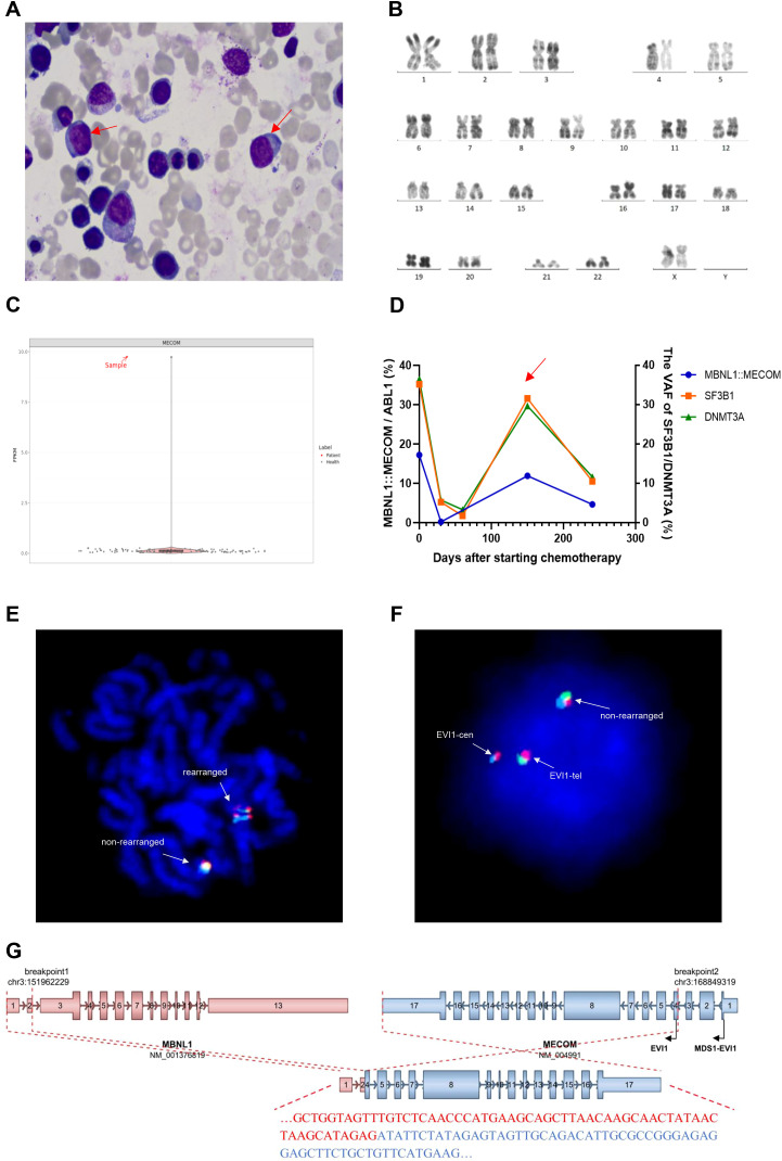 Figure 1
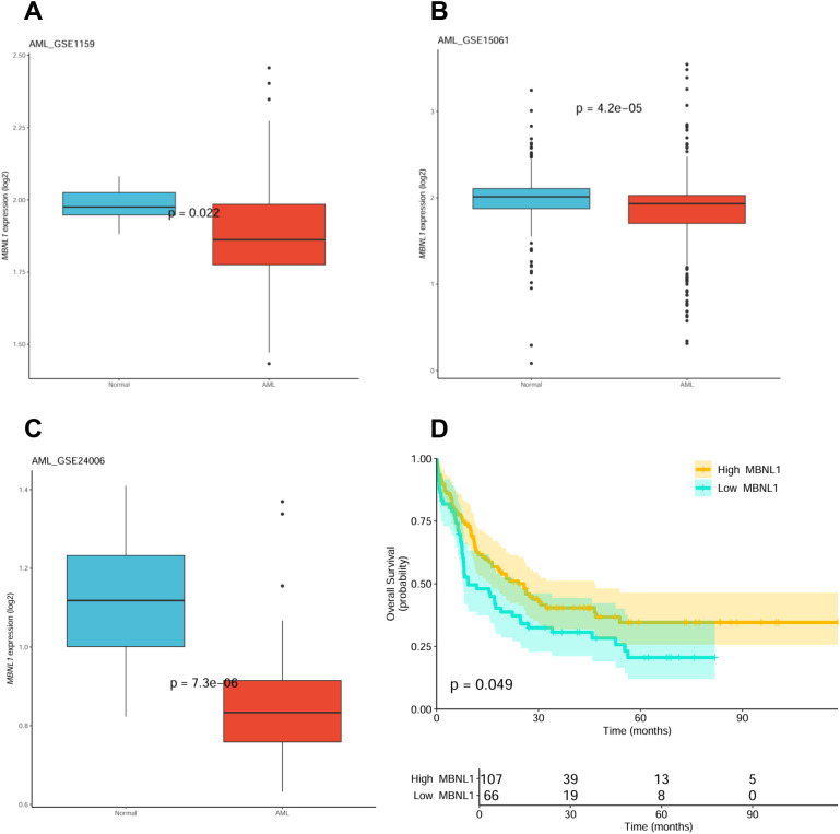 Figure 2
Peer Reviews
No public reviews on file for this paper yet. If you reviewed it on a platform where reviews are public (OpenReview, ICLR, NeurIPS, ICML), you can paste yours below so the community can read it here.
Videos
No videos yet. Explain this paper in a talk, walkthrough, or lecture? Add one.
Taxonomy
TopicsAcute Myeloid Leukemia Research · Blood disorders and treatments · Multiple Myeloma Research and Treatments
Introduction
Acute myeloid leukemia (AML) with MECOM (the MDS1 and EVI1 complex locus) rearrangement is recognized as a distinct entity in the 2008 World Health Organization (WHO) classification of hematolymphoid tumors (1). Patients with this subtype exhibit rapid disease progression and poor prognosis, achieving complete remission (CR) rates of only 15-20% with conventional chemotherapy, a median survival time of 4–6 months, and a 5-year overall survival rate of less than 15% (2). The classical MECOM rearrangements, derived from inv(3)(q21;q26) or t(3;3)(q21;q26), lead to the GATA2 enhancer (3q21) adjacent to MECOM (3q26), driving EVI1 overexpression and monoallelic expression of GATA2 (3). EVI1, first identified in retrovirus-induced murine AML models, plays a crucial role in hematopoiesis and leukemogenesis. Its aberrant overexpression is frequently observed in myeloid malignancies, including AML (4). Functionally, EVI1 regulates erythroid, granulocytic, and megakaryocytic differentiation. Studies demonstrate that it contributes to hematopoietic neoplasia through transcriptional repression, epigenetic dysregulation, and interactions with pathways such as TGF-β/SMAD and PI3K/AKT (5). EVI1 exists normally in the form of MDS1-EVI1 and is lowly expressed in normal bone marrow. Rearrangements often disrupt the region between exon 1 of MDS1 and exon 1 of EVI1, leading to activated transcription of EVI1 (6, 7). Beyond the classical inv(3)/t(3;3), a series of atypical MECOM rearrangements have been identified. Chromosomal 3q26 translocations in these AML cases frequently involve super-enhancer hijacking genes active in myeloid development (e.g., CD164, PROM1, CDK6, MYC), thereby driving EVI1 overexpression and potential GATA2 involvement (8, 9). These patients also showed poor treatment response, with EVI1 overexpression but no or low MDS1-EVI1 levels (8, 9). Consequently, the latest WHO classification revises this subtype as AML with MECOM rearrangement even if peripheral blood and/or bone marrow blasts < 20% (10). To date, more than 30 additional 3q26 rearrangements have been identified (11). Here, we report a new cytopenic case with an MBNL1::MECOM rearrangement detected by next-generation sequencing (NGS).
Case report
A 60-year-old woman was admitted to the Affiliated People’s Hospital of Jiangsu University with progressive fatigue over three months. Physical examination revealed sternal tenderness and scattered purpura on the dorsolateral aspect of the right hand. Splenomegaly was present, palpable 6 cm below the costal margin, with a spleen volume of 268.68 cm^3^ measured by color Doppler ultrasonography. Hepatomegaly and lymphadenopathy were not detected. Peripheral blood examination demonstrated pancytopenia: white blood cell 2.32 × 10^9^/L, hemoglobin 8.1 g/dL, and platelet count 18 × 10^9^/L with 1% blasts, 3% promyelocytes and 2% normoblasts. Bone marrow smear showed 3% myeloblasts (Figure 1A). The percentage of erythroid cells increased to 64% accompanying megaloblastoid changes, scattered trinuclear erythrocytoblasts and internuclear bridging. Ring sideroblasts increased to 28%. Hypercellularity was found by bone marrow biopsy in which blasts were revealed as 2%~3% by CD34 immunohistochemistry. Cytogenetic analysis demonstrated a normal karyotype (Figure 1B). NGS on 318 genes associated with hematologic malignancies identified SF3B1 R625L mutation with 35.2% of variant allele frequency (VAF) and DNMT3A S894Efs27 mutation (VAF 36.5%). Whole-transcriptome RNA sequencing (RNA-seq) of bone marrow sample identified a rare MBNL1::MECOM fusion gene, accompanied by upregulated EVI1 expression (Figure 1C). Thus, this patient was diagnosed with AML with MECOM rearrangement according to the latest WHO classification (10, 11). Following two cycles of azacitidine (AZA) and venetoclax (VEN) therapy, the patient achieved complete remission with incomplete hematologic recovery (CRi). The MBNL1::MECOM transcript level decreased to 0.22%, and the VAFs of SF3B1 and DNMT3A mutations were both reduced to approximately 5% (Figure 1D). After four additional months of AZA+VEN treatment, the MBNL1::MECOM transcript level increased to 11.93%, and the VAFs of SF3B1 and DNMT3A mutations rose to around 30%, despite the absence of increased blast counts in peripheral blood or bone marrow. Persistent pancytopenia was present also the doses of AZA and VEN were reduced and deferred. The patient is still alive dependently on red blood cell transfusion.
(A) Wright-Giemsa staining of the bone marrow aspirate smear showed 3% myeloblasts (indicated by red arrows; ×1000 magnification). The percentage of erythroid cells was increased to 64% with dysplastic features, including megaloblastoid changes, scattered trinucleated erythroblasts, and internuclear bridging. Ring sideroblasts accounted for 28%. According to the World Health Organization (WHO) classification, these findings support a diagnosis of myelodysplastic syndrome with ring sideroblasts (MDS-RS). (B) R-banded karyotype analysis of the bone marrow specimen demonstrating a normal karyotype. (C) Comparison of MECOM expression levels between the patient and the healthy controls. (D) Minimal residual disease (MRD) levels were monitored in the patient’s bone marrow samples, quantifying the MBNL1::MECOM fusion transcript by Real-time Quantitative PCR (RQ-PCR) (ABL1 as the reference gene) and the SF3B1 and DNMT3A mutations by droplet digital PCR (ddPCR). At the time point indicated by the red arrow, the MRD levels showed a significant increase compared to preceding measurements, indicating the relapse of acute myeloid leukemia (AML). (E, F) Fluorescence in situ hybridization (FISH) was performed on the patient’s bone marrow sample using a tri-color break-apart MECOM (EVI1) probe (Kanglu Biotech Co., Ltd.). The blue, green and red probes label the centromeric region, the telomeric region, and the MECOM (EVI1) locus respectively. FISH confirmed MECOM rearrangement in both metaphase (E) and interphase (F) nuclei. This abnormal pattern was detected in 18% of the scored interphase cells. (G) Schematic diagram illustrating the structure and predicted functional consequence of the MBNL1::MECOM fusion. The breakpoints are located between exons 2 and 3 of MBNL1 and between exons 3 and 4 of MECOM. This disrupts the translation of the full-length MDS1-EVI1 transcript and permits translation initiation from the native EVI1 start codon, indicating that the fusion transcript does not encode a novel chimeric protein. Similar to other atypical MECOM rearrangements, the MBNL1::MECOM fusion likely drives leukemogenesis by hijacking the MBNL1 promoter to cause aberrant EVI1 overexpression.
Discussion
This is the third reported case harboring MBNL1::MECOM rearrangement. The two previously reported cases include one with chronic myeloid leukemia (CML) in blast crisis (BC) phase (CML-BC) and one with AML secondary to myelodysplastic neoplasm (MDS) (9, 12). Both cases were identified only by NGS, as fluorescence in situ hybridization (FISH) was not performed for these two patients. Moreover, detailed clinical data were not described either. This recurrent fusion of MBNL1::MECOM is derived from the juxtaposition of MBNL1 gene located on chromosome 3q25.1 and MECOM. The subtle inversion between 3q25.1 and 3q26.2 is difficult to detect by conventional karyotype analysis due to the adjacency of these two bands. The disease of this patient would have been misdiagnosed as MDS with SF3B1 mutation had RNA-seq not been performed, underscoring the critical role of RNA-seq in identifying such cryptic fusions. FISH assay, using the three-color break-apart probes spanning the MECOM gene, revealed a break-apart signal pattern in 18% of cells (Figures 1E, F), which confirms the MECOM rearrangement. The breakpoints were located between exons 2 and 3 of MBNL1 and between exons 3 and 4 of MECOM (Figure 1G). Analysis of the spliced sequence using the Expasy Translate tool (https://web.expasy.org/translate/) indicated that the fusion transcript does not encode a novel chimeric protein. Instead, it disrupts the translation of the full-length MDS1-EVI1 transcript, permitting translation initiation from the native EVI1 start codon (c.665–667 ATG, p.M189), effectively utilizing the MBNL1 promoter. Consistent with this mechanism, EVI1 overexpression was detected (Figure 1C). Therefore, we propose that similar to other atypical MECOM rearrangements, the MBNL1::MECOM fusion likely drives leukemogenesis by hijacking the MBNL1 promoter to cause aberrant EVI1 overexpression. It’s a pity that this study lacks direct functional evidence, such as assay for transposase-accessible chromatin with sequencing (ATAC-seq) or luciferase reporter assays, to conclusively prove that the MBNL1 promoter directly drives EVI1 expression in this context. Future experimental validation is required to clearly determine the mechanistic basis of this fusion.
In AML, the recurrent SF3B1 mutations are most frequently found in the subtypes of myelodysplasia-related AML (MDS-MR) (11). Several previous studies have shown that SF3B1 mutation is one of the most frequent co-mutations in myeloid neoplasms (9, 13–15). An outstanding biological question is the clonal evolutionary relationship between these two events. A recent study has further confirmed the strong association of SF3B1 mutation with MECOM rearrangements in AML (16). Among 41 AML patients with SF3B1 mutations, GATA2::MECOM was found in 10 cases (24%). In their 2 patients, the VAFs of SF3B1 mutation (both approximately 40%) and the percentages of MECOM rearrangement (both approximately 80%) suggest in the prior MDS stage that they exist in the same leukemic clone. In our patient, the proportion of cells harboring the SF3B1 mutation (35.2% VAF, corresponding to approximately 70.4% of cells) was significantly higher than the proportion of cells with the MECOM rearrangement (only 18% as detected by FISH). This finding suggests that the MBNL1::MECOM rearrangement may be a secondary event. Although the MBNL1::MECOM transcript level was dynamically monitored, concurrent FISH analysis was not performed at each time point. Consequently, two independent clones cannot be excluded definitely. Future studies involving serial sampling to track both the VAF and the percentage of rearranged cells, or to perform single-cell sequencing, are required to ultimately establish the relevance and time sequence of these two clones, thereby providing deeper mechanistic insights into disease progression.
SF3B1 mutation enhances the leukemogenicity of hematopoietic cells in inv(3) mice (14). Although the coexistence of SF3B1 mutation does not affect the prognosis of AML patients (17), elucidating the potential synergistic mechanisms between these two events is crucial, given the strong association between SF3B1 mutation and MECOM rearrangement as well as the adverse prognosis associated with MECOM rearrangement. These two abnormalities likely jointly exacerbate erythroid differentiation blockade and may form a self-amplifying positive feedback loop, thereby synergistically driving the initiation and progression of leukemia. First, both are closely associated with erythroid differentiation blockade. The SF3B1 mutation disrupts erythropoiesis by inducing mis-splicing of genes involved in ineffective erythropoiesis (18) and by causing premature downregulation of GATA1, which impairs terminal erythroid differentiation and leads to anemia (19). In contrast, EVI1 overexpression blocks normal erythropoiesis through its interaction with the transcription factor GATA1 (20). Second, the SF3B1 mutation can lead to aberrant splicing of EVI1 itself and accelerate leukemia development in vivo (14). The convergence of these mechanisms may have contributed to the aggressive disease biology in this case, which suggests a promising direction for future basic research.
The asynchronous changes of gene mutations, MECOM transcript and blasts suggest that SF3B1 mutation MECOM rearrangement are the successive events at preleukemic stage. However, MECOM FISH at relapse was not performed to compare the size of MECOM rearranged clone with blast percentage, unable to completely rule out the relevance of MECOM rearrangement with blasts if the percentage of positive FISH signals is also low.
Analysis of MBNL1 expression across AML datasets (GSE1159, GSE15061, GSE24006) reveals its significant downregulation compared to controls (Figures 2A-C), and The Cancer Genome Atlas (TCGA) data associates low MBNL1 expression with poor prognosis (Figure 2D). MBNL1 functions as a tumor suppressor and RNA metabolism regulator (21, 22). Its disruption via rearrangement, coupled with SF3B1 mutation, could synergistically exacerbate splicing errors, providing a compounded proliferative advantage to leukemic cells (23).
(A-C) Analysis of MBNL1 expression across three independent public datasets. Gene expression data from GSE1159 (A), GSE15061 (B), and GSE24006 (C) were analyzed separately. Box plots show MBNL1 expression in AML bone marrow samples compared to healthy donor (HD) controls, revealing significant downregulation of MBNL1 in AML. Data were normalized and log2-transformed within each dataset. Due to the independent nature of these datasets and the exploratory purpose of this cross-validation, no cross-dataset batch effect correction was applied. (D) Low MBNL1 expression is associated with inferior overall survival in AML. The Kaplan-Meier survival curve was generated from The Cancer Genome Atlas (TCGA) AML cohort (n=173). Patients were stratified by the median expression level of MBNL1. The log-rank p-value of 0.049 suggests a trend toward inferior prognosis with low MBNL1 expression in this univariate model.
Conclusion
In summary, we reported a rare recurrent MBNL1::MECOM rearrangement derived from inv(3)(q25.1; q26.2) in a cytopenic patient, accompanying SF3B1 mutation. Given the adverse prognosis associated with MECOM rearrangements, it is crucial to actively search for them in SF3B1-mutated patients using appropriate FISH or RNA-seq, even in the absence of cytogenetic clues or excessive blasts. As a single case report with inherent limitations and relatively short follow-up to date, we will continue monitoring this patient’s disease course. Furthermore, we anticipate that additional cases of MBNL1::MECOM or other rare MECOM partner rearrangements will be identified and reported, enhancing our understanding of this high-risk AML subgroup. Meanwhile, this case underscores the need for further research into the synergistic biological role of spliceosome mutations and MECOM rearrangements in driving leukemia.
The reference list from the paper itself. Each links out to its DOI / PubMed record.
- 1Swerdlow SH Campo E Harris NL Jaffe ES Pileri SA Stein H . WHO Classification of Tumours of Haematopoietic and Lymphoid Tissues. 4th. Lyon: IARC Press (2008).
- 2Gröschel S Lugthart S Schlenk RF Valk PJ Eiwen K Goudswaard C . High EVI 1 expression predicts outcome in younger adult patients with acute myeloid leukemia and is associated with distinct cytogenetic abnormalities. J Clin Oncol. (2010) 28:2101–7. doi: 10.1200/JCO.2009.26.0646, PMID: 20308656 · doi ↗ · pubmed ↗
- 3Gröschel S Sanders MA Hoogenboezem R de Wit E Bouwman BAM Erpelinck C . A single oncogenic enhancer rearrangement causes concomitant EVI 1 and GATA 2 deregulation in leukemia. Cell. (2014) 157:369–81. doi: 10.1016/j.cell.2014.02.019, PMID: 24703711 · doi ↗ · pubmed ↗
- 4Vriend J Delwel R Pastoors D . Mechanisms of enhancer-driven oncogene activation. Int J Cancer. (2026) 158:333–41. doi: 10.1002/ijc.35330, PMID: 39853740 PMC 12628035 · doi ↗ · pubmed ↗
- 5Liang B Wang J . EVI 1 in leukemia and solid tumors. Cancers (Basel). (2020) 12:2667. doi: 10.3390/cancers 12092667, PMID: 32962037 PMC 7564095 · doi ↗ · pubmed ↗
- 6Soderholm J Kobayashi H Mathieu C Rowley JD Nucifora G . The leukemia-associated gene MDS 1/EVI 1 is a new type of GATA-binding transactivator. Leukemia. (1997) 11:352–8. doi: 10.1038/sj.leu.2400584, PMID: 9067573 · doi ↗ · pubmed ↗
- 7Hinai AA Valk PJM . Review: Aberrant EVI 1 expression in acute myeloid leukaemia. Br J Haematol. (2016) 172:870–8. doi: 10.1111/bjh.13898, PMID: 26729571 · doi ↗ · pubmed ↗
- 8Ottema S Mulet-Lazaro R Beverloo HB Erpelinck C van Herk S van der Helm R . Atypical 3q 26/MECOM rearrangements genocopy inv(3)/t(3;3) in acute myeloid leukemia. Blood. (2020) 136:224–34. doi: 10.1182/blood.2019003701, PMID: 32219447 · doi ↗ · pubmed ↗