Mapping cellular vulnerability in Parkinson’s disease using retro-AAVs and preformed α-synuclein fibrils
Fanni F. Geibl, Ahmed A. S. Musa, Leo Dietrich, Helena Wolter, David L. Wokosin, Sharof Khudayberdiev, Marco B. Rust, Rong Chen, Valina L. Dawson, Ted M. Dawson, Wolfgang H. Oertel, D. James Surmeier, Martin T. Henrich

TL;DR
This study explores why some brain cells are vulnerable to Parkinson's disease pathology while others are not, using a mouse model and viral tracing techniques.
Contribution
The study identifies cell-autonomous factors influencing vulnerability to α-synuclein pathology in Parkinson's disease.
Findings
α-synuclein pathology spreads along anatomical pathways but does not affect all connected neurons equally.
Neurons with larger axonal arbors and higher mitochondrial oxidation levels are more vulnerable to α-synuclein accumulation.
Certain brain regions act as 'super-seeders', promoting widespread pathology propagation.
Abstract
Parkinson disease (PD) is characterized by progressive neuronal loss within defined brain regions, accompanied by α-synuclein (αSyn)-rich inclusions, termed Lewy pathology (LP). However, it is unclear which cellular factors render certain neuronal populations vulnerable, while others stay devoid of LP throughout the course of disease. This study aimed to identify and compare the cellular architecture of vulnerable and non-vulnerable neurons exposed to αSyn pathology by using a projection-based retro-AAV approach in combination with an in vivo α-synucleinopathy mouse model. To do so, a set of viral genetic, immunohistochemical, and optical tools was used in combination with the preformed αSyn fibril (PFF) model. αSyn pathology propagated robustly into the input connectome of the pedunculopontine nucleus (PPN). However, we observed a marked mismatch between the anatomically expected and…
Genes, proteins, chemicals, diseases, species, mutations and cell lines named across the full text — each resolved to its canonical identifier and authoritative record.
Click any figure to enlarge with its caption.
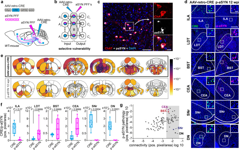 Figure 1
 Figure 2
 Figure 3
 Figure 4
Peer Reviews
No public reviews on file for this paper yet. If you reviewed it on a platform where reviews are public (OpenReview, ICLR, NeurIPS, ICML), you can paste yours below so the community can read it here.
Videos
No videos yet. Explain this paper in a talk, walkthrough, or lecture? Add one.
Taxonomy
TopicsParkinson's Disease Mechanisms and Treatments · Neurological disorders and treatments · Transcranial Magnetic Stimulation Studies