Circulating Tumor Cells in Uveal Melanoma: Multi-Marker Detection and Association With Disease State
Daniel P. de Bruyn, Fabiana Lucia Bassil, Mike Wu, Aaron B. Beasley, Jolanda Vaarwater, Mai N. Van, Jaco Kraan, Robert M. Verdijk, Dion Paridaens, Caroline M. van Rij, Nicole C. Naus, Annelies de Klein, Elin S. Gray, Erwin Brosens, Emine Kiliç

TL;DR
This study explores the use of circulating tumor cells in uveal melanoma patients as a non-invasive way to monitor disease progression and treatment response.
Contribution
The study introduces a multi-marker approach for detecting circulating tumor cells in uveal melanoma and shows its feasibility across disease stages.
Findings
CTC detection rates were higher in metastatic patients compared to those with localized disease.
CTC counts increased significantly during fractionated stereotactic radiotherapy.
Baseline CTC counts were not associated with tumor size or molecular risk class.
Abstract
Uveal melanoma (UM) is primarily treated with eye-sparing radiotherapy, leaving limited tumor tissue for molecular analysis. Circulating tumor cells (CTCs) may offer a minimally invasive alternative for genomic tumor profiling. This pilot study evaluated the feasibility of a multi-marker CTC capture approach in UM patients at diagnosis, during fractionated stereotactic radiotherapy (fSRT), and at metastatic progression. Patients with localized or metastatic UM were prospectively enrolled. Peripheral blood samples were collected at baseline, during fSRT, and on detection of metastases. CTCs were captured and enumerated using a multi-marker approach and fluorescence microscopy. A total of 76 patients were included: 68 with localized disease and eight with metastatic disease. Four patients presented initially with localized disease but developed metastasis during follow-up: for…
Genes, proteins, chemicals, diseases, species, mutations and cell lines named across the full text — each resolved to its canonical identifier and authoritative record.
Click any figure to enlarge with its caption.
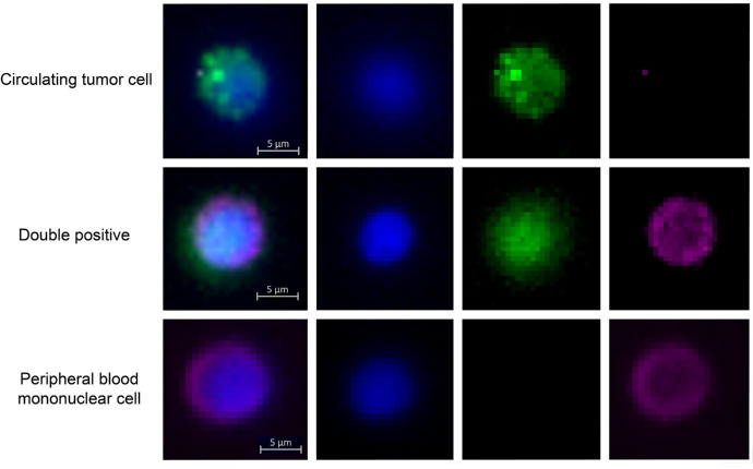 Figure 1
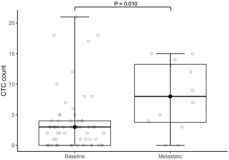 Figure 2
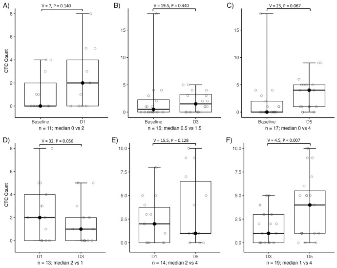 Figure 3
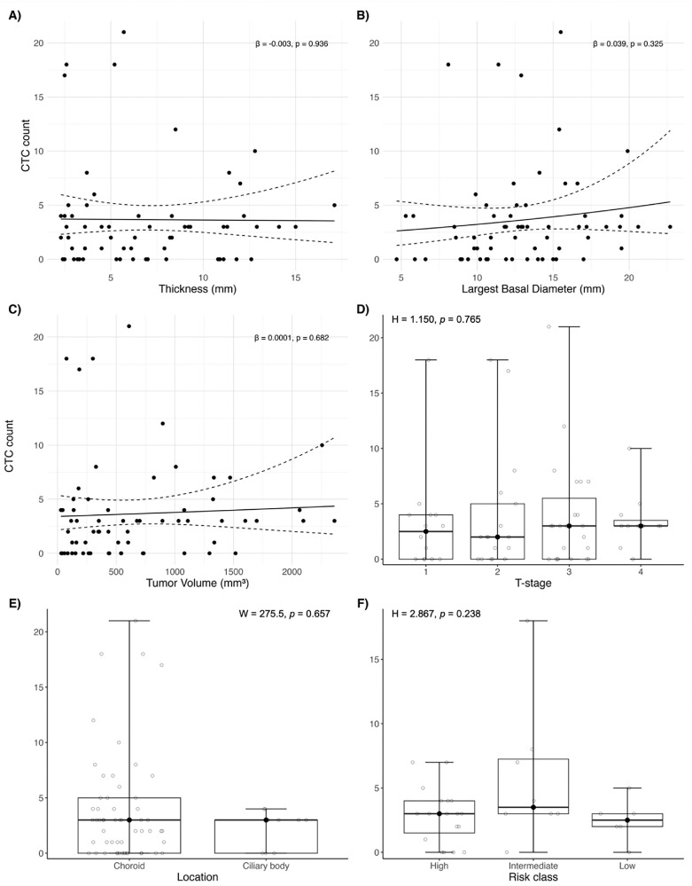 Figure 4
Peer Reviews
No public reviews on file for this paper yet. If you reviewed it on a platform where reviews are public (OpenReview, ICLR, NeurIPS, ICML), you can paste yours below so the community can read it here.
Videos
No videos yet. Explain this paper in a talk, walkthrough, or lecture? Add one.
Taxonomy
TopicsOcular Oncology and Treatments · Retinal Development and Disorders · Veterinary Oncology Research