Non‐Invasive, High‐Resolution (1H2O) Metabolic Activity Diffusion Imaging [MADI] of Rat Glioma
Joshua W. Schlegel, Samantha M. Holland, Felice D. Kelly, Eric M. Baker, Jared Stoller, William Packwood, Xin Li, Ramon F. Barajas, Charles S. Springer, Martin M. Pike

TL;DR
A new MRI technique called MADI measures water movement in rat brain tumors without contrast agents, offering high-resolution metabolic insights.
Contribution
MADI introduces a non-invasive method to quantify cellular water efflux and related metabolic parameters in brain tumors.
Findings
Tumor kioV values were significantly lower than contralateral regions, indicating reduced water efflux per cell.
MADI showed higher resolution and contrast compared to 18FDG-PET in detecting tumor metabolic activity.
Tumor glycolysis contributed minimally to overall energy production based on the water-glucose index (WGI).
Abstract
We have recently developed a metabolic activity imaging approach entitled Metabolic Activity Diffusion Imaging [MADI] which utilizes diffusion weighted MRI to quantify kio, the homeostatic cellular H2O efflux rate constant without the use of contrast agents, thus enabling measurement in both normal and tumor brain regions. Importantly, kio quantifies transmembrane water cycling, a significant proportion of which is coupled to Na+/K+ ATPase activity and associated cellular energy utilization, hence constituting a key metabolic biomarker. MADI also quantifies the cell volume (V), and cell density (ρ); these enable quantification of the kioV and kioVρ products, which convert the kio rate constant to rates of water efflux per cell (units: pL/s/cell) and per tissue (units: pL/s/uL [tissue]), respectively. Representing its first application to brain cancer, MADI was comprehensively evaluated…
Genes, proteins, chemicals, diseases, species, mutations and cell lines named across the full text — each resolved to its canonical identifier and authoritative record.
Click any figure to enlarge with its caption.
 Figure 1
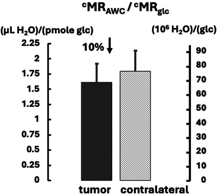 Figure 2
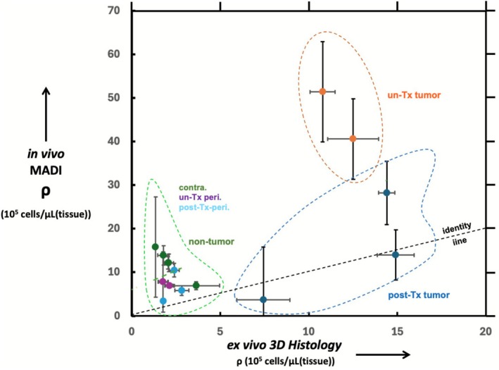 Figure 3
 Figure 4
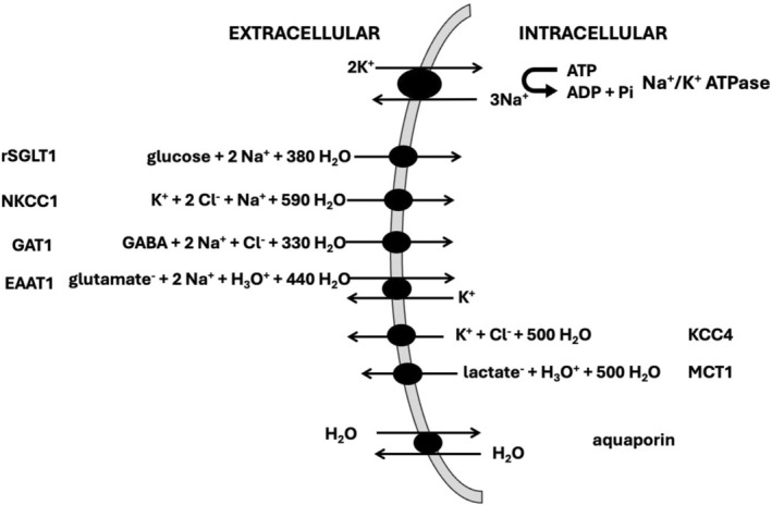 Figure 5
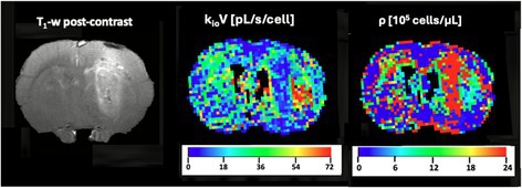 Figure 6
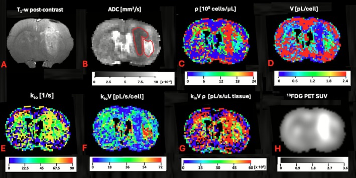 Figure 7
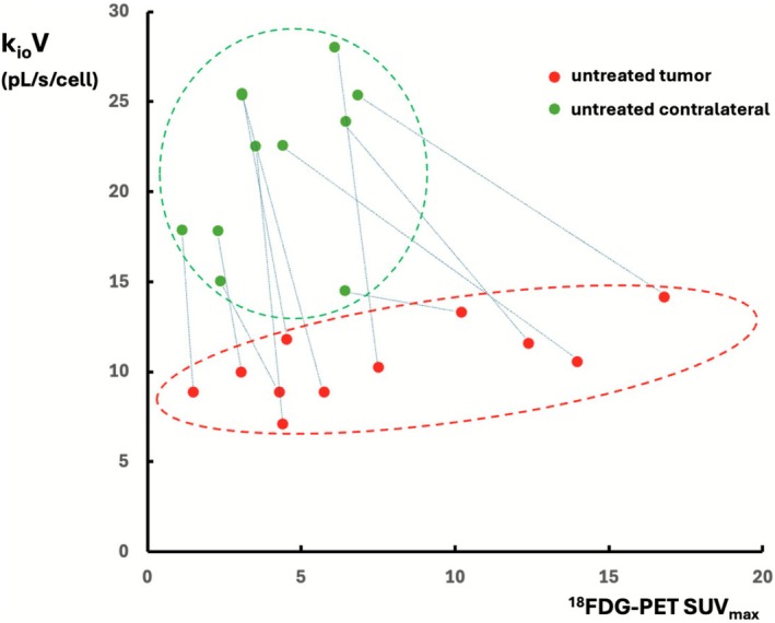 Figure 8
 Figure 9
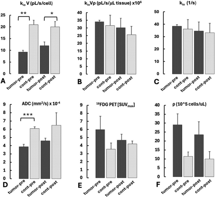 Figure 10
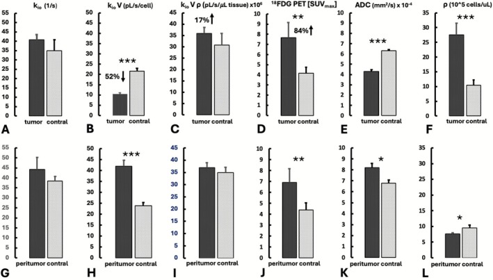 Figure 11
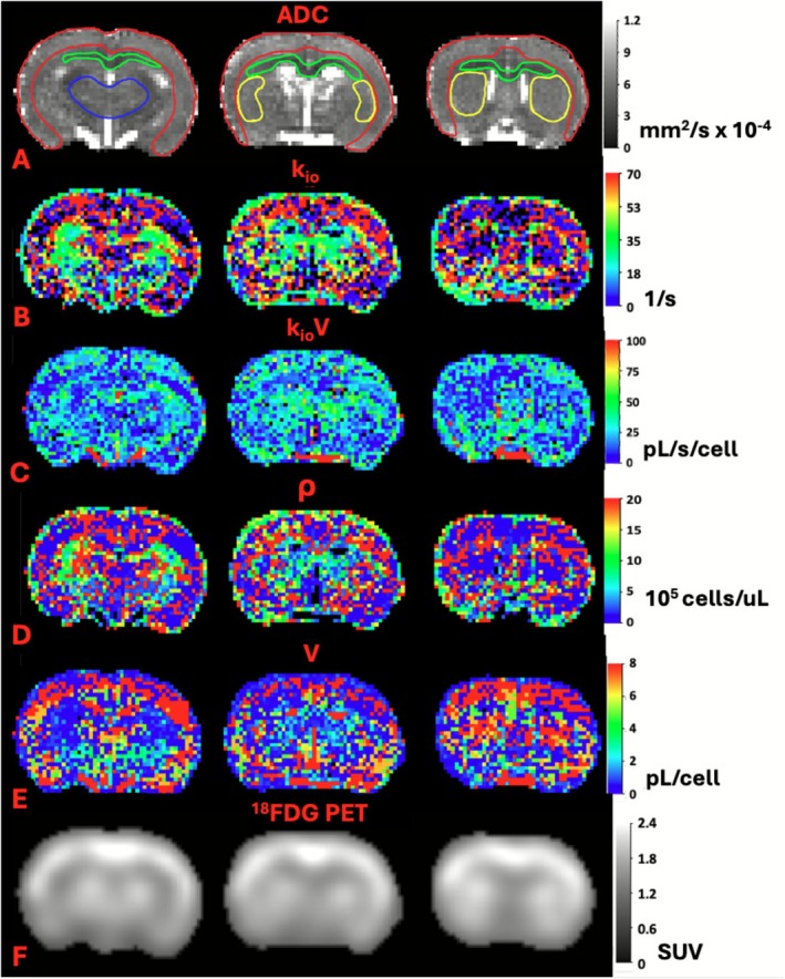 Figure 12
Peer Reviews
No public reviews on file for this paper yet. If you reviewed it on a platform where reviews are public (OpenReview, ICLR, NeurIPS, ICML), you can paste yours below so the community can read it here.
Videos
No videos yet. Explain this paper in a talk, walkthrough, or lecture? Add one.
Taxonomy
TopicsMRI in cancer diagnosis · Advanced Neuroimaging Techniques and Applications · Glioma Diagnosis and Treatment