sCellST predicts single-cell gene expression from H& E images
Loïc Chadoutaud, Marvin Lerousseau, Daniel Herrero-Saboya, Julian Ostermaier, Jacqueline Fontugne, Emmanuel Barillot, Thomas Walter

TL;DR
This paper introduces a deep learning model that predicts single-cell gene expression from H&E images, enabling detailed molecular insights from standard histological slides.
Contribution
The novel approach uses a Multiple Instance Learning framework to infer single-cell gene expression from full-slide morphology, improving resolution and biological relevance.
Findings
The model matches patch-based methods in spot-level prediction tasks while capturing fine-grained morphological variation.
It recovers biologically meaningful gene expression patterns across two cancer datasets.
The approach distinguishes fine cell populations and enables molecular-level interpretation of histological slides at scale.
Abstract
Understanding the spatial organization of individual cell types within tissue and how this organization is disrupted in disease, is a central question in biology and medicine. Hematoxylin and eosin-stained slides are widely available and provide detailed morphological context, while spatial gene expression profiling offers complementary molecular insights, though it remains costly and limited in accessibility. Predicting gene expression directly from histological images is therefore an attractive goal. However, existing approaches typically rely on small image patches, limiting resolution and the ability to capture fine-grained morphological variation. Here, we introduce a deep learning approach that predicts single-cell gene expression from morphology, matching patch-based methods on spot level prediction tasks. The model recovers biologically meaningful expression patterns across two…
Genes, proteins, chemicals, diseases, species, mutations and cell lines named across the full text — each resolved to its canonical identifier and authoritative record.
Click any figure to enlarge with its caption.
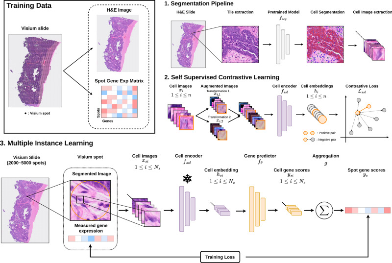 Figure 1
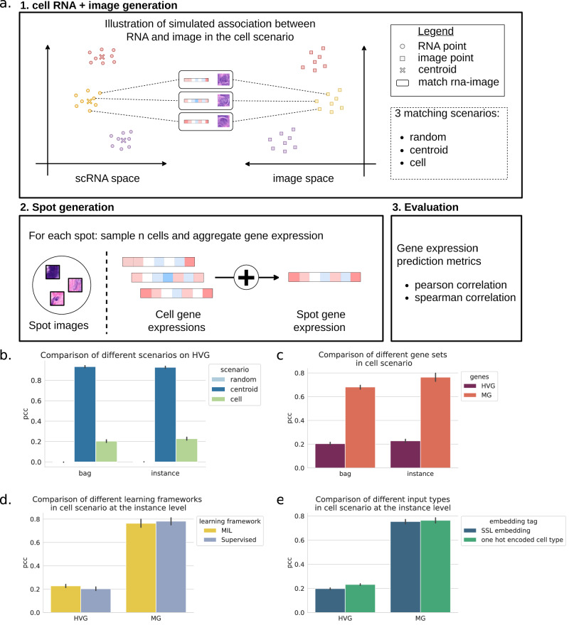 Figure 2
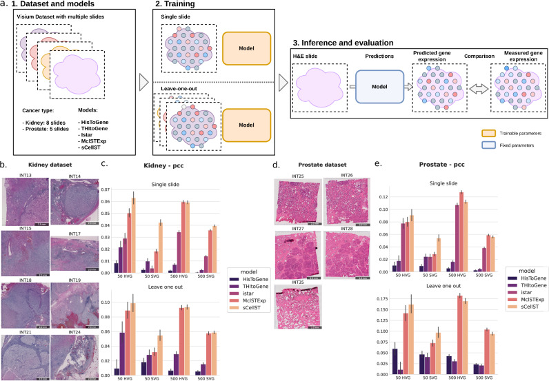 Figure 3
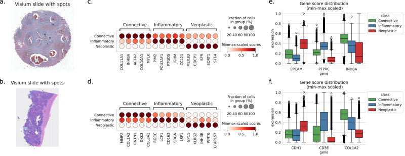 Figure 4
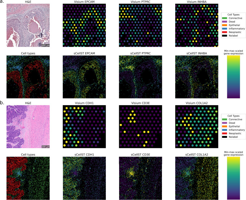 Figure 5
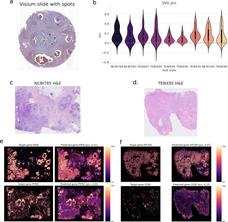 Figure 6
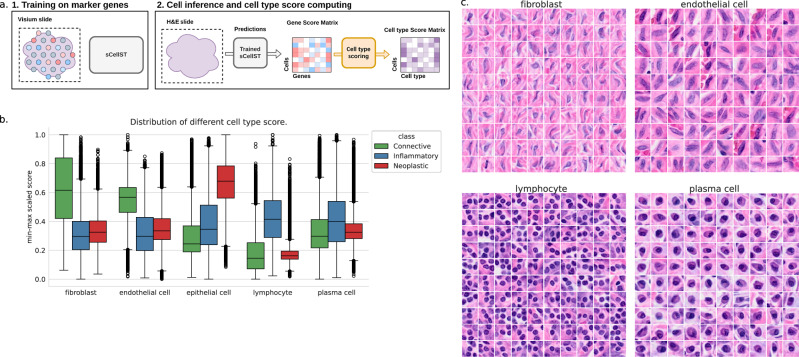 Figure 7
Peer Reviews
No public reviews on file for this paper yet. If you reviewed it on a platform where reviews are public (OpenReview, ICLR, NeurIPS, ICML), you can paste yours below so the community can read it here.
Videos
No videos yet. Explain this paper in a talk, walkthrough, or lecture? Add one.
Taxonomy
TopicsSingle-cell and spatial transcriptomics · Cell Image Analysis Techniques · AI in cancer detection