A novel endoscopic ultrasound system assisted by artificial intelligence for the recognition of pancreatic parenchyma and the detection of solid/cystic lesions
Sho Takahashi, Tomoya Takahashi, Toshio Fujisawa, Ippei Ikoma, Yasuhisa Jimbo, Ko Tomishima, Hiroyuki Isayama

Abstract
Click any figure to enlarge with its caption.
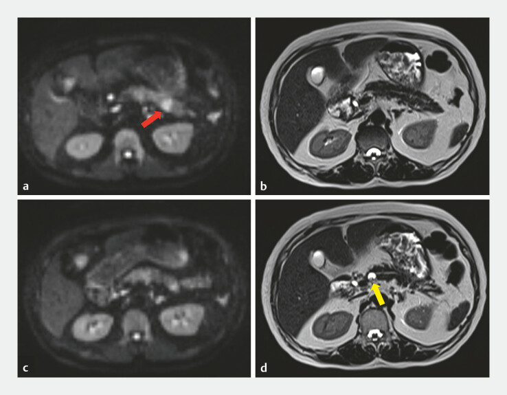 Fig. 1
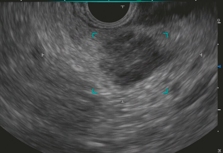 Fig. 2
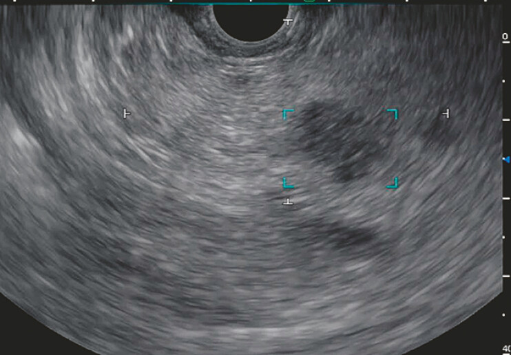 Fig. 3
Peer Reviews
No public reviews on file for this paper yet. If you reviewed it on a platform where reviews are public (OpenReview, ICLR, NeurIPS, ICML), you can paste yours below so the community can read it here.
Videos
No videos yet. Explain this paper in a talk, walkthrough, or lecture? Add one.
Taxonomy
TopicsPancreatic and Hepatic Oncology Research · Pancreatitis Pathology and Treatment · Gallbladder and Bile Duct Disorders
Endoscopic ultrasound (EUS) is an essential modality for detecting pancreatic solid and cystic lesions. However, EUS skill acquisition remains difficult for trainee endoscopists 1 . To facilitate the skill acquisition of trainee endoscopists, an EUS system assisted by artificial intelligence (EUS-AI), EW10-US01 (CAD EYE; FUJIFILM Corporation, Tokyo, Japan), has recently been developed and released for clinical use 2 3 . This system provides two functions: recognition of the pancreatic parenchyma, visualized as a white cross, and detection of solid and cystic lesions, visualized as a blue box. These outputs are overlaid in real time on live EUS images with optional acoustic alerts. We report a case in which this novel system contributed to the detection of pancreatic solid lesions that had not been identified via magnetic resonance imaging (MRI).
An 81-year-old woman presented with worsening control of type 2 diabetes mellitus, which prompted further MRI evaluation. The examination revealed a 15 mm hyperintense lesion in the pancreatic tail in diffusion-weighted imaging ( Fig. 1 a, b ). In addition, a 13 mm cystic lesion in the pancreatic body was identified in heavy T2 weighted images ( Fig. 1 c, d ). For diagnostic confirmation, EUS-guided tissue acquisition (EUS-TA) was performed with the assistance of EUS-AI ( Video 1 ). After the previously identified lesion in the pancreatic tail had been confirmed ( Fig. 2 ), subsequent screening with EUS-AI identified another a solid 12 mm lesion adjacent to the cyst in the pancreatic body, which had not been detected via MRI ( Fig. 3 ). EUS-TA of both lesions was performed, and pathological examination confirmed adenocarcinoma. This patient underwent pancreaticoduodenectomy.
a Diffusion-weighted imaging (DWI) demonstrated a 15 mm hyperintense lesion in the pancreatic tail (red arrow). b Heavily T2-weighted imaging did not demonstrate a corresponding lesion. c DWI did not demonstrate a solid lesion with diffusion restriction in the pancreatic body. d Heavily T2-weighted imaging demonstrated a 13 mm cystic lesion in the pancreatic body (yellow arrow).
Using the EUS-AI system, the 15 mm lesion in the pancreatic tail previously detected via MRI was enclosed by the blue box. EUS-AI, EUS system assisted by artificial intelligence; MRI, magnetic resonance imaging.
EUS-AI detected the 12 mm lesion in the pancreatic body adjacent to the cyst that had not been detected via MRI. EUS-AI, endoscopic ultrasound system assisted by artificial intelligence; MRI, magnetic resonance imaging.
Real-time recognition of the pancreatic parenchyma and detection of solid/cystic lesions by an artificial intelligence-assisted EUS system. EUS, endoscopic ultrasound.Video 1
This case highlights the clinical feasibility of this commercially available EUS-AI system that provides the real-time recognition of pancreatic parenchyma and detection of solid and cystic lesions in routine practice. EUS-AI may contribute to more accurate examinations by facilitating the skill acquisition of trainee endoscopists.
Endoscopy_UCTN_Code_CCL_1AF_2AZ
The reference list from the paper itself. Each links out to its DOI / PubMed record.
- 1Delsa H Khannoussi W Ghoneem E Endoscopic ultrasound training: Current state, challenges, and the path to proficiency World J Gastrointest Endosc 20251710745810.4253/wjge.v 17.i 8.10745840838151 PMC 12362506 · doi ↗ · pubmed ↗
- 2Das A Nguyen CC Li F Digital image analysis of EUS images accurately differentiates pancreatic cancer from chronic pancreatitis and normal tissue Gastrointest Endosc 20086786186718179797 10.1016/j.gie.2007.08.036 · doi ↗ · pubmed ↗
- 3Huang J Fan X Liu W Applications and Prospects of Artificial Intelligence-Assisted Endoscopic Ultrasound in Digestive System Diseases Diagnostics (Basel)202313281510.3390/diagnostics 1317281537685350 PMC 10487217 · doi ↗ · pubmed ↗