Cranial Defect Reconstruction With Custom 3D‐Printed Hydroxyapatite Scaffolds Augmented With rhBMP‐2 or Dipyridamole in a Nonhuman Primate Model
Griffin P. Bins, Heather A. Burkart, William Molair, Samuel Kogan, Dominic A. Massary, Angel Cabrera Pereira, Adem Aksu, Frank Reinauer, Daniel A. Couture, Lukasz Witek, Christopher M. Runyan

TL;DR
This study shows that 3D-printed hydroxyapatite scaffolds with rhBMP-2 can effectively repair large cranial bone defects in nonhuman primates, outperforming other treatments.
Contribution
Demonstrates clinical readiness of rhBMP-2-augmented 3D-printed scaffolds for cranial defect repair in a nonhuman primate model.
Findings
rhBMP-2-treated scaffolds showed significantly greater bone bridging (∼90%) compared to DIPY (∼9%) and uncoated scaffolds (10%).
Bone volume in rhBMP-2-treated scaffolds was significantly higher than in DIPY and uncoated groups at 12 months.
rhBMP-2 scaffolds exhibited superior mechanical properties and bone ingrowth compared to other groups.
Abstract
Reconstruction of critical‐sized bone defects, particularly in the cranio‐maxillofacial region, presents unique challenges due to the need for integration with adjacent well‐vascularized tissue and the absence of significant load‐bearing requirements. This study evaluated the clinical readiness of bone tissue engineering (BTE) for critically sized cranial defects using custom 3D‐printed hydroxyapatite scaffolds augmented with either recombinant human bone morphogenetic protein‐2 (rhBMP‐2) or dipyridamole (DIPY) in a highly translational nonhuman primate model. Identical 5 × 5‐cm vertex guided craniotomies were created in 12 macaques: Three cynomolgus macaques served as negative controls to validate the critical size nature of the defect, while nine rhesus macaques underwent scaffold reconstruction. Subjects were divided into three groups: uncoated scaffolds (n = 3), scaffolds augmented…
Genes, proteins, chemicals, diseases, species, mutations and cell lines named across the full text — each resolved to its canonical identifier and authoritative record.
Click any figure to enlarge with its caption.
 Figure 1
 Figure 2
 Figure 3
 Figure 4
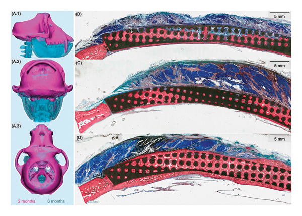 Figure 5
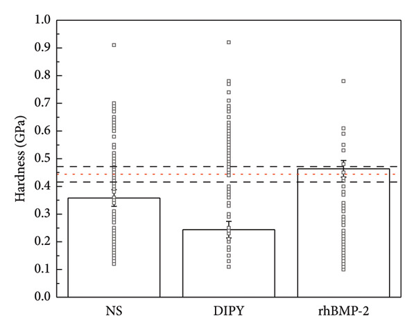 Figure 6
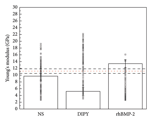 Figure 7
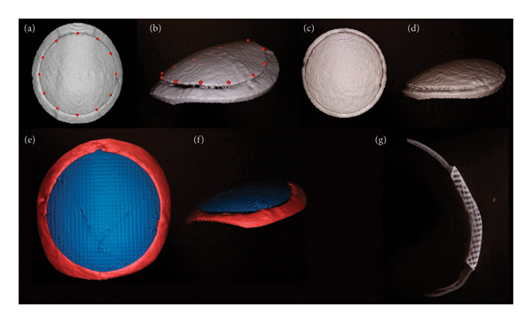 Figure 8
Peer Reviews
No public reviews on file for this paper yet. If you reviewed it on a platform where reviews are public (OpenReview, ICLR, NeurIPS, ICML), you can paste yours below so the community can read it here.
Videos
No videos yet. Explain this paper in a talk, walkthrough, or lecture? Add one.
Taxonomy
TopicsBone Tissue Engineering Materials · Craniofacial Disorders and Treatments · Facial Trauma and Fracture Management