Lung Density Measurement in Fontan Patients Using Zero Echo Time Sequences
Alessia Callegari, Konstantinos Zeimpekis, Barbara E. U. Burkhardt, Emanuela R. Valsangiacomo Buechel, Fraser M. Callaghan, Jakob Usemann, Martin Hersberger, Christian J. Kellenberger, Julia Geiger

TL;DR
This study explores using MRI to measure lung density in patients who have undergone a Fontan procedure, linking lung density to heart and lung health indicators.
Contribution
The study introduces lung density measurement via zero echo time MRI as a potential diagnostic tool for Fontan patients.
Findings
Patients with higher lung-to-background ratio (LBR) showed increased extracellular volume and decreased TIMP-1 and sST-2 levels.
Elevated LBR was associated with a history of higher pulmonary arterial pressure and collateral embolization.
LBR did not correlate with most cardiovascular or pulmonary parameters, suggesting lung density is a unique indicator.
Abstract
Pulmonary complications are known to occur in patients after Fontan palliation. Cardiac MRI is performed in the follow-up of Fontan patients to assess single ventricular function, hemodynamics and potential collateral flow. To date, pulmonary function tests have been used to detect functional lung impairment, but lung MRI has not been integrated into imaging follow-up. In this study, we measured lung-to-background ratio (LBR) on zero echo time (ZTE) MRI sequences in 32 children with Fontan palliation. Patients were divided into 2 groups: LBR > 1.5 and < 1.5 and assessed for associations between LBR and ventricular function, fibrosis, hemodynamics as well as biomarkers, spirometry and previous catheter results. We observed significantly increased extracellular volume (ECV) and decreased tissue inhibitor of metalloproteinase 1 (TIMP-1), soluble suppression of tumorigenicity 2 (sST-2) and…
Genes, proteins, chemicals, diseases, species, mutations and cell lines named across the full text — each resolved to its canonical identifier and authoritative record.
Click any figure to enlarge with its caption.
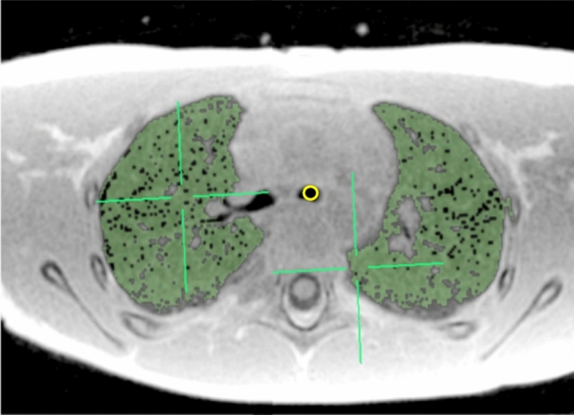 Figure 1
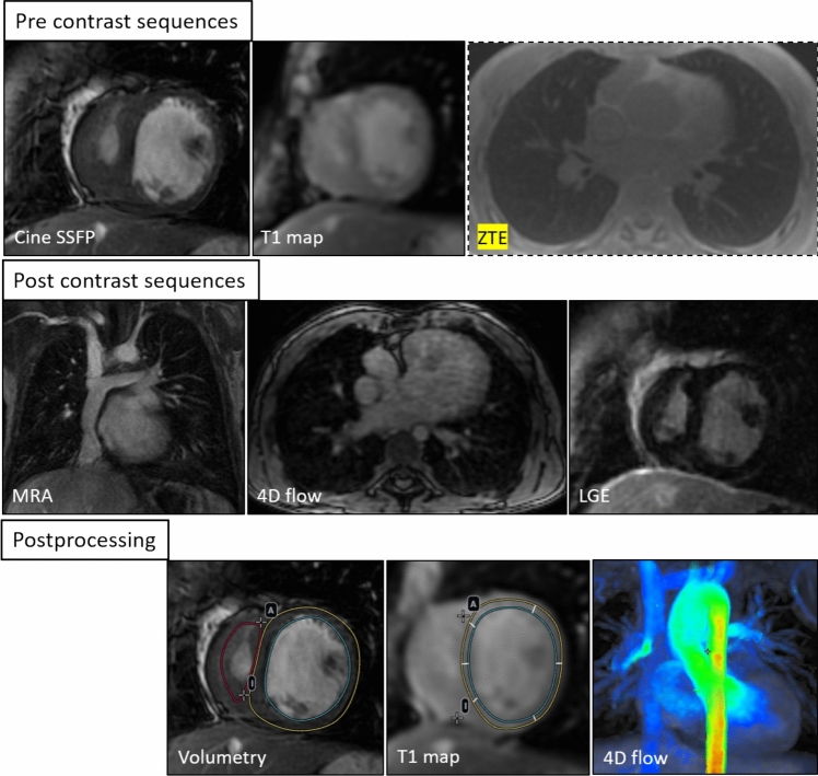 Figure 2
 Figure 3
 Figure 4
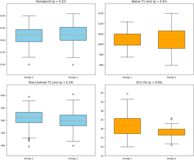 Figure 5
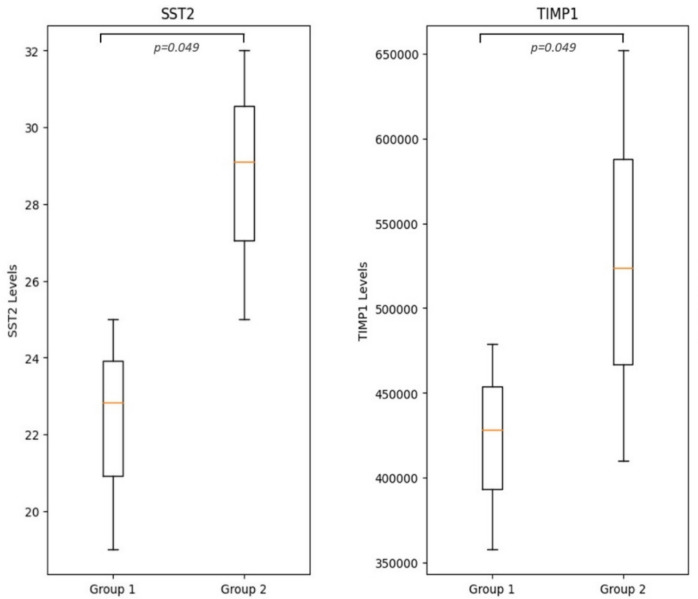 Figure 6
Peer Reviews
No public reviews on file for this paper yet. If you reviewed it on a platform where reviews are public (OpenReview, ICLR, NeurIPS, ICML), you can paste yours below so the community can read it here.
Videos
No videos yet. Explain this paper in a talk, walkthrough, or lecture? Add one.
Taxonomy
TopicsCongenital Heart Disease Studies · Ultrasound in Clinical Applications · Congenital Diaphragmatic Hernia Studies