A practical tip for estimating skeletal maturation in children aged < 3 years using humeral ossification on chest radiographs: A retrospective study
Tomohiro Tsuru, Shota Inoue, Hiromi Edo, Shuichi Suzuki, Kohsuke Imai, Taiki Nozaki, Hiroshi Shinmoto

TL;DR
This study introduces a new method to estimate bone age in children under 3 years old using chest X-rays, avoiding the need for extra imaging.
Contribution
The study demonstrates a strong linear relationship between humeral ossification and age, and provides sex-specific formulas for rapid skeletal maturation assessment.
Findings
A strong positive correlation (r > 0.88) was found between humeral ossification center diameter and chronological age.
Sex-specific formulas (0.5 × age + 4 for males, 0.6 × age + 3 for females) were proposed for practical clinical use.
Reference ranges (±2 SD) were established for six 6-month age groups to identify deviations in skeletal maturation.
Abstract
Bone age assessment in children aged < 3 years is difficult owing to the limited visibility of carpal ossification centres. We aimed to evaluate skeletal maturation patterns in relation to chronological age using humeral ossification on chest radiographs. We retrospectively reviewed the chest radiographs of 187 children aged 0– < 3 years. Three readers independently measured the longitudinal diameter of the humeral head epiphyseal ossification centre. Interobserver agreement was assessed with the intraclass correlation coefficient (ICC). Pearson’s correlation and linear regression analyses were performed. Reference ranges (± 1 standard deviation [SD] and ± 2 SD) were established for six 6-month age groups. A strong positive correlation was observed between ossification centre diameter and age (r > 0.88 for all observers). The ICC was 0.96 (p < 0.001), indicating excellent interobserver…
Genes, proteins, chemicals, diseases, species, mutations and cell lines named across the full text — each resolved to its canonical identifier and authoritative record.
Click any figure to enlarge with its caption.
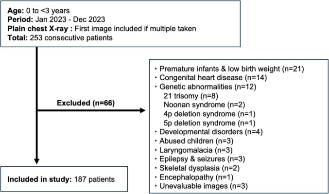 Figure 1
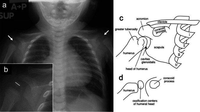 Figure 2
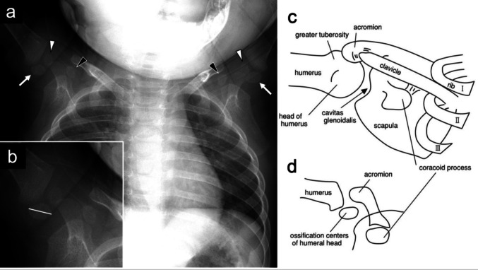 Figure 3
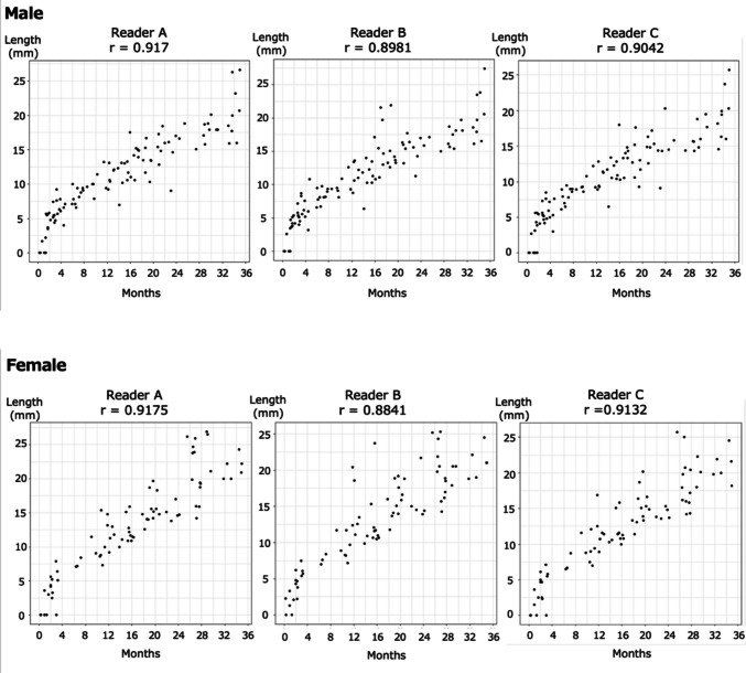 Figure 4
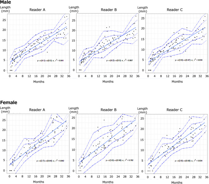 Figure 5
Peer Reviews
No public reviews on file for this paper yet. If you reviewed it on a platform where reviews are public (OpenReview, ICLR, NeurIPS, ICML), you can paste yours below so the community can read it here.
Videos
No videos yet. Explain this paper in a talk, walkthrough, or lecture? Add one.
Taxonomy
TopicsForensic Anthropology and Bioarchaeology Studies · Orthopedic Infections and Treatments · Medical Imaging and Pathology Studies