Opportunistic osteoporosis screening on FDG PET/CT scans in breast carcinoma: a comparison with DXA
Nitin Gupta, Manpreet Kaur

TL;DR
Breast cancer patients undergoing PET/CT scans can be screened for osteoporosis and fracture risk using CT attenuation values, reducing the need for additional DXA scans.
Contribution
This study demonstrates that FDG PET/CT scans can effectively detect osteopenia, osteoporosis, and fracture risk with high sensitivity and specificity.
Findings
Post-chemo ± hormonal therapy increases osteopenia, osteoporosis, and fracture risk in breast cancer patients.
CT attenuation values ≤174.6 HU and ≤117.2 HU detect osteopenia and osteoporosis with high sensitivity and specificity.
PET/CT scans can identify patients needing osteoporosis treatment without requiring additional DXA referrals.
Abstract
Reduced bone mineral density is often observed in breast cancer patients. Routine PET/CT scans can be used for detection of low bone mineral density. To evaluate prevalence of osteopenia, osteoporosis and fracture risk in pre and post-therapy breast carcinoma patients undergoing 18F-FDG PET/CT scans. In this retrospective study L1-L4 vertebral and femoral neck CT mean Hounsfield unit attenuation and their corresponding SUVmax values from initial staging and end of treatment FDG PET/CT scans performed in breast carcinoma patients were compared. Post chemo ± hormonal therapy FDG PET/CT HU values were also compared to DXA scan T- scores. Significant increase in prevalence of post chemo ± hormonal therapy osteopenia, osteoporosis and fractures (62%, 18% and 16% vs baseline of 35%, 4% and 9% respectively). CECT mean attenuation values of ≤174.6 HU and ≤117.2 HU for detection of osteopenia…
Genes, proteins, chemicals, diseases, species, mutations and cell lines named across the full text — each resolved to its canonical identifier and authoritative record.
Click any figure to enlarge with its caption.
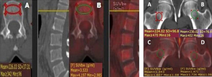 Figure 1
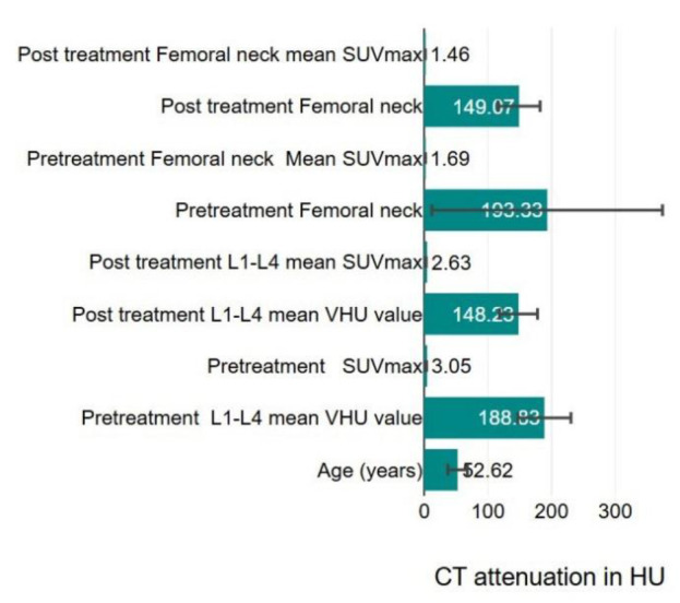 Figure 2
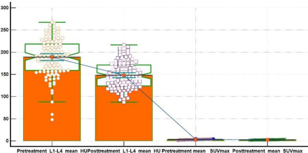 Figure 3
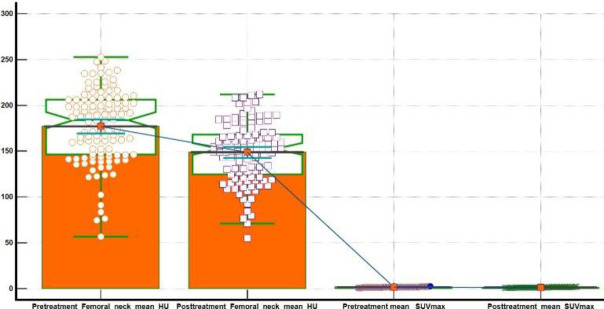 Figure 4
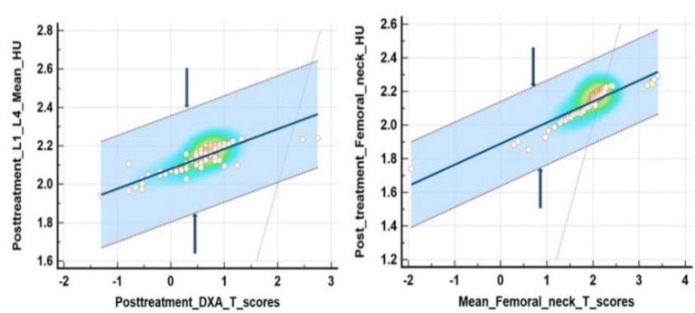 Figure 5
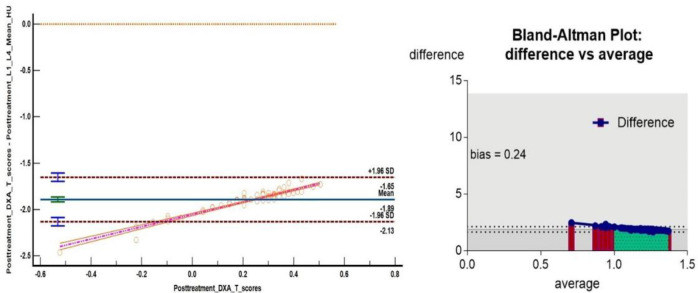 Figure 6
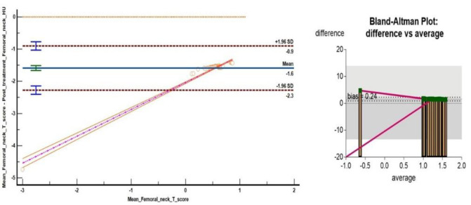 Figure 7
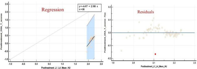 Figure 8
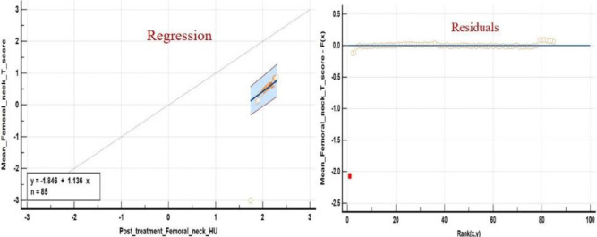 Figure 9
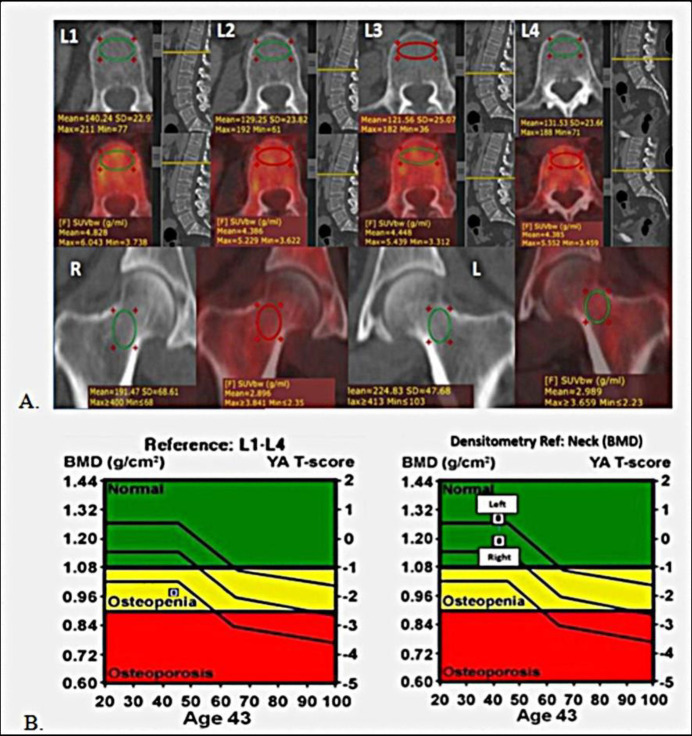 Figure 10
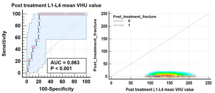 Figure 11
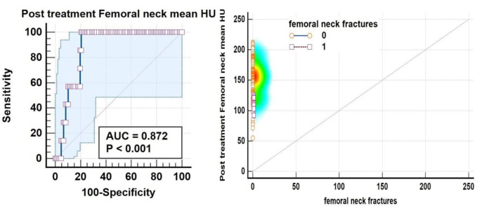 Figure 12
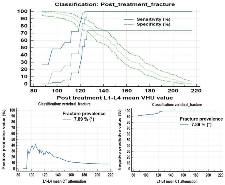 Figure 13
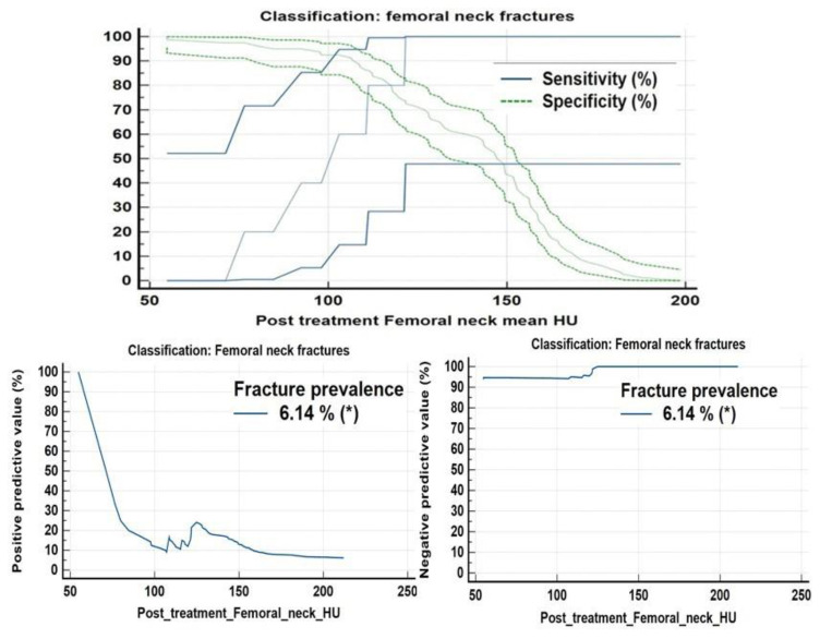 Figure 14
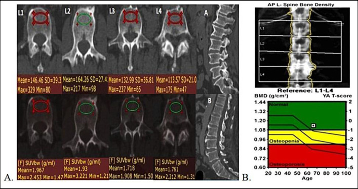 Figure 15
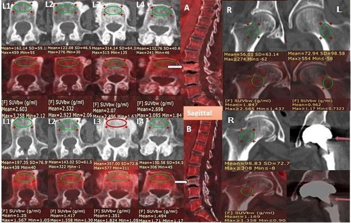 Figure 16
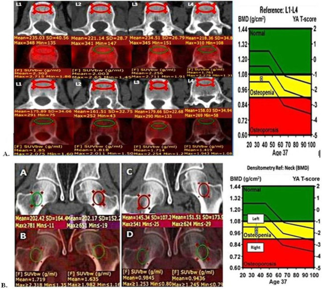 Figure 17
Peer Reviews
No public reviews on file for this paper yet. If you reviewed it on a platform where reviews are public (OpenReview, ICLR, NeurIPS, ICML), you can paste yours below so the community can read it here.
Videos
No videos yet. Explain this paper in a talk, walkthrough, or lecture? Add one.
Taxonomy
TopicsMedical Imaging Techniques and Applications · Bone health and treatments · Cancer Risks and Factors