Early and progressive retinal microglial changes in APPNL-F/NL-F mouse model of Alzheimer’s disease revealed by an automated image analysis software
Lidia Sánchez-Puebla, Inés López-Cuenca, Miguel A. Sánchez-Puebla, Ana Granados, Ana I. Ramírez, Juan Llorens, Takaomi C. Saido, Takashi Saito, Carmen Nieto-Vaquero, María A. Moro, Valentín Moreno, José M. Ramírez, Rosa de Hoz

TL;DR
This study shows that retinal microglia in a mouse model of Alzheimer's disease show early and progressive changes, suggesting the retina could be a non-invasive biomarker for early detection.
Contribution
The study introduces an automated image analysis system, MorphoSomas, to detect early retinal microglial changes in Alzheimer's disease models.
Findings
APPNL-F/NL-F mice showed early microglial activation at 6 months, with increased cell number and soma size.
Progressive microglial dysfunction was observed through reduced arborization and skeletonization in APPNL-F/NL-F mice.
Automated analysis revealed significant differences in retinal microglial morphology between AD and control mice.
Abstract
Alzheimer’s disease (AD) is characterized by the accumulation of misfolded proteins that trigger neuroinflammation and neuronal loss. The retina, as an extension of the central nervous system, mirrors these pathological processes and represents a potential biomarker. Microglial activation, a key component of neuroinflammation, can be morphologically assessed through automated image analysis. This study performed a quantitative and morphological analysis of retinal microglia in the APPNL-F/NL-F mouse model of AD across aging (6–20 months) and comparing them with age-matched C57BL/6 J controls using an automated image analysis software. A cross-sectional design was applied to 72 mice (36 APPNL-F/NL-F and 36 WT). Retinas samples were processed by Iba-1 immunohistochemistry. Quantified parameters included cell number, soma size, arborization area, skeletonization, fluorescence intensity,…
Genes, proteins, chemicals, diseases, species, mutations and cell lines named across the full text — each resolved to its canonical identifier and authoritative record.
Click any figure to enlarge with its caption.
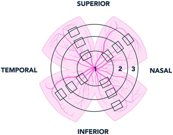 Figure 1
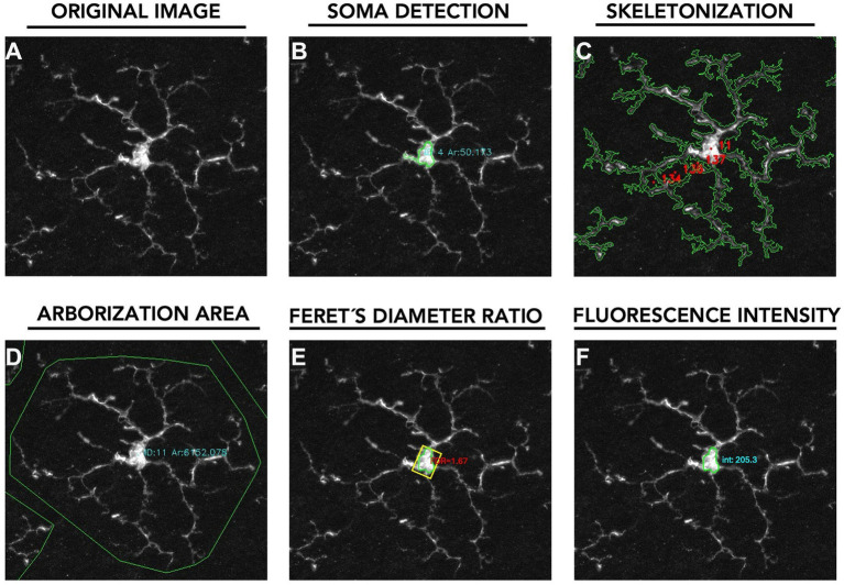 Figure 2
 Figure 3
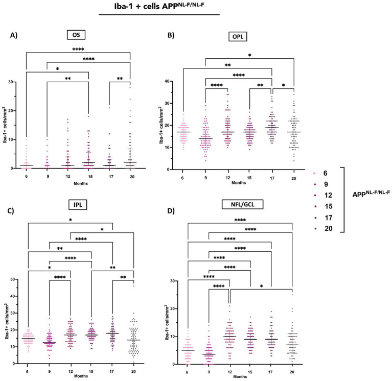 Figure 4
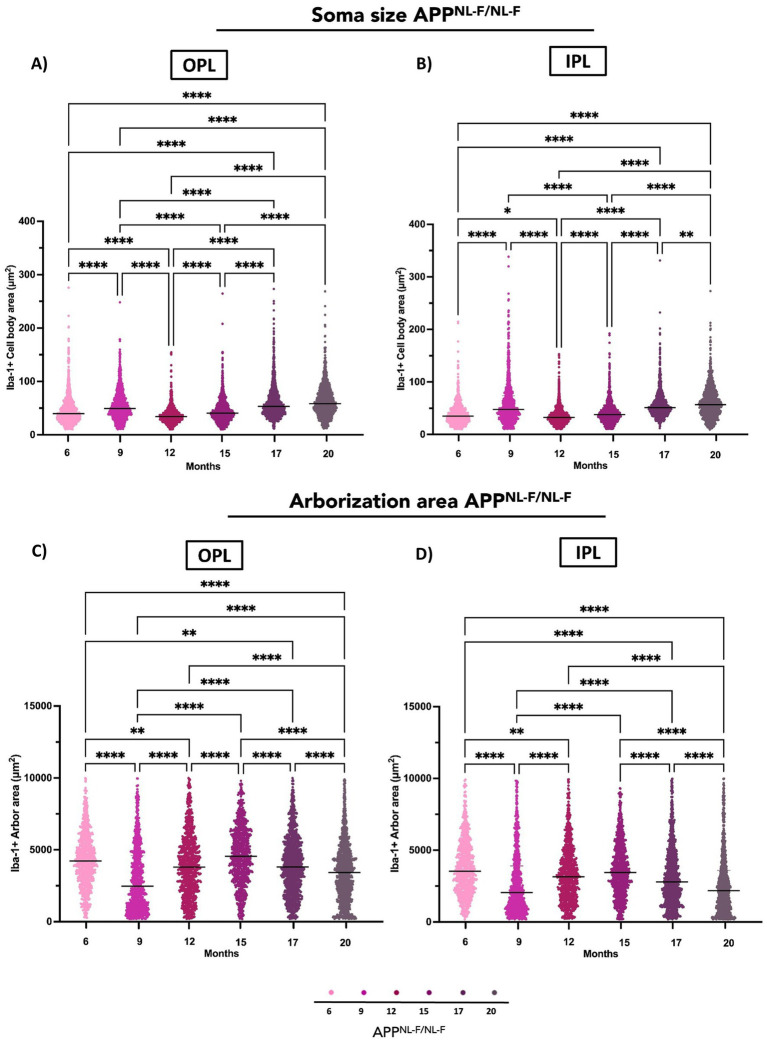 Figure 5
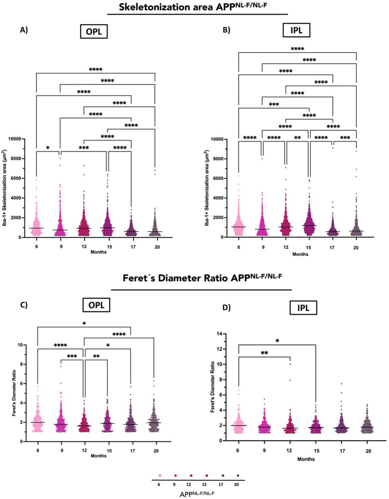 Figure 6
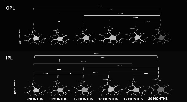 Figure 7
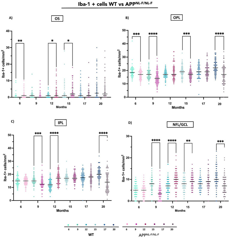 Figure 8
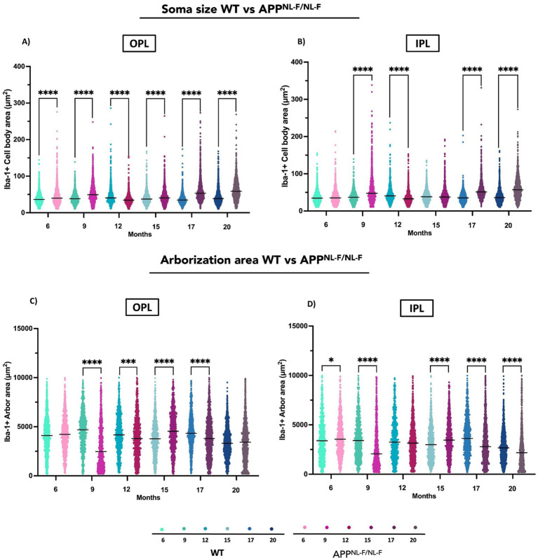 Figure 9
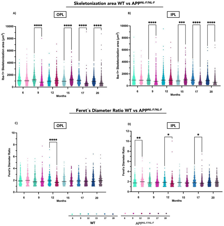 Figure 10
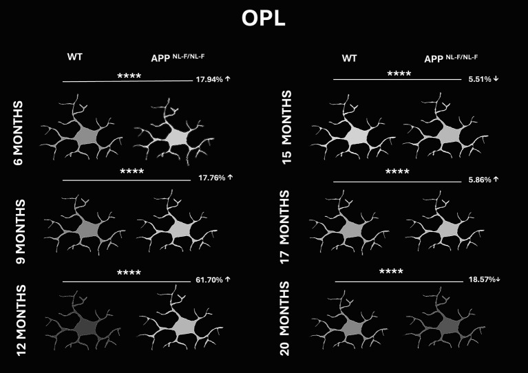 Figure 11
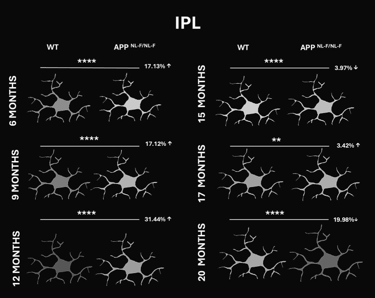 Figure 12
Peer Reviews
No public reviews on file for this paper yet. If you reviewed it on a platform where reviews are public (OpenReview, ICLR, NeurIPS, ICML), you can paste yours below so the community can read it here.
Videos
No videos yet. Explain this paper in a talk, walkthrough, or lecture? Add one.
Taxonomy
TopicsRetinal Imaging and Analysis · Alzheimer's disease research and treatments · Retinal Diseases and Treatments