Impact of Contrast-Enhanced Mammography on Breast Imaging Reporting and Data System (BI-RADS) Reclassification: Correlation With Histopathology and Clinical Outcomes
Mariam Malik, Umal Baneen Zahra, Faisal Ehsan Cheema, Aamena Irfan Shami, Amira Shami, Muhammad Imran

TL;DR
Contrast-enhanced mammography improves breast lesion classification by revealing hidden cancers and reducing uncertainty in diagnosis.
Contribution
The study demonstrates how contrast-enhanced mammography modifies BI-RADS classifications and correlates with histopathology outcomes.
Findings
CEM correctly upgraded 12 lesions and correctly downgraded six lesions based on histopathology.
False-positive upgrades occurred in fibroadenoma and sclerosing adenosis cases.
CEM reduced diagnostic uncertainty in dense breast tissue and indeterminate findings.
Abstract
Purpose: This study aims to evaluate how contrast-enhanced mammography (CEM) modifies the Breast Imaging Reporting and Data System (BI-RADS) classification initially assigned on digital mammography (DM), and to assess the accuracy of CEM-driven upgrades, downgrades, and confirmations using histopathology as reference. Methods: This was a retrospective observational study that included symptomatic patients who underwent both DM and subsequent CEM within a short interval. The initial BI-RADS grading was assigned based on DM findings, while CEM findings were categorized as either correct or incorrect upgrades, downgrades, or confirmations, based on final histopathology or follow-up results. The study included 60 patients across all grades of BI-RADS. Results: Among patients initially assigned BI-RADS 0-5 on DM, CEM correctly upgraded 12 lesions and correctly downgraded six lesions. CEM…
Genes, proteins, chemicals, diseases, species, mutations and cell lines named across the full text — each resolved to its canonical identifier and authoritative record.
Click any figure to enlarge with its caption.
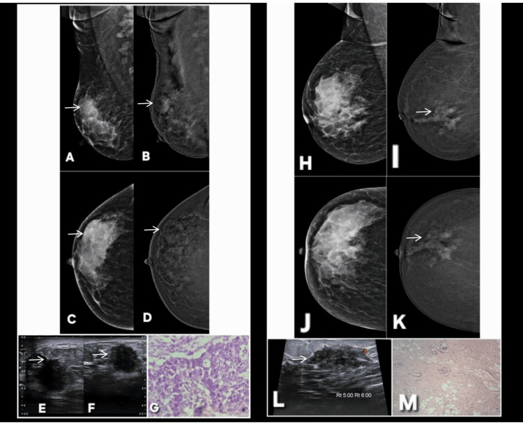 Figure 1
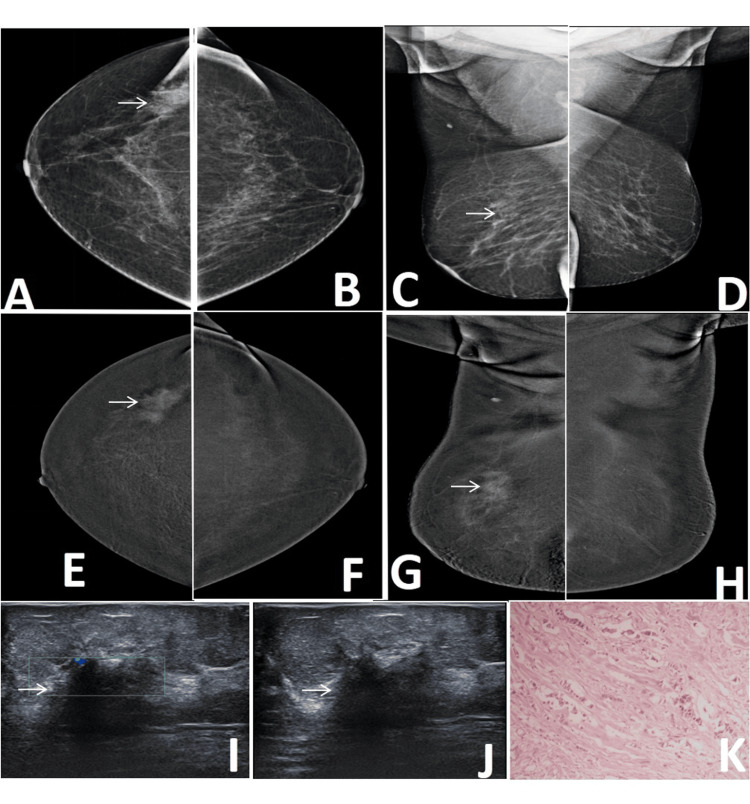 Figure 2
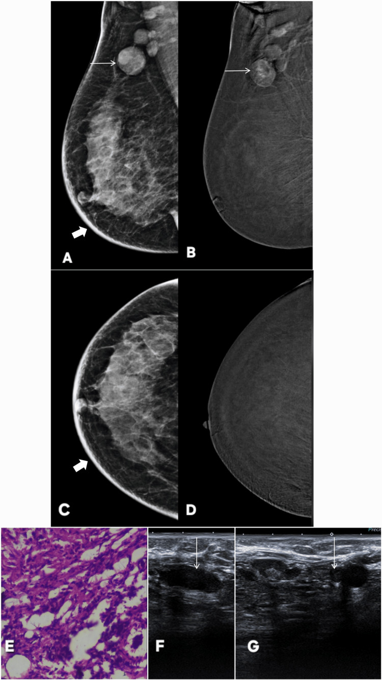 Figure 3
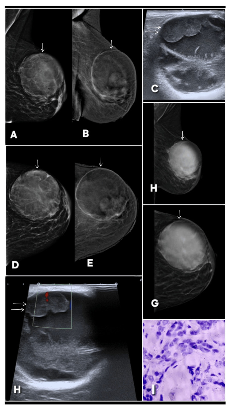 Figure 4
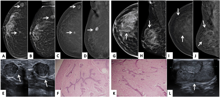 Figure 5
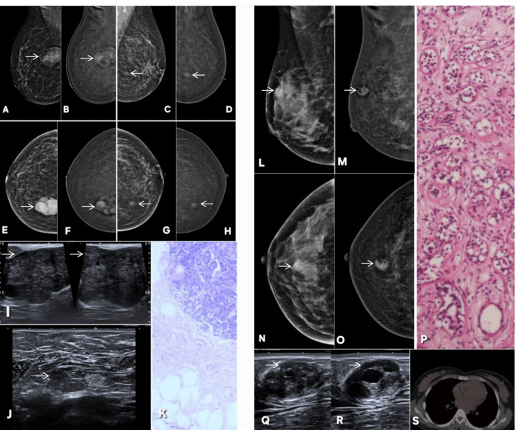 Figure 6
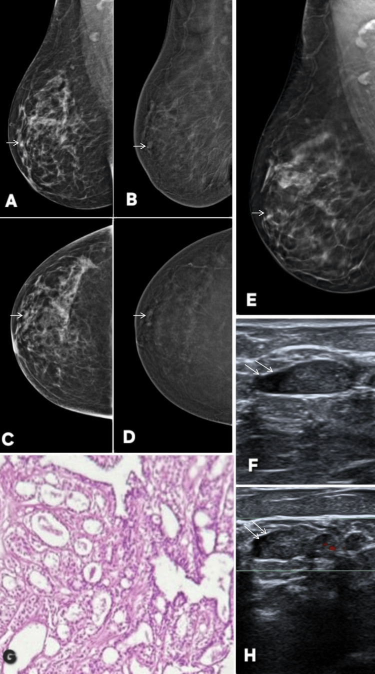 Figure 7
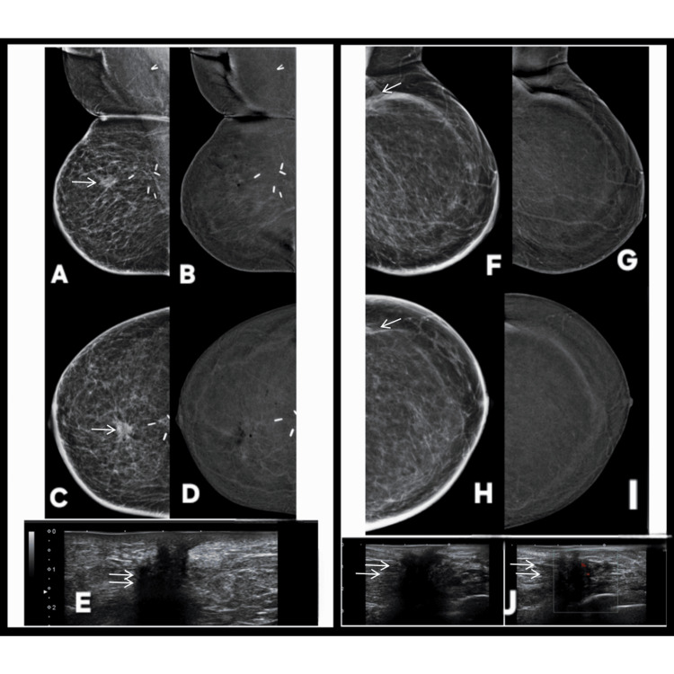 Figure 8
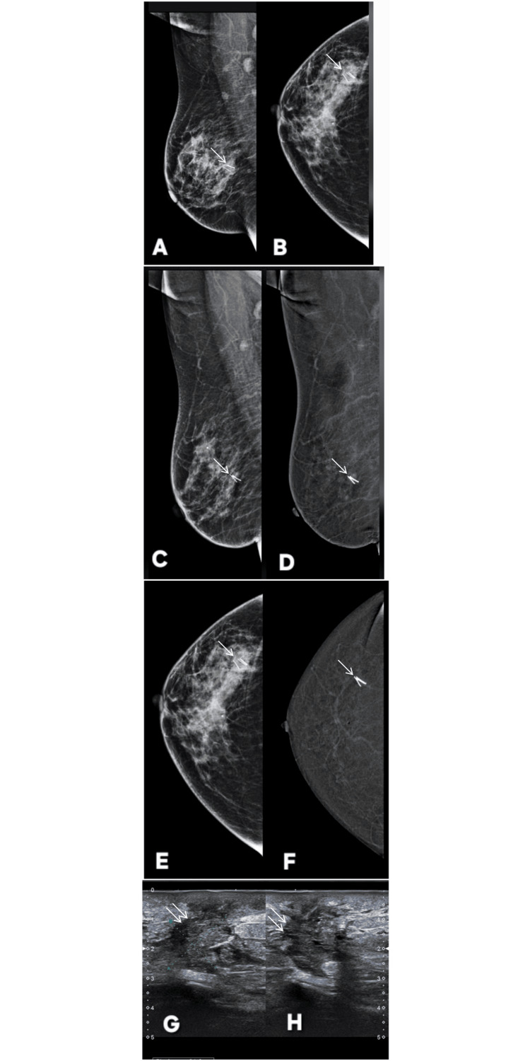 Figure 9
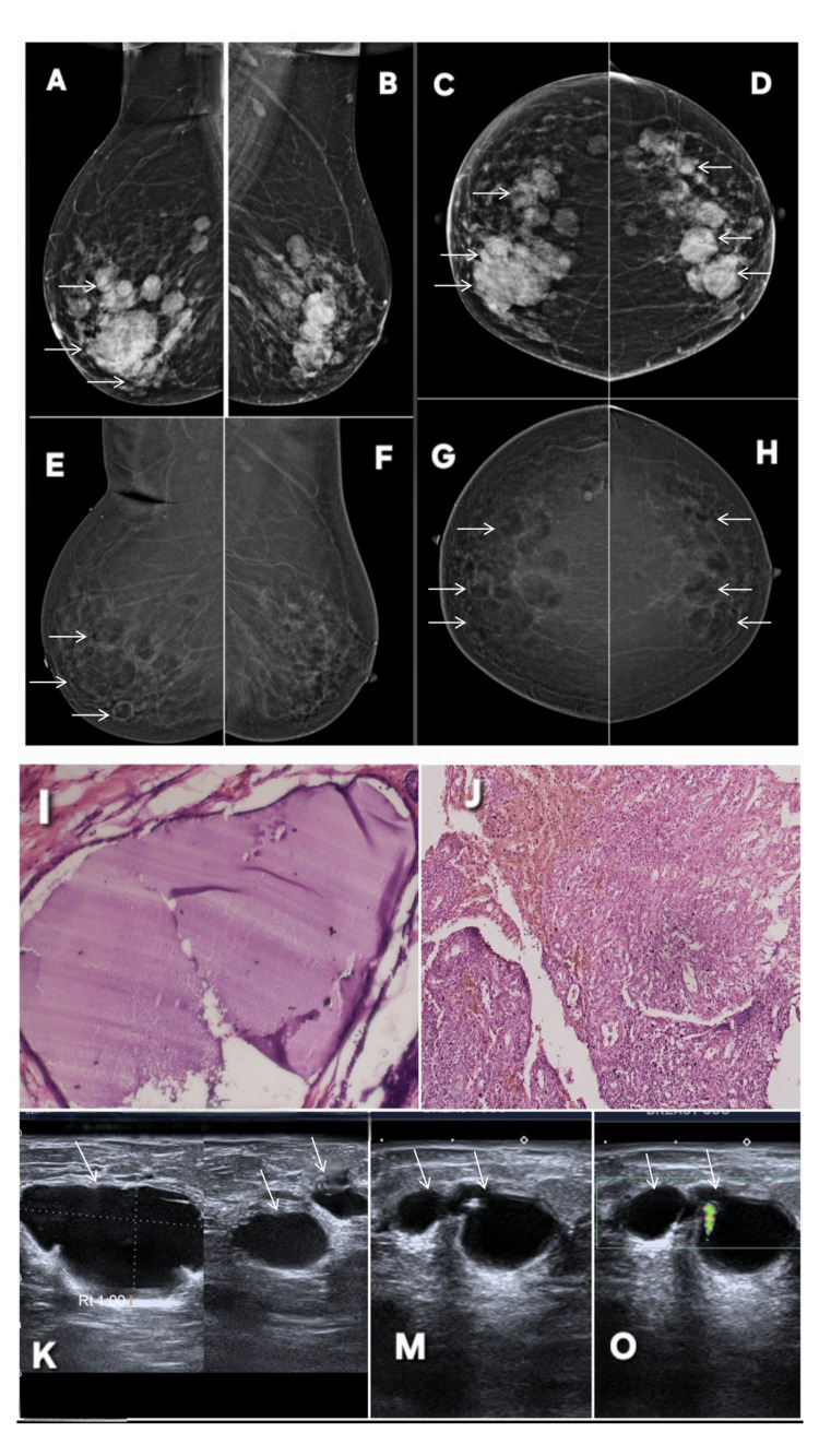 Figure 10
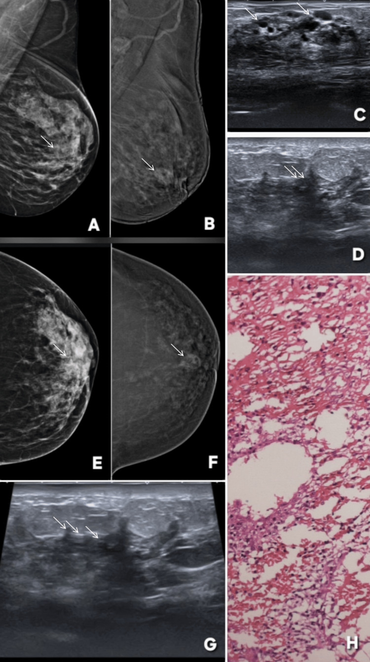 Figure 11
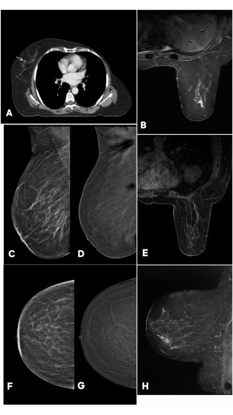 Figure 12
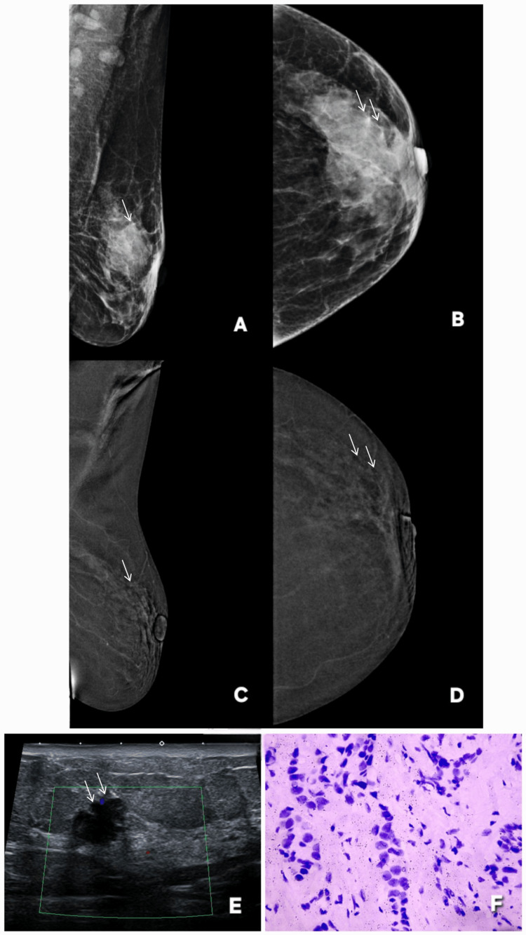 Figure 13
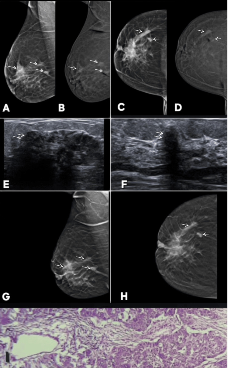 Figure 14
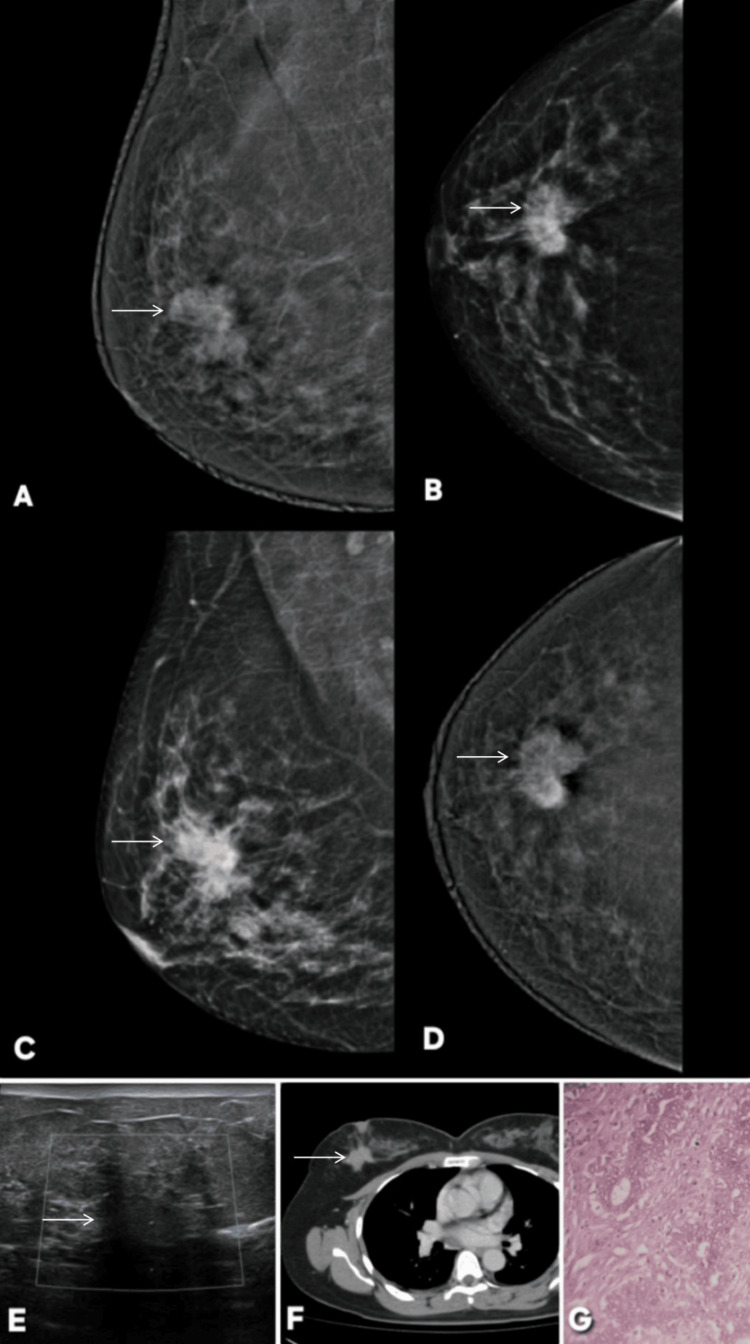 Figure 15
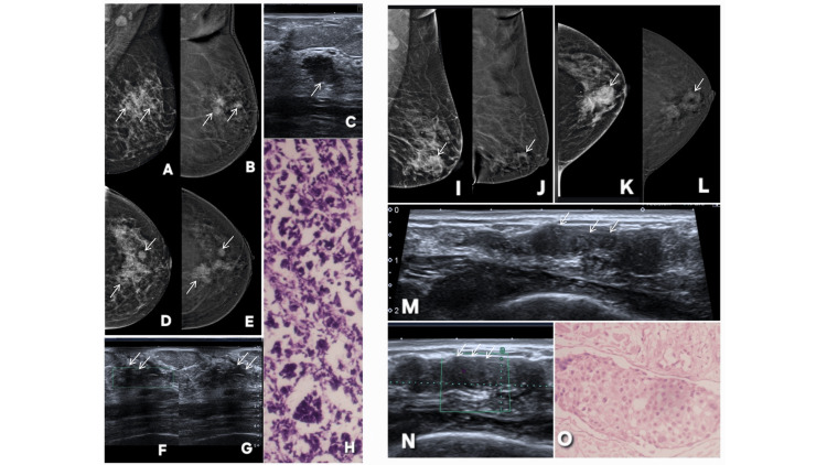 Figure 16
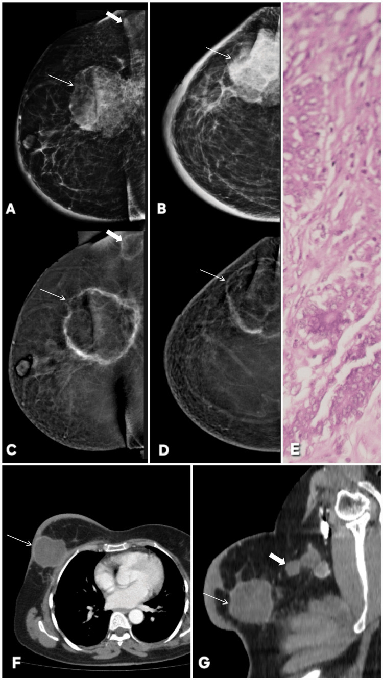 Figure 17
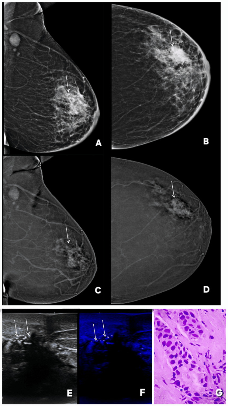 Figure 18
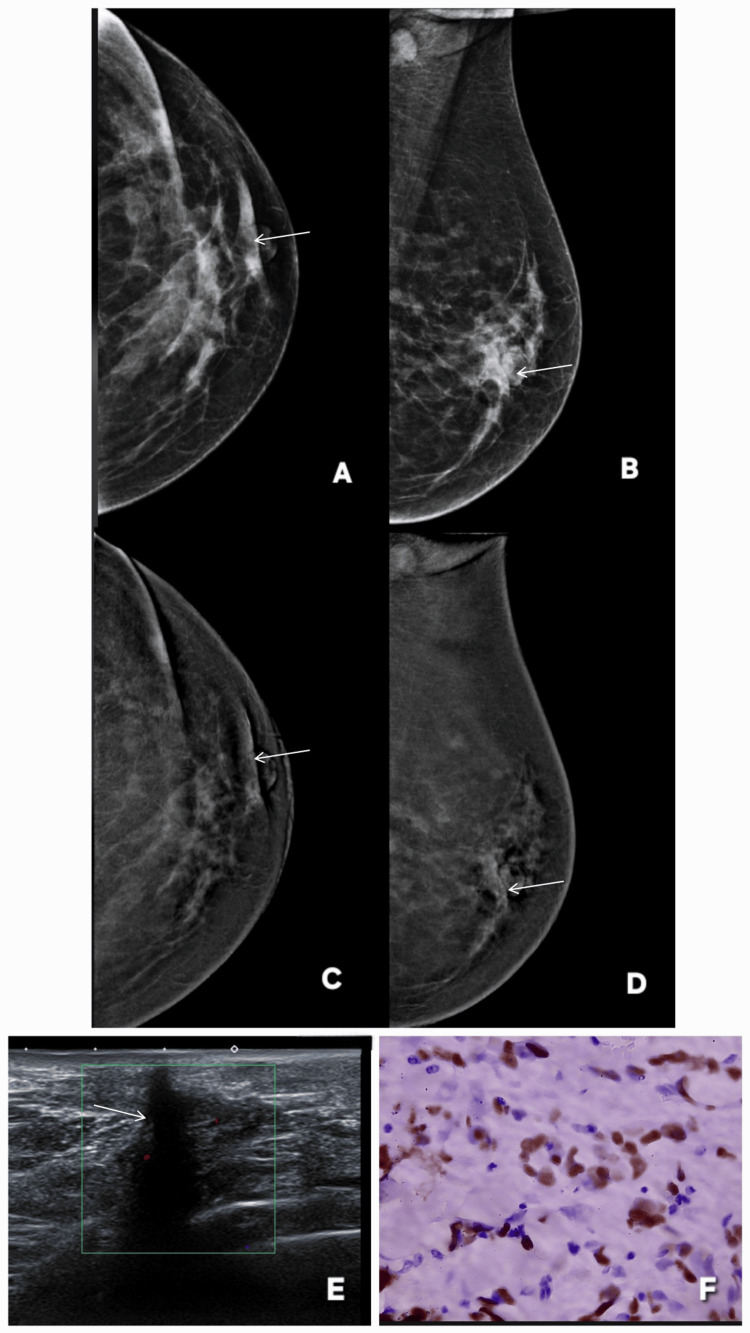 Figure 19
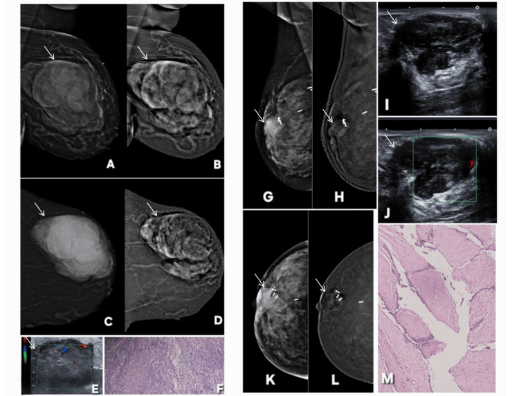 Figure 20
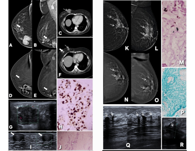 Figure 21
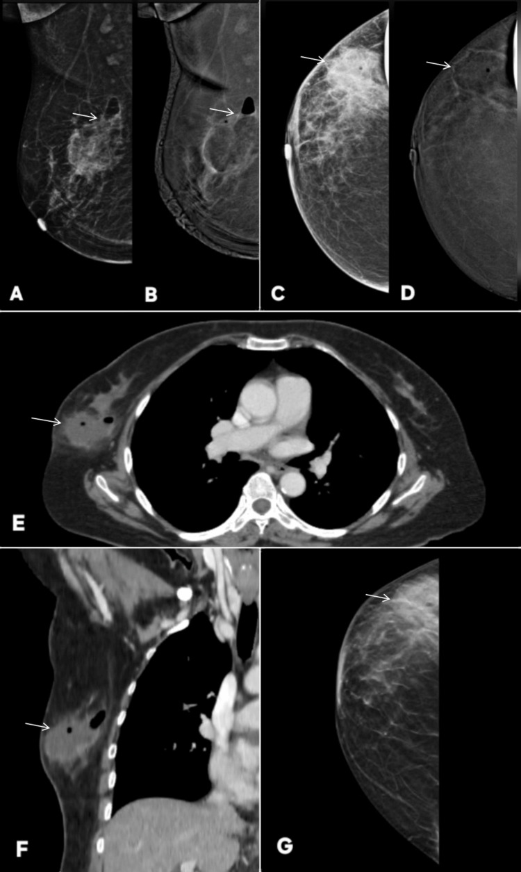 Figure 22
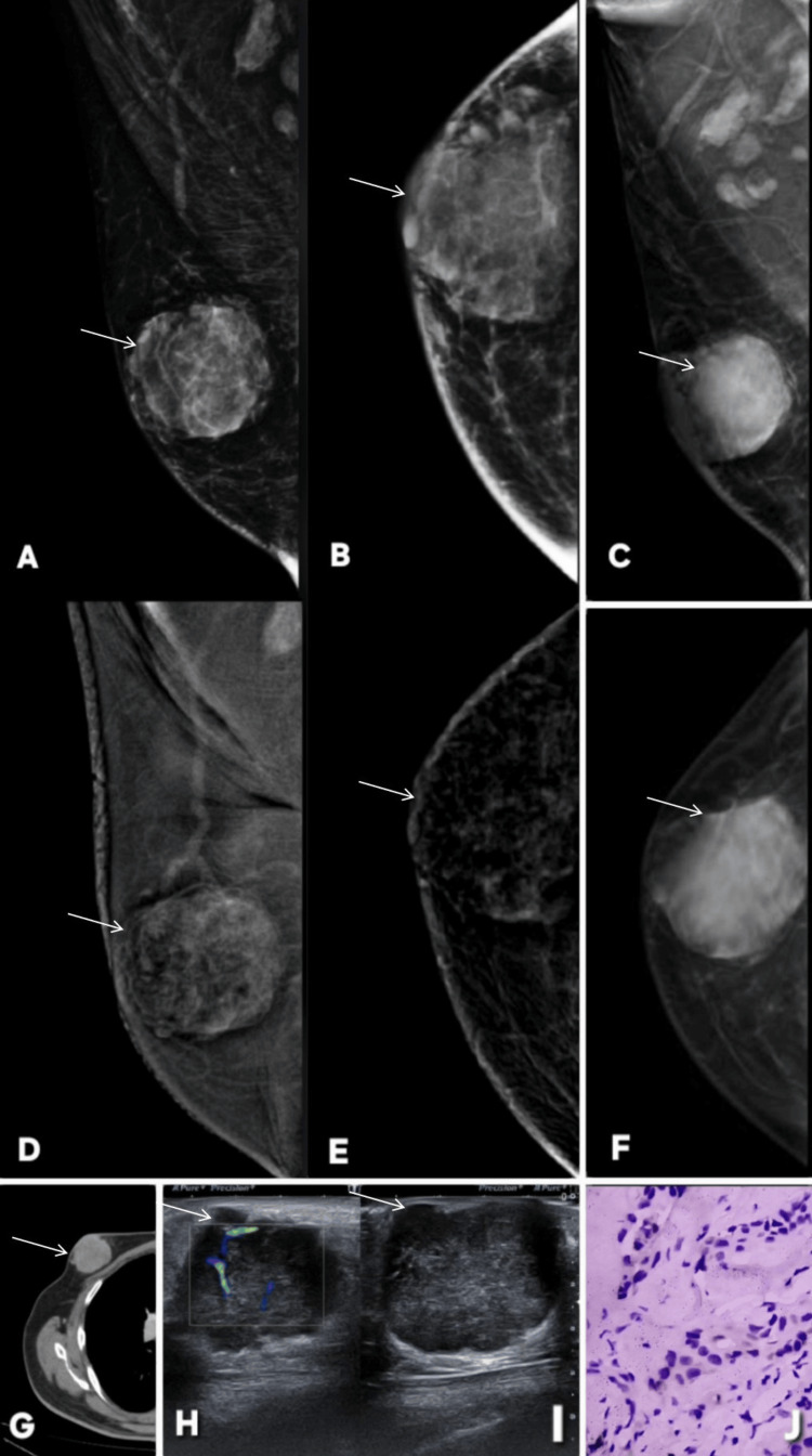 Figure 23
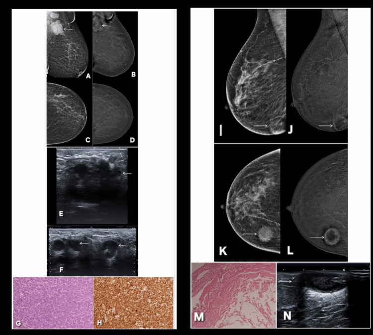 Figure 24
Peer Reviews
No public reviews on file for this paper yet. If you reviewed it on a platform where reviews are public (OpenReview, ICLR, NeurIPS, ICML), you can paste yours below so the community can read it here.
Videos
No videos yet. Explain this paper in a talk, walkthrough, or lecture? Add one.
Taxonomy
TopicsDigital Radiography and Breast Imaging · Breast Lesions and Carcinomas · AI in cancer detection