Evaluation of Partial Volume Correction Techniques for Sodium MRI of the Achilles Tendon
Rika Möller, Benedikt Kamp, Paula Leja, Thomas A. Thiel, Eric Bechler, Hans‐Jörg Wittsack, Gerald Antoch, Armin M. Nagel, Lena M. Wilms, Miriam Frenken, Anja Müller‐Lutz

TL;DR
This paper compares different methods to correct for partial volume effects in sodium MRI scans of the Achilles tendon, finding that a new method called eSTC gives the most accurate results.
Contribution
The novel eSTC method for partial volume correction in sodium MRI is introduced and shown to outperform existing techniques.
Findings
All PVC methods improved accuracy compared to no correction, with eSTC showing the smallest difference in simulated data.
In vivo results showed eSTC and GTM reduced resolution-related aTSC differences to less than 8 mM, compared to 38.8 mM without correction.
The eSTC method produced the most accurate apparent tissue sodium content calculations for Achilles tendon imaging.
Abstract
To evaluate partial volume correction (PVC) techniques for sodium MRI of the Achilles tendon in situ and in vivo. Five PVC methods were evaluated including a volume ratio of the proton and sodium segmentations (PSSR), a modified least trimmed square (3D‐mLTS) linear regression, a geometric transfer matrix (GTM) approach, a single target correction (STC), and a novel estimated single target correction (eSTC). Their performance was tested using simulated data and 3 T MR data of two volunteers' Achilles tendons acquired at different resolutions: 1.5, 2.0, 3.0, and 4.5 mm3. Since there was no in vivo ground truth, the highest‐resolution apparent tissue sodium contents (aTSC) were used. In the simulation, all PVC methods reduced the difference between the actual and calculated concentrations and were 11.69 ± 6.17 mM without PVC, 4.90 ± 5.40 mM with the PSSR, 4.86 ± 5.19 mM with the mLTS,…
Genes, proteins, chemicals, diseases, species, mutations and cell lines named across the full text — each resolved to its canonical identifier and authoritative record.
Click any figure to enlarge with its caption.
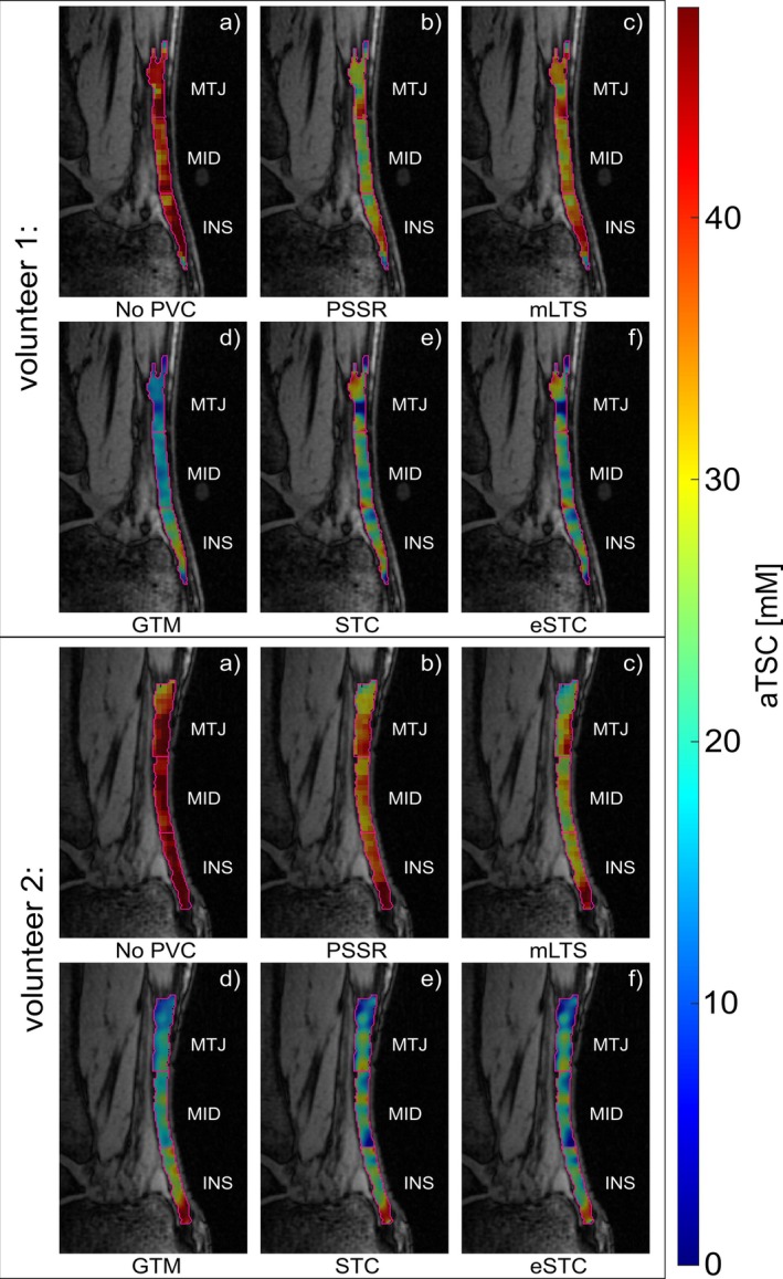 Figure 1
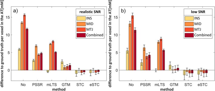 Figure 2
 Figure 3
 Figure 4
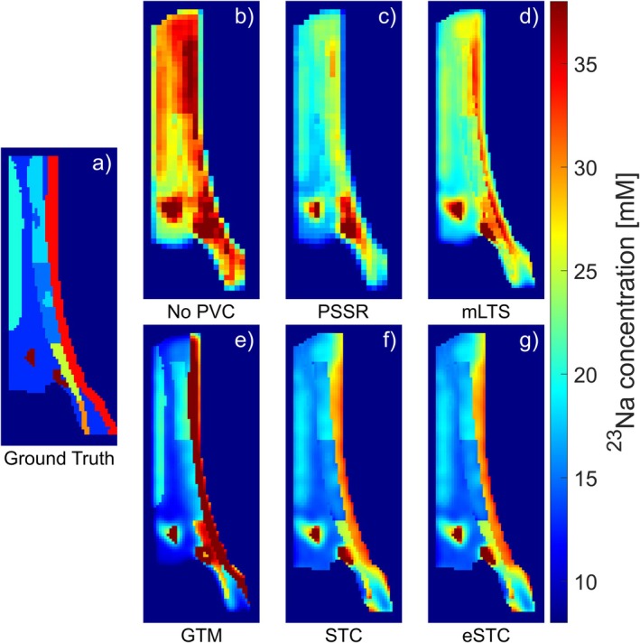 Figure 5
Peer Reviews
No public reviews on file for this paper yet. If you reviewed it on a platform where reviews are public (OpenReview, ICLR, NeurIPS, ICML), you can paste yours below so the community can read it here.
Videos
No videos yet. Explain this paper in a talk, walkthrough, or lecture? Add one.
Taxonomy
TopicsTendon Structure and Treatment · Shoulder Injury and Treatment · Advanced MRI Techniques and Applications