Investigation of Endogenous Renal CEST Contrast and the Influence of Respiratory Motion on a Clinical 3 Tesla MRI: An In Vivo and In Vitro Study
Patrik Jan Gallinnis, Benedikt Kamp, Karl Ludger Radke, Rika Möller, Anna‐Katharina Juric, Julia Stabinska, Vít Herynek, Gerald Antoch, Hans‐Jörg Wittsack, Alexandra Ljimani, Anja Müller‐Lutz

TL;DR
This study shows that timed breathing reduces motion artifacts in kidney MRI scans and reveals distinct chemical signals in different kidney regions.
Contribution
The study introduces timed breathing as a method to reduce motion artifacts in renal CEST MRI and identifies compartment-specific CEST effects.
Findings
Timed breathing significantly reduces motion artifacts in both phantom and in vivo CEST MRI data.
CEST effects vary significantly between renal cortex, medulla, and pelvis at different ppm frequencies.
MTRasym values in a patient with ccRCC differ significantly from those in healthy volunteers.
Abstract
The aim is to evaluate the effectiveness of timed breathing in reducing respiratory motion artifacts in renal chemical exchange saturation transfer (CEST) MRI and to assess potential differences in CEST effects between renal compartments. An electro‐pneumatic phantom with a kidney CEST model simulated variable respiratory motion and sequence‐synchronized breathing. Motion‐induced deviations from a static reference were quantified using the mean absolute error (MAE). Ten healthy volunteers (six females, four males; 25.2 ± 1.9 years) and one patient (47 years) with ccRCC (3.0 × 2.2 × 2.2) cm3 were examined on a 3 T MRI system using a multi‐echo gradient echo sequence with 15 Gaussian‐shaped saturation pulses (B1 = 1.5 μT, t p = t ipd = 100 ms). CEST effects in cortex, medulla, and pelvis at 1.0, 2.0, and 3.5 ppm were quantified by MTRasym analysis under timed and free‐breathing…
Genes, proteins, chemicals, diseases, species, mutations and cell lines named across the full text — each resolved to its canonical identifier and authoritative record.
Click any figure to enlarge with its caption.
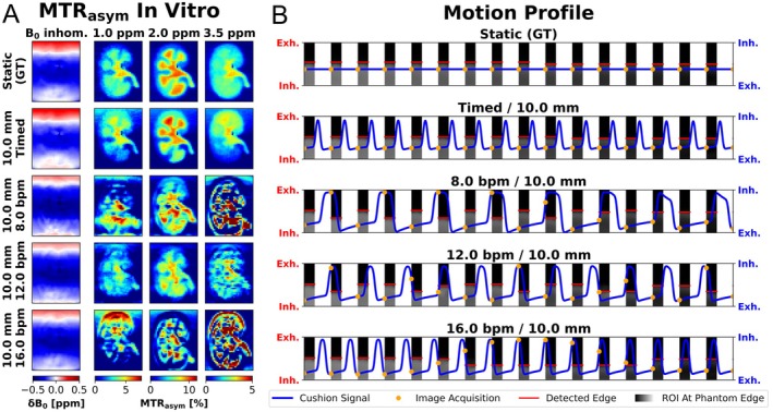 Figure 1
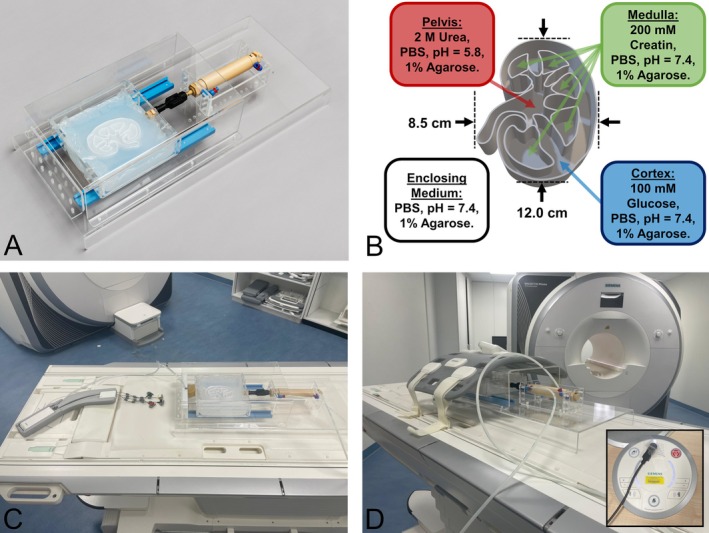 Figure 2
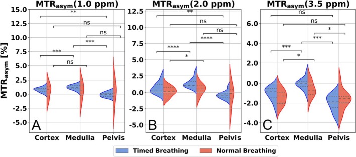 Figure 3
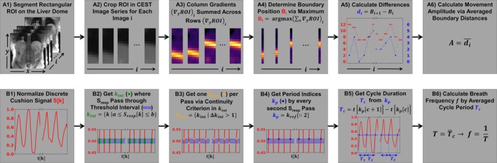 Figure 4
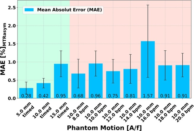 Figure 5
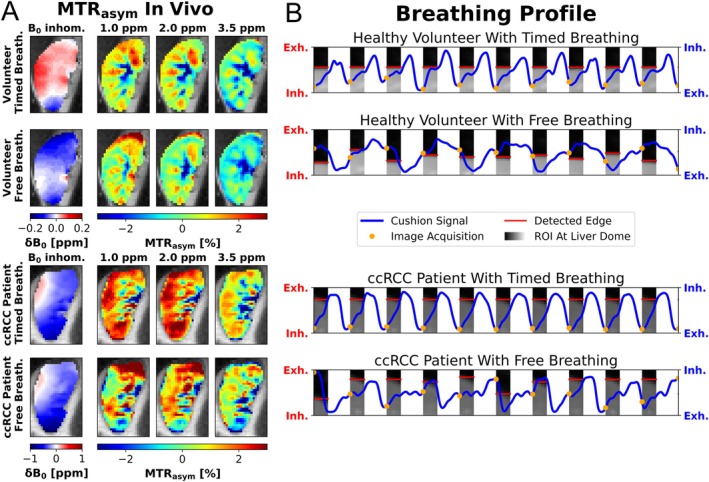 Figure 6
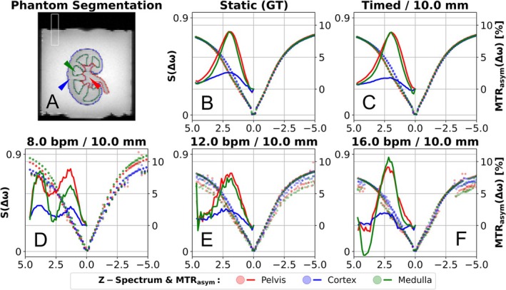 Figure 7
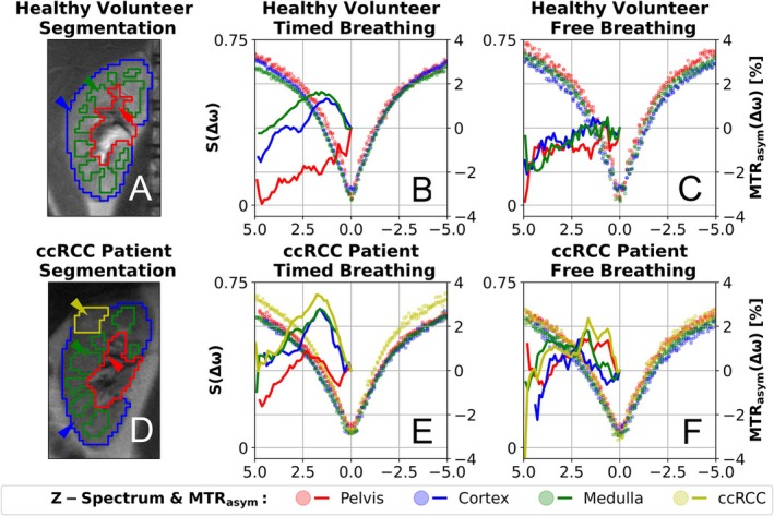 Figure 8
Peer Reviews
No public reviews on file for this paper yet. If you reviewed it on a platform where reviews are public (OpenReview, ICLR, NeurIPS, ICML), you can paste yours below so the community can read it here.
Videos
No videos yet. Explain this paper in a talk, walkthrough, or lecture? Add one.
Taxonomy
TopicsAdvanced MRI Techniques and Applications · MRI in cancer diagnosis · Lanthanide and Transition Metal Complexes