Quantitative Susceptibility Mapping in Skull Base Chordoma: In Silico Analysis and In Vivo Application Towards Indirect Hypoxia Assessment
P. Fenech, L. Morelli, G. Parrella, S. Imparato, A. Iannalfi, S. Lillo, E. Orlandi, G. Baroni, C. Paganelli

TL;DR
This study explores using QSM to assess hypoxia in skull base chordomas through simulations and patient data, showing promising results for non-invasive tumor characterization.
Contribution
The study introduces an optimized QSM pipeline for skull base chordomas and demonstrates its potential for indirect hypoxia assessment.
Findings
The optimized QSM pipeline achieved a phase unwrapping error of 38.36 ppm and background field removal error between 49 and 53 Hz.
QSM features correlated significantly with Ki-67 in SBC patients, with Spearman's coefficients of 0.8 and -0.8.
A binary classifier based on QSM features achieved 85.7% accuracy in distinguishing low- and high-proliferation tumors.
Abstract
To evaluate quantitative susceptibility mapping (QSM) beyond the brain through realistic simulations and to explore preliminary evidence that may be indicative of hypoxia in skull base chordomas (SBC). Each step of the QSM pipeline was optimized within an in silico framework consisting of (i) phase unwrapping, (ii) background field removal, and (iii) dipole field inversion, which were tested on a realistic phantom to generate accurate susceptibility maps. The optimized pipeline was then applied to seven SBC patients, analyzing tumor heterogeneity and correlating QSM features with the proliferation index (Ki‐67), towards hypoxia assessment. A binary classifier was developed to distinguish low‐ and high‐proliferation tumors based on first‐order QSM features. The optimal phase unwrapping method combined with dipole inversion provided an error of 38.36 ppm. The best strategy for…
Genes, proteins, chemicals, diseases, species, mutations and cell lines named across the full text — each resolved to its canonical identifier and authoritative record.
Click any figure to enlarge with its caption.
 Figure 1
 Figure 2
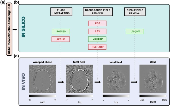 Figure 3
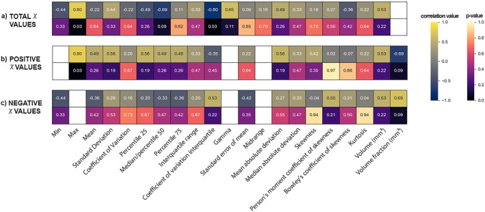 Figure 4
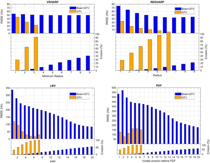 Figure 5
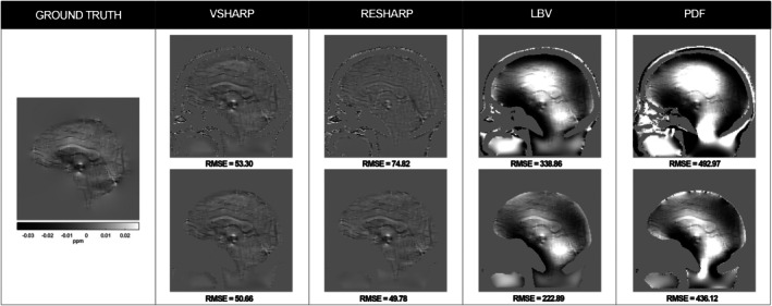 Figure 6
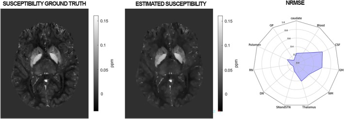 Figure 7
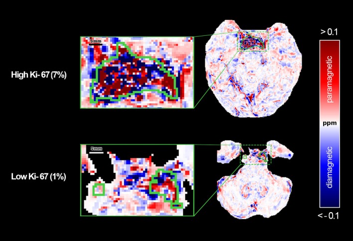 Figure 8
Peer Reviews
No public reviews on file for this paper yet. If you reviewed it on a platform where reviews are public (OpenReview, ICLR, NeurIPS, ICML), you can paste yours below so the community can read it here.
Videos
No videos yet. Explain this paper in a talk, walkthrough, or lecture? Add one.
Taxonomy
TopicsBone Tumor Diagnosis and Treatments · Head and Neck Surgical Oncology · Sarcoma Diagnosis and Treatment