Occurrence of Tick‐Borne Pathogens in Rhipicephalus sanguineus Sensu Lato From Domestic Dogs in Kumasi, Ghana
Sandra Abankwa Kwarteng, Jubin Osei Mensah, Patrick Kwasi Obuam, Enoch Ago Odenteh, Priscilla Denkyira Foriwaah, Anne Ifunanya Mbelede, Edwin Dziwornu, Ewurabena Oduma Duker, Jessica Dufie Boakye, Gayheart Deladem Agbotse, Jennifer Nyamekye Yanney, Millie-Cindy Aba Aude Koffi

TL;DR
This study identifies tick-borne pathogens in dog ticks from Kumasi, Ghana, highlighting the presence of Rickettsia, Ehrlichia, and Anaplasma with implications for human and animal health.
Contribution
The study provides the first baseline data on tick-borne pathogens in Rhipicephalus sanguineus ticks in Ghana.
Findings
36 out of 88 tick pools tested positive for pathogen DNA.
Uncultured Anaplasma sp. was the most prevalent pathogen detected.
No association was found between pathogen detection and tick or dog characteristics.
Abstract
Tick‐borne pathogens, transmitted by ticks, infect humans and animals worldwide. The brown dog tick, Rhipicephalus sanguineus sensu lato, is a significant vector of a number of pathogens, including Ehrlichia canis, Rickettsia and Anaplasma species. In Ghana, there is limited information on the pathogens carried by Rh. sanguineus s.l. As such, Rh. sanguineus ticks taken from domestic dogs in Kumasi were screened for tick‐borne pathogens, including Coxiella burnetii, Rickettsia, Babesia, Theileria, Anaplasma, Ehrlichia and Hepatozoon species. A total of 204 ticks collected from 56 infested dogs were morphologically identified as Rh. sanguineus s.l. From the 88 pools screened, 36 (40.9%) were positive for pathogen DNA. The pathogens identified were Rickettsia africae (5 pools), Ehrlichia canis (10 pools) and uncultured Anaplasma sp. (21 pools) with maximum likelihood estimates as 2.48%…
Genes, proteins, chemicals, diseases, species, mutations and cell lines named across the full text — each resolved to its canonical identifier and authoritative record.
Click any figure to enlarge with its caption.
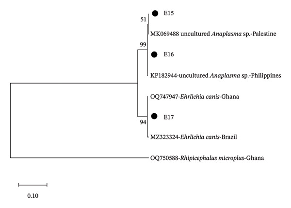 Figure FIGURE 1
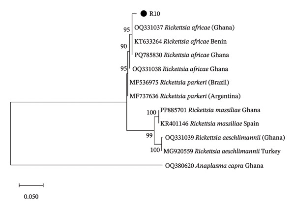 Figure FIGURE 2
| Characteristics |
|
|---|---|
| Location, | |
| Adagya | 31 (55) |
| Catena Veterinary Clinic | 25 (45) |
| Dog breed, | |
| Boerboel | 1 (1.8) |
| Cane Corso | 2 (3.6) |
| Caucasian Shepherd | 1 (1.8) |
| French Bulldog | 1 (1.8) |
| German Shepherd | 2 (3.6) |
| Husky | 1 (1.8) |
| Maltese | 3 (5.4) |
| Mongrel | 40 (71) |
| Pitbull | 1 (1.8) |
| Poodle | 1 (1.8) |
| Rottweiler | 3 (5.4) |
| Dog sex, | |
| Female | 22 (39) |
| Male | 34 (61) |
| Dog age (months), median (IQR) | 8 (4–24) |
| Species | Pathogen | Positives ( | Pool infection rate (%) | Minimum infection rate (%) | Maximum likelihood |
|---|---|---|---|---|---|
|
|
| 10/88 | 11.36 (5.59–19.91) | 4.93 (2.39–8.87) | 5.22 (2.69–9.15) |
|
|
| 5/88 | 5.68 (1.87–12.76) | 2.46 (0.80–5.65) | 2.48 (0.93–5.38) |
|
| Uncultured | 21/88 | 23.86 (15.42–34.14) | 10.34 (6.52–15.38) | 11.20 (7.32–16.29) |
| Location | Species | Pathogen | Positives ( | Pool infection rate (%) | Minimum infection rate (%) | Maximum likelihood |
|---|---|---|---|---|---|---|
| Adagya |
|
| 2/42 | 4.76 (0.58–16.16) | 2.60 (0.32–9.07) | 2.61 (0.47–8.29) |
| Adagya |
|
| 5/42 | 11.90 (3.98–25.63) | 6.49 (2.14–14.51) | 6.67 (2.53–14.14) |
| Adagya |
| Uncultured | 8/42 | 19.05 (8.60–34.12) | 10.39 (4.59–19.45) | 11.08 (5.32–20.07) |
| Catena Veterinary Clinic |
|
| 8/46 | 17.39 (7.82–31.42) | 6.35 (2.78–12.13) | 6.92 (3.29–12.81) |
| Catena Veterinary Clinic |
|
| 0/46 | 0.00 (0.00–7.71) | 0.00 (0.00–2.89) | 0.00 (0.00–2.96) |
| Catena Veterinary Clinic |
| Uncultured | 13/46 | 28.26 (15.99–43.46) | 10.32 (5.61–17.00) | 11.19 (6.51–17.81) |
| Characteristics |
|
| Uncultured | ||||||
|---|---|---|---|---|---|---|---|---|---|
| N | IRR (95% CI) |
| N | IRR (95% CI) |
| N | IRR (95% CI) |
| |
| Breed (dog) | 88 | 88 | 88 | ||||||
| Mongrel | — | — | — | ||||||
| Other | 2.63 (0.68–10.2) | 0.16 | 0.00 (0.00 to Inf) | > 0.99 | 1.07 (0.44–2.59) | 0.88 | |||
| Sex (dog) | 88 | 88 | 88 | ||||||
| Female | — | — | — | ||||||
| Male | 0.49 (0.12–1.95) | 0.31 | 157, 143, 015, 689, 921 (0.00 to Inf) | 0.97 | 0.98 (0.41–2.33) | 0.96 | |||
| Age (months) | 88 | 1.00 (0.94–1.06) | 0.97 | 88 | 0.97 (0.89–1.05) | 0.48 | 88 | 1.02 (0.98–1.05) | 0.36 |
| Sex (tick) | 88 | 88 | 88 | ||||||
| Female | — | — | — | ||||||
| Male | 3, 119, 900, 944, 862, 863 (0.00 to Inf) | 0.94 | 0.00 (0.00 to Inf) | > 0.99 | 1.40 (0.58–3.38) | 0.45 | |||
Peer Reviews
No public reviews on file for this paper yet. If you reviewed it on a platform where reviews are public (OpenReview, ICLR, NeurIPS, ICML), you can paste yours below so the community can read it here.
Videos
No videos yet. Explain this paper in a talk, walkthrough, or lecture? Add one.
Taxonomy
TopicsVector-borne infectious diseases · Viral Infections and Vectors · Zoonotic diseases and public health
1. Introduction
Ticks are obligatory hematophagous vectors that transmit numerous pathogens that negatively impact people and animals worldwide [1]. Due to its widespread distribution and intimate relationship with domestic dogs, which are common companion animals in both urban and rural areas, Rhipicephalus sanguineus s.l., often known as the brown dog tick, is of special veterinary and public health significance [2]. Numerous pathogens that cause substantial morbidity and mortality in domestic animals and, occasionally, in humans are spread by Rh. sanguineus s.l. [2, 3]. A typical example is Ehrlichia canis, which causes canine ehrlichiosis in dogs [4]. Other pathogens, such as Anaplasma and Rickettsia species, have also been reported in Rh. sanguineus s.l. ticks collected from dogs [5]. In Ghana, dogs in Kumasi have been reported as infected with pathogens including Ehrlichia canis, Hepatozoon canis and Anaplasma platys [6]. Furthermore, Rh. sanguineus s.l. collected from dogs were found infected with Rickettsia spp. [7]. These findings indicate the importance of dog ticks and the need to include them in surveillance efforts to prevent infection spread. The second‐largest city in Ghana, Kumasi, is known for its high human population and close contact with domestic animals, especially dogs. Humans, domestic animals and ticks frequently interact in the urban and peri‐urban environment, which makes it perfect for the spread of diseases carried by ticks. Despite this, little is known about the variety and frequency of tick‐borne pathogens in Rh. sanguineus s.l. ticks in dogs in this area. Knowing the distribution and abundance of these tick‐borne pathogens is essential for determining the risk of infection for humans and dogs and developing effective control measures.
This study was therefore carried out to molecularly screen Rh. sanguineus ticks obtained from domestic dogs in Kumasi, Ghana, for the presence of tick‐borne pathogens of zoonotic and veterinary importance. The results add to the expanding body of information on West African tick‐borne diseases and draw attention to the possible veterinary and public health consequences of infections in an urban African environment.
2. Methods
In this cross‐sectional study, ticks were collected from 56 infested domestic dogs from April to May 2025 at two locations in urban Kumasi: Catena Veterinary Clinic (6.67687, −1.61158) and Adagya (6.62026, −1.57909). At Adagya, tick collection was carried out during a community dog vaccination programme, where ticks were removed from visibly infested dogs. Each dog was physically examined by a veterinarian, and ticks were removed using blunt forceps and stored in 15 mL Falcon tubes containing 70% ethanol. Information such as location, dog breed, age and sex was recorded for each infested dog. In the laboratory, the ticks were identified morphologically using available taxonomic keys [8] and pooled (1–6) based on the sex of the tick species. Total nucleic acid was extracted from the pools using the QIAamp Extraction Kit (Qiagen, Valencia, CA, USA) according to the manufacturer’s instructions [9].
Ethical clearance for this study was obtained from the Animal Research Ethics Committee (AREC) of the Kwame Nkrumah University of Science and Technology (KNUST 0087).
2.1. Pathogen Detection
The samples were screened for Coxiella burnetii by the amplification of 687 bp using an assay targeting the IS1111 fragment, a transposon‐like repetitive region [10, 11]. Primers that target the Rickettsia rOmpA gene (ompA) were used to perform a 632 bp amplification to detect Rickettsia DNA in ticks [12]. The samples were also screened for Babesia/Theileria DNA using primers that target the 560 bp portion of the Babesia and Theileria ssrRNA gene [13]. Primers targeting the 345 bp region of the Ehrlichia genus 16S rRNA gene were used to detect the presence of Ehrlichia/Anaplasma DNA in the ticks [14]. An assay that amplifies the 18S rRNA gene of Hepatozoon species at 666 bp was used to screen the ticks for Hepatozoon species [15].
The positive PCR products were subsequently purified and sequenced using Sanger sequencing.
2.2. Phylogenetic and Statistical Analysis
Following a review of the nucleotide sequences produced in this investigation using Chromas (Version 2.6.6), a consensus sequence was edited, cleaned and created using MEGA (Version 12.0.11). Next, each sequence was contrasted with other sequences (https://blast.ncbi.nlm.nih.gov/Blast.cgi) in the NCBI database. MEGA’s Clustal Omega programme was used for sequence alignment, and the maximum likelihood approach was used to build phylogenetic trees [16]. A thousand bootstrap replicates were used to calculate the confidence indices inside the phylogenetic trees, and the findings were shown as percentages on the branches. For the GenBank sequences used in the phylogenetic analysis, the different accession numbers and countries of origin have been specified. We used descriptive statistics, such as counts and proportions, to summarise the data. Next, we determined each pathogen’s minimum infection rate, providing the precise 95% CIs as well as the point estimate. We also used the PooledInfRate tool (https://github.com/CDCgov/PooledInfRate) to compute the pooled prevalence estimate. Tick species and tick species within each location were used to stratify these estimations. Next, we used Poisson regression and clustered errors to evaluate those characteristics linked to higher prevalence ratios (PRs) for each detected pathogen. We adjusted for animal identification and pool size using an offset. R Version 4.3.3 was used for all analysis.
3. Results
A total of 56 dogs were examined from Catena Veterinary Clinic (25) and Adagya (31) (Table 1). Most of the dogs were mongrels (71%), male (61%) and had a median age of 8 months. All the 204 adult ticks collected from the dogs (average of 3.64 ticks per dog) were identified as Rhipicephalus sanguineus s.l. The males were 109 (53.43%), while the females were 95 (46.57%).
From the 88 pools screened, pathogen DNA was detected in 36 (40.9%) tick pools. The pathogens identified were R. africae in 5 (5.68%) tick pools, Ehrlichia canis in 10 (11.36%) pools and uncultured Anaplasma sp. in 21 (23.86%) pools. Generally, the MLE of the identified pathogens was 2.48% (95% CI: 0.93, 5.38%), 5.22% (95% CI: 2.69, 9.15%) and 11.20% (95% CI: 7.32, 16.29%) for R. africae, E. canis and uncultured Anaplasma sp., respectively (Table 2). The MLE of uncultured Anaplasma sp. in the tick pools was 11.08% (95% CI: 5.32, 20.07%) from Adagya and 11.19% (95% CI: 6.51, 17.81%) from Catena Veterinary Clinic (Table 3). The MLE of the zoonotic pathogen R. africae was 6.67% (95% CI: 2.53, 14.14%) in tick pools obtained from Adagya. There was no association between the detection of a pathogen and the tick sex or dog breed, age or sex (Table 4). None of the tick pools were positive for C. burnetii, Babesia, Theileria or Hepatozoon DNA.
3.1. Basic Local Alignment Search Tool and Phylogenetic Analysis
It was observed that the sequences E15 and E16 were 100% similar to uncultured Anaplasma sp. from Palestine (MK069488). The sequence E17 was also 100% similar to E. canis from Brazil (MZ323324), and the sequence R10 was 98% similar to R. africae from Benin (KT633264). From the phylogenetic tree, it was seen that the sequences E15 and E16 were related to uncultured Anaplasma sp. reported from Palestine and the Philippines, while the sequence E17 was related to E. canis from Brazil (Figure 1). Sequence R10 was found to be closely related to R. africae reported from Ghana and Benin (Figure 2).
Phylogenetic analysis of the identified Ehrlichia and Anaplasma based on the 16S rRNA gene. The sequences from this study are E15, E16 and E17.
Phylogenetic analysis of the identified Rickettsia based on the OmpA gene. The sequence ID from this study is designated as R10.
The sequences generated in this study have been submitted to GenBank with accession numbers: uncultured Anaplasma sp. (PV861966 and PV861967), E. canis (PV861968) and R. africae (PV870140).
4. Discussion
Rhipicephalus sanguineus s.l. ticks collected from domestic dogs in Kumasi were infected with tick‐borne pathogens of zoonotic and veterinary importance.
Ehrlichia canis was identified in 11.36% of the tick pools in this study. This finding is higher than the E. canis prevalence of 6.7% reported previously in Ghana [17] but lower than 23.7% reported in a study from Nigeria [5]. This pathogen causes the disease canine ehrlichiosis, a common and frequently underdiagnosed disease in tropical Africa that affects dogs [4, 18]. Its clinical symptoms can range from mild to severe and include fever, anaemia, bleeding abnormalities and immunosuppression [18]. The fact that E. canis was found in the sampled ticks suggests that domestic dogs are involved in transmission cycles and could act as infection reservoirs. Reports of seropositivity in humans exposed to Rh. sanguineus s.l. ticks and reported instances in immunocompromised persons and blood donors underscore a possible zoonotic danger, notwithstanding the rarity of human ehrlichiosis caused by E. canis [19–22]. This emphasises how crucial it is for veterinarians and public health professionals in Kumasi to be aware of integrated tick management strategies.
In this study, uncultured Anaplasma sp. was also detected in the Rh. sanguineus s.l. ticks. Their intracellular nature and the absence of culture methods make it difficult to characterise these uncultured strains [23, 24], but their identification in ticks that parasitise domestic dogs raises questions regarding their potential to be pathogenic. Thrombocytopenia, lethargy and fever are common symptoms of Anaplasma infections in dogs, which can make veterinary diagnosis and treatment more difficult [25, 26]. Furthermore, caution is required due to the zoonotic potential of some Anaplasma species [23], particularly in areas where tick exposure is common. To determine the role of the identified uncultured Anaplasma sp. in animal and human disease, additional molecular characterisation and epidemiological research are necessary.
This study reports the detection of R. africae in Rh. sanguineus s.l. The detection of R. africae in 5.68% of the tick pools from this study is higher than the 2.2% reported by a previous study in Ghana [17]. Rickettsia africae, the causative agent of African tick‐bite fever (ATBF), is primarily transmitted by Amblyomma variegatum and Amblyomma hebraeum [27]. However, Rhipicephalus and Hyalomma ticks that cofeed with Amblyomma ticks can be infected with R. africae [28]. Among travellers and rural people in sub‐Saharan Africa, ATBF is a major cause of febrile illness [29]. Studies in Ghana focussed on livestock ticks have reported the occurrence of R. africae [30–33]. With the frequent interactions among domestic animals in Ghana, there is the risk of tick or tick‐borne pathogen exchange [34]. Given the presence of R. africae in ticks that infest domestic dogs, dogs may serve as amplifying hosts or reservoirs, which could raise the danger of human exposure in urban settings. Since Rh. sanguineus frequently infests dogs in homes, there is a chance that humans could become infected via tick bites, particularly in areas where people are not well informed or take precautions against tick bites [2]. Increased surveillance, prevention of tick bites and the inclusion of R. africae infection in the differential diagnosis of feverish diseases in the region are all recommended by this finding.
The complicated epidemiology of tick‐borne illnesses in urban African environments is highlighted by the codetection of all three pathogens in Rh. sanguineus ticks from Kumasi. Domestic dogs facilitate the maintenance and spread of several tick‐borne diseases in humans by acting as significant hosts and reservoirs [35]. Tick bites and associated pathogen transmission are more likely in Kumasi due to the proximity of people and dogs. The findings of this study bring out two challenges: effectively diagnosing and managing tick‐borne infections in dogs to better protect animal health and reducing the risk of zoonotic infections to safeguard human health. Regular tick control for dogs, public education campaigns on preventing tick bites and enhanced diagnostic tools for tick‐borne illnesses in the veterinary and human healthcare sectors are all examples of effective control measures. Furthermore, continuous molecular surveillance will be an effective method for detecting circulating pathogens and monitoring the risk of emerging threats.
5. Conclusion
This study reports the occurrence of E. canis, R. africae and uncultured Anaplasma sp. in Rh. sanguineus s.l. collected from domestic dogs. The identification of these pathogens in tick species that are closely linked to humans and dogs highlights the possible danger of tick‐borne diseases in veterinary and public health settings. There is an urgent need for integrated surveillance and control strategies to reduce the risk posed to the health of both domestic animals and humans.
Funding
No funding was received for this manuscript.
Conflicts of Interest
The authors declare no conflicts of interest.
The reference list from the paper itself. Each links out to its DOI / PubMed record.
- 1Brites-Neto J. , Duarte K. M. R. , and Martins T. F. , Tick-Borne Infections in Human and Animal Population Worldwide, Veterinary World. (2015) 8, no. 3, 301–315, 10.14202/vetworld.2015.301-315, 2-s 2.0-84924935134.27047089 PMC 4774835 · doi ↗ · pubmed ↗
- 2Dantas-Torres F. , Biology and Ecology of the Brown Dog Tick, Rhipicephalus sanguineus , Parasites & Vectors. (2010) 3, no. 1, 1–11, 10.1186/1756-3305-3-26, 2-s 2.0-77950674814.20377860 PMC 2857863 · doi ↗ · pubmed ↗
- 3Shih C. M. , Ko P. Y. , and Chao L. L. , Molecular Survey and Genetic Analysis of Ehrlichia canis in Rhipicephalus sanguineus Ticks Infesting Dogs in Northern Taiwan, Microorganisms. (2025) 13, no. 6, 10.3390/MICROORGANISMS 13061372.PMC 1219628740572260 · doi ↗ · pubmed ↗
- 4Aziz M. U. , Hussain S. , Song B. , Ghauri H. N. , Zeb J. , and Sparagano O. A. , Ehrlichiosis in Dogs: A Comprehensive Review About the Pathogen and Its Vectors With Emphasis on South and East Asian Countries, Veterinary Sciences. (2022) 10, no. 1, 10.3390/VETSCI 10010021.PMC 986337336669021 · doi ↗ · pubmed ↗
- 5Kamani J. , Baneth G. , Mumcuoglu K. Y. et al., Molecular Detection and Characterization of Tick-Borne Pathogens in Dogs and Ticks From Nigeria, P Lo S Neglected Tropical Diseases. (2013) 7, no. 3, 10.1371/JOURNAL.PNTD.0002108, 2-s 2.0-84875973662.PMC 359132523505591 · doi ↗ · pubmed ↗
- 6Clarke L. L. , Ballweber L. R. , Allen K. , Little S. E. , and Lappin M. R. , Prevalence of Select Vector-Borne Disease Agents in Owned Dogs of Ghana, Journal of the South African Veterinary Association. (2014) 85, no. 1, 10.4102/jsava.v 85i 1.996, 2-s 2.0-84931393042.25686301 · doi ↗ · pubmed ↗
- 7Nimo-Paintsil S. C. , Mosore M. , Addo S. O. et al., Ticks and Prevalence of Tick-Borne Pathogens From Domestic Animals in Ghana, Parasites & Vectors. (2022) 15, no. 1, 10.1186/s 13071-022-05208-8.PMC 891778435279200 · doi ↗ · pubmed ↗
- 8Walker A. R. , Bouattour A. , Camicas J.-L. et al., Ticks of Domestic Animals in Municipal Abattoir for Their Technical Support, Africa: A Guide to Identification of Tick Species, 2003.