Constitutive NF-kB Activation Is Amplified by VSV in Aggressive PC3 Prostate Cancer Cells That Resist Viral Oncolysis
Alaa A. Abdelmageed, Jack F. Smerczynski, Mukul Kandwal, Lute J. Douglas, Tori L. Russell, Matthew C. Morris, Stephen Dewhurst, Maureen C. Ferran

TL;DR
This study shows that aggressive prostate cancer cells resist a virus therapy due to increased NF-kB signaling, which could lead to new treatment strategies.
Contribution
The study identifies constitutive NF-κB activation as a mechanism of resistance to VSV oncolysis in PC3 prostate cancer cells.
Findings
NF-κB localized to the nucleus in VSV-infected PC3 cells but not in LNCaP cells.
PC3 cells showed higher levels of total and phosphorylated IκB-α compared to LNCaP cells.
VSV infection increased phosphorylated NF-κB p65 in PC3 cells and upregulated NF-κB-dependent genes like IL12 and IL6.
Abstract
Cancer cells often have defects in antiviral pathways, making them susceptible to oncolytic viruses like vesicular stomatitis virus (VSV). However, some cancer cells resist viral infection through the constitutive expression of interferon-stimulated genes. This study examined whether NF-κB activation and NF-κB-dependent antiviral signaling contribute to resistance to VSV infection in the PC3 cell line, derived from an aggressive metastatic prostate cancer (PrCa) tumor. We found that NF-κB localized to the nucleus in VSV-infected PC3 cells, but not in the VSV-susceptible LNCaP PrCa cell line. Analysis of the upstream NF-κB inhibitor IκB-α revealed higher levels of both total and phosphorylated IκB-α in PC3 cells compared to LNCaP cells, indicating constitutive activation of the NF-κB pathway via an IκB-α-dependent mechanism. Notably, VSV infection did not alter IκB-α phosphorylation in…
Genes, proteins, chemicals, diseases, species, mutations and cell lines named across the full text — each resolved to its canonical identifier and authoritative record.
Click any figure to enlarge with its caption.
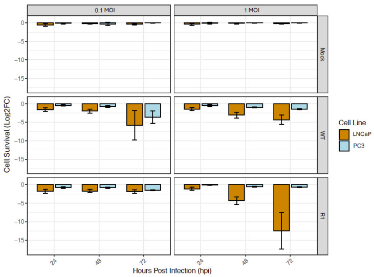 Figure 1
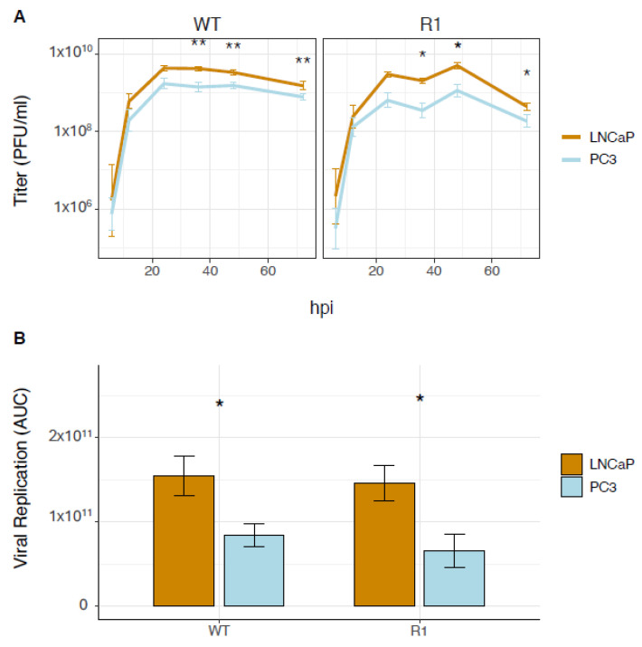 Figure 2
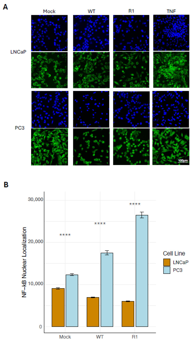 Figure 3
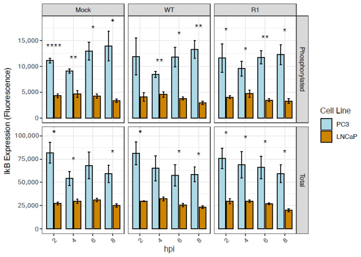 Figure 4
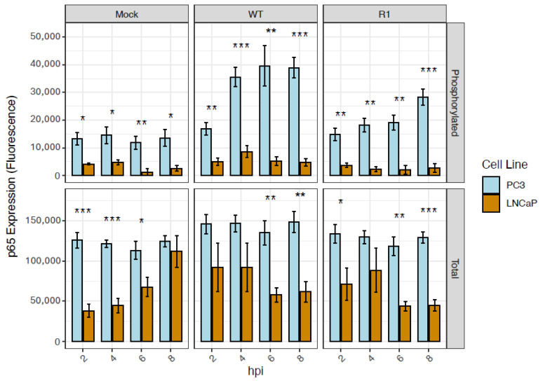 Figure 5
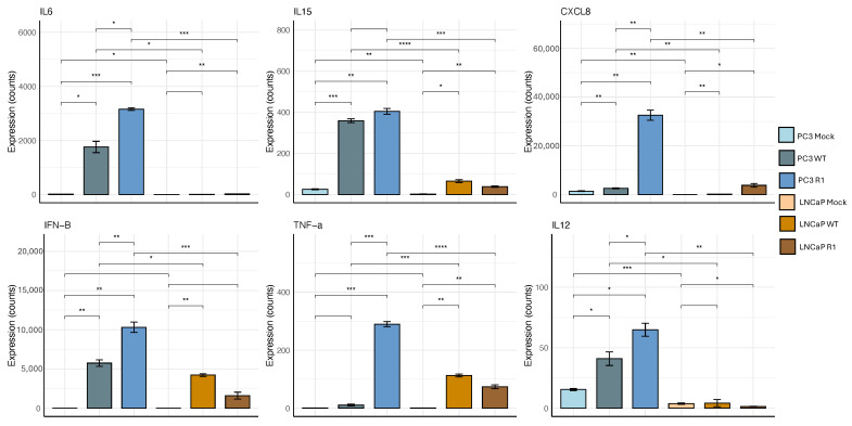 Figure 6
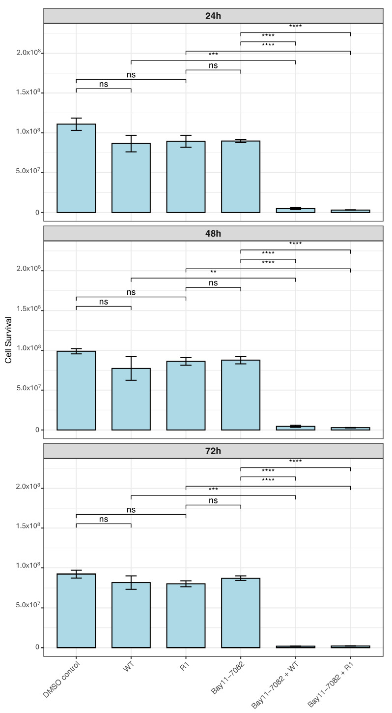 Figure 7
- —NIH
- —Dewhurst lab at University of Rochester
- —Ferran lab at Rochester Institute of Technology
Peer Reviews
No public reviews on file for this paper yet. If you reviewed it on a platform where reviews are public (OpenReview, ICLR, NeurIPS, ICML), you can paste yours below so the community can read it here.
Videos
No videos yet. Explain this paper in a talk, walkthrough, or lecture? Add one.
Taxonomy
Topicsinterferon and immune responses · NF-κB Signaling Pathways · Virus-based gene therapy research
1. Introduction
Genetic abnormalities can accumulate in tumors, driving uncontrolled cell proliferation and survival. These mutations often perturb antiviral responses, including the interferon (IFN) pathway, leaving malignant cells susceptible to oncolytic (cancer-killing) viruses such as VSV [1,2,3,4]. In contrast, healthy cells retain intact antiviral pathways that restrict viral replication and prevent cytotoxicity. Therefore, oncolytic viruses selectively replicate in cancer cells, causing direct cytolysis and the release of tumor-associated antigens, which trigger immunogenic cell death. This process enhances anti-tumor immunity by activating both innate and adaptive immune responses, promoting systemic tumor clearance and potential long-term therapeutic benefits [5].
Preclinical studies indicate that vesicular stomatitis virus (VSV) exhibits strong anti-tumor activity against various cancers, including lung [6,7,8], breast [9,10,11], and prostate cancers (PrCa) [12,13,14]. Early-phase clinical trials have demonstrated that VSV-based oncolytic virotherapy is generally well-tolerated and holds promise as a therapeutic strategy, especially when combined with immune checkpoint inhibitors [15,16,17]. Common approaches to improve safety and enhance tumor specificity of VSV include the use of recombinant viruses that are unable to suppress antiviral pathways in infected cells, a function of the viral matrix (M) protein. These include viruses that express IFN type-I from the viral genome or contain a deletion or substitution at position 51 of M [18,19,20]. In addition, efforts to increase targeting of the virus to cancer cells include combining VSV infection with different classes of chemotherapeutic drugs [20].
Despite these encouraging results, challenges remain. Resistance to oncolytic virus-mediated cytotoxicity has been observed across various cancer types and is considered a main challenge to developing viruses as oncolytic agents. Due to tumor heterogeneity, both virus-sensitive and virus-resistant cells can coexist in the same tumor [4]. Resistant tumor cells exhibit reduced susceptibility to viral infection and replication; however, a comprehensive understanding of the molecular mechanisms that drive resistance is lacking.
Retention of antiviral pathways is believed to be a key factor in resistance to oncolytic viruses. This has been observed in the PC3 PrCa cell line, which was derived from an aggressive metastatic PrCa tumor [21]. PC3 cells exhibit reduced susceptibility to killing by both wild-type (WT) [13,18,22,23,24] and mutant VSV strains [13,22,25]. Comparatively, the LNCaP cell line is sensitive to VSV infection [4,13,26] and was derived from a lymph node that was proximal to a prostate tumor [27]. Another study demonstrated that multiple early stages of the VSV replication cycle are delayed in the PC3 cell line, but not in LNCaP cells, likely as a consequence of preserved antiviral signaling mechanisms [4].
Constitutive activation of NF-kB, a pivotal transcription factor involved in the induction of antiviral responses, has been observed in many aggressive and metastatic tumors [28]. This constitutive activation is implicated in promoting several oncogenic processes, including accelerated cell cycle progression, increased cellular proliferation [29,30], resistance to apoptosis, enhanced metastatic potential in prostate cancer (PrCa) [31], and reduced responsiveness to conventional therapies [32]. Given these roles, the present study aimed to investigate whether NF-kB activation and NF-kB-dependent antiviral signaling contribute to the resistance of PC3 cells to oncolytic viral infection. The prognosis for patients with advanced metastatic PrCa remains poor, with a five-year survival rate of approximately 30% [13]. Moreover, therapeutic options for late-stage disease are limited, and treatments effective in other solid tumors have shown minimal efficacy in this context. These challenges underscore an urgent need to develop novel therapeutic strategies for the management of advanced PrCa.
Our findings confirm that PC3 cells are resistant to VSV-mediated killing [13], using both wild-type VSV (WT) and a mutant virus containing a substitution at position 51 of the M protein (R1; see Methods section for a full description of this virus). We also show that PC3 cells exhibit constitutive activation of NF-kB through an IkB-a-dependent pathway [28,33]. In contrast, LNCaP cells are susceptible to VSV infection [4,13,26] and do not exhibit constitutive NF-kB activity [26]. In PC3 cells, phosphorylation of IkB-a at serine residue 32 was unaffected by VSV infection, suggesting that the constitutive activity of NF-kB is not further modulated through canonical IkB-a phosphorylation. Notably, PC3 cells displayed higher levels of the NF-kB p65 protein subunit relative to LNCaP cells, with the most elevated levels of phosphorylated p65 (serine 276) following VSV infection.
NF-kB translocation (activation) in PC3 cells was found to be significantly higher than in LNCaP cells in the absence of viral infection, confirming constitutive activation of NF-kB in the resistant cells [34,35]. This was associated with high baseline levels of both total and phosphorylated IkB-a. Following VSV infection in PC3 cells, nuclear translocation of NF-kB was further increased, but IkB-a phosphorylation was unchanged, suggesting that VSV may amplify NF-kB signaling in PC3 cells through an IkB-a–independent/non-canonical pathway. In contrast, little NF-kB activation was noted in LNCaP cells, regardless of VSV infection. To evaluate downstream functional consequences of this differential NF-kB signaling, we assessed the expression of NF-kB-dependent effector genes in response to VSV infection. Baseline expression of several genes encoding antiviral cytokines and proinflammatory mediators was higher in uninfected PC3 cells compared to LNCaP cells, and VSV infection further amplified their expression.
Collectively, these findings suggest that constitutive and VSV-enhanced NF-kB activation contribute to the resistance of the highly metastatic PC3 PrCa cell line to VSV infection, likely through the upregulation of NF-kB-dependent antiviral and inflammatory signaling pathways.
2. Methods
2.1. Cells, Viruses, and Infections
The LNCaP and PC3 human PrCa cell lines were obtained from the American Type Culture Collection (ATCC) (CRL-1740 and CRL-1435, respectively) and were grown in RPMI-1640 containing 10% FBS (ATCC). The heat-resistant (HR-C) strain of the Indiana serotype of VSV was used as the WT virus. Mutant T1026R1 (R1), a temperature-stable revertant of T1026, was derived from the Indiana HR-C strain of VSV by chemical mutagenesis (supplied by C. P. Stanners). The R1 mutant, which harbors a single amino acid substitution at position 51 of the M protein (M51R), was used as a representative example of a virus strain that is attenuated relative to the WT virus—and thus has a more favorable safety profile for potential use as an oncolytic agent in humans [18,19,20]. All viruses were grown on Vero cells as described [36] in EMEM growth medium (ATCC). Cells were infected with each virus at multiplicities of infection (MOI) ranging from 0.1, 1, 5, 10, to 50 plaque-forming units (PFUs)/cell (see Figure legends). Viruses were allowed to adsorb to target cells in serum-free culture medium for 1 h at 37 °C, after which complete (serum-containing) culture medium was added to the cells.
2.2. PrestoBlue Viability Assay
PC3 and LNCaP cells were seeded in Santa Cruz Biotech black frame/clear bottom cell culture 96-well plates (sc-204468). After 24 h, cells were infected with either WT VSV or the R1 mutant at an MOI of 0.1 or 1. At 24, 48 and 72 h post infection (hpi), supernatants were removed and 100 µL of master mix was added, composed of 10% PrestoBlue HS Cell Viability Reagent (Thermo Fisher Sci, Waltham, MA, USA) in serum-free, phenol red-free RPMI medium. This viability reagent is reduced by viable cells and converted into a red fluorescent dye. Plates were then incubated in the dark at 37 °C for 40 min and a fluorescence reading was taken on a SpectraMax iD3 Plate Reader (Molecular Devices, San Jose, CA, USA) using an Em/Ex 590/550 nm wavelength range.
2.3. Plaque Assay
6-well plates were infected with WT or R1 VSV strains at an MOI of 0.1. After each corresponding hpi, a serial dilution of the supernatant was performed (between 1 × 10^−2^ and 1 × 10^−9^). A plaque quantification assay was then performed using the serial dilutions and according to the Cell Biology Protocols workflow. For the readout, the number of plaques for each sample was counted and the area under the curve (AUC) was calculated by the trapezoidal rule in the pracma CRAN package (Version 2.4.6).
2.4. Immunofluorescence
Immunofluorescence analysis was conducted as described [36,37] except that cells were blocked with UltraCruz^®^ Blocking Reagent (sc-516214, Santa Cruz Biotechnology, Dallas, TX, USA) for 30 min followed by incubation with an NF-kB-(p65) antibody conjugated to Alexa Fluor^®^ 488 (sc-8008 AF488, Santa Cruz Biotechnology) for 90 min at room temperature. Images were then taken using a Leica TCS SP5 II confocal microscope, from Leica Microsystems, Mannheim, Germany. Nuclear localization of p65 was quantified using ImageJ2 version 2.0.0-rc-3. Locations of cell nuclei and density of NF-kB staining in those regions were determined by automated analysis of DAPI and FITC channel images. Each DAPI channel image was subjected to a Gaussian blur filter with a sigma value of 2. Next, the image was thresholded using the default algorithm, and the watershed tool was used to separate adjoining nuclei. Finally, nuclei were defined as regions of interest ranging from 200–20,000 pixels in size, with nuclei on the edge of each image excluded. The intensity of NF-kB staining in each nuclear region was then quantified. The background fluorescence times the area of each nuclear region was subtracted from the integrated fluorescent density of NF-kB staining in each nucleus to yield the corrected total cell fluorescence (CTCF) for each cell. Median CTCF among all cells in each image was taken as representative.
2.5. IkB-a LUMIT Assay
LNCaP or PC3 cells in Corning white 96-well cell culture plates were treated with 20 µM (30 µL) of the proteasome inhibitor MG132 for 1 h before infection. Next, PC3 cells were infected with WT or R1 VSV at an MOI of 50 PFU/cell for 1 h, while LNCaP cells were infected at an MOI of 10 PFU/cell. After adsorption, the cells were re-fed with complete media containing 20 µM (40 µL) of MG132. At the indicated hpi, the amount of total and phosphorylated IkB-a was determined using the LUMIT Immunoassay Cellular Systems Kit from Promega, Maddison, WI, USA according to the manufacturer’s directions. The following antibodies were purchased from Cell Signaling Technology Inc., Danvers, MA, USA and were used at 150 ng/mL: rabbit anti-phospho-IκBα (#2859), mouse anti-phospho-IκBα (#9246), mouse anti-IκBα (#4814), and rabbit anti-IκBα (#4812). Briefly, the cells were lysed, and the appropriate combination of primary and secondary antibodies was added to the cell lysates. Luminescence substrate was introduced to the antibody lysate mixture and luminescence values of phosphorylated and total IkB-a protein were quantified using a SpectraMax iD3 Plate Reader (Molecular Devices, San Jose, CA, USA).
2.6. P65 LUMIT Assay
LNCaP or PC3 cells were seeded in a Corning white 96-well cell culture plate 24 h prior to infection. Cells were infected with WT or R1 VSV at an MOI of 50 PFU/cell for PC3 cells and an MOI of 10 PFU/cell for LNCaP cells. At the indicated hpi, the amount of total and phosphorylated p65 protein was determined using the LUMIT Immunoassay Cellular Systems Kit from Promega according to the manufacturer’s directions. The following antibodies were purchased from Cell Signaling Technology: mouse anti-phospho-p65 (#13346), rabbit anti-p65 (#8242), and mouse anti-p65 (#6956). Briefly, the cells were lysed, and the appropriate combination of primary and secondary antibodies was added to the cell lysates. Luminescence substrate was introduced to the antibody lysate mixture and luminescence values of phosphorylated and total p65 protein were quantified using a SpectraMax iD3 Plate Reader (Molecular Devices).
2.7. RNA Sequencing
Cells were grown in 30 mm tissue culture dishes coated with poly-D-lysine (PDL). Infection was carried out using VSV at an MOI of 50 and 10 PFU/cell for PC3 and LNCaP, respectively. At 6 hpi for LNCaP and 8 hpi for PC3 cells, cells were lysed in plates using RIPA lysis buffer (Thermo Scientific, Waltham, MA, USA; RIPA Lysis and Extraction Buffer). Samples were sent to the University of Rochester Genomic Center for library preparation and sequencing. Analysis was performed on count data using the Bioconductor DESeq2 package (Version 1.44.0) to calculate differential expression (DE) levels. Contrasts were made on the DE data by comparing virus treatments to mock treatment for each cell line.
2.8. Bay11-7082
Cells were treated with 2.5 µM of Bay11-7082, purchased from Med Chem Express, Monmouth Junction, NJ, USA; cat#: HY-13453, for 24, 48 and 72 h. 6 h prior to each timepoint, LNCaP cells were infected with an MOI of 10, while at 8 h prior to each timepoint, PC3 cells were infected with an MOI of 50. Following infection, samples were treated with PrestoBlue, and readout was measured as fluorescence with a SpectraMax iD3 Plate Reader (Molecular Devices).
2.9. Statistical Analysis
Statistical analysis throughout this paper was performed in RStudio (Version R version 4.4.0 (2024-04-24) –“puppy cup”) using either the Student’s t test or the one-way ANOVA test and an asterisk indicates a significant change (p < 0.05). Unless otherwise noted, results are expressed as means and error bars indicate the ±standard error of the means (SEM).
3. Results
3.1. PC3 Cells Are More Resistant to VSV-Mediated Killing than LNCaP Cells
To assess cellular resistance to VSV, cell death in response to infection with WT VSV or the mutant VSV strain, R1, at an MOI of either 0.1 or 1 MOI was determined using a PrestoBlue assay. PC3 cells exhibited lower levels of cell killing, as compared to LNCaP cells at 24, 48, and 72 hpi (Figure 1). This was especially evident at 72 hpi with WT VSV at an MOI of 0.1, and at 72 hpi with R1 VSV at an MOI of 1. These findings confirm the greater resistance of PC3 cells to VSV-mediated cell killing versus LNCaP cells.
3.2. VSV Replicates More Efficiently in LNCaP Cells than in PC3
To assess VSV replication in both cell lines, a multiple-step growth curve assay was performed. Both cell lines were infected with an MOI of 0.1 and supernatants were isolated for plaque assays. Marginal differences in replication between the strains were observed over 72 hpi; however, such differences were insignificant (Figure 2A). On the other hand, significant differences in WT and R1 replication were observed between PC3 and LNCaP throughout 72 h of infection (Figure 2B). These results suggest that infection with the same MOI of VSV in both cell lines leads to differential replication, confirming the importance of establishing a synchronous infection.
3.3. Baseline NF-kB Nuclear Translocation Is Elevated in PC3 Cells Relative to LNCaP Cells, and Is Further Increased by VSV Infection
To test whether VSV infection of the two cell lines leads to activation of the proinflammatory and antiviral NF-kB pathway, an immunofluorescence (IF) assay of NF-kB nuclear localization was performed. As a positive control, cells were treated with TNF-a, a potent activator of NF-kB which resulted in NF-kB nuclear localization in virtually all PC3 cells, but in relatively few LNCaP cells (Figure 3A)—suggesting that both cell types are capable of responding to signals that induce NF-kB activation, but that PC3 cells do so more strongly than LNCaP cells. Consistent with this, nuclear localization of the NF-kB subunit, p65, was significantly higher in mock-treated PC3 cells than in mock-treated LNCaP cells (Figure 3B), indicating elevated baseline levels of NF-kB activation in PC3 cells versus LNCaP cells.
To address the effect of VSV infection on NF-kB nuclear localization, cells were infected with either WT VSV or the R1 VSV mutant. For these experiments, a lower MOI of virus (MOI 10) and a slightly shorter infection time (6 hpi) were used to infect the highly susceptible LNCaP cells versus the more resistant PC3 cells (MOI 50, 8 hpi), in order to achieve synchronous viral infection [13] while avoiding excessive cell death over the course of the experiment. The levels of NF-kB nuclear localization were significantly elevated in PC3 cells following infection with both WT VSV and the R1 VSV mutant, as compared to uninfected cells (mock; Figure 3B). In contrast, NF-kB nuclear localization in LNCaP cells was unaffected (or even modestly reduced) by VSV infection (Figure 3B).
3.4. Total and Phosphorylated Levels of the NF-kB-Inhibitor, IkB-a, Are Higher in PC3 Cells than in LNCaP Cells, Under All Conditions Tested (Including Baseline)
To understand the underlying basis for the higher levels of NF-kB nuclear translocation in PC3 cells, we examined the levels of expression and phosphorylation of IkB-a, an inhibitory protein that binds to NF-kB and retains it in the cytoplasm. The levels of both total and phosphorylated IkB-a were measured by LUMIT assays. In all conditions (including baseline), and at all time points examined, total levels of IkB-a were higher in PC3 cells than in LNCaP cells (Figure 4). Phosphorylated levels of IkB-a were also significantly higher in PC3 cells under all conditions (including baseline). This is important because phosphorylation of IkB-a is associated with accelerated degradation of this inhibitory protein—and thus higher levels of NF-kB activation.
3.5. Total Levels of the NF-kB Subunit, p65, Are Higher at Baseline in PC3 Cells than in LNCaP Cells
The contribution of the NF-kB subunit p65, or RelA, to the differential activation of the NF-kB pathway in PC3 versus LNCaP cells was determined by using LUMIT assays to measure total and phosphorylated levels of p65 in both cell types, both at baseline and in response to VSV infection. This assay focused on phosphorylation of serine 276 of p65, since this is known to induce a conformational change that increases its interaction with transcription coactivators like CREB binding protein (CBP); thereby enhancing the transcriptional activity of NF-kB [38]. At baseline, total levels of p65 were higher in PC3 cells than in LNCaP cells (Figure 5). Moreover, levels of phosphorylated p65 were elevated in VSV-infected PC3 cells compared to VSV-infected LNCaP cells, especially in response to infection with WT VSV. Therefore, the constitutive NF-kB activation observed in PC3 cells (Figure 3) is likely due to increased IkB-a phosphorylation (Figure 4) and elevated total expression of the p65 subunit of NF-kB (Figure 5).
3.6. PC3 Cells Express Higher Levels of Representative Antiviral Genes in Response to VSV Infection than LNCaP Cells
The data in Figure 3 and Figure 4 indicate that baseline levels of NF-kB activation are elevated in PC3 cells versus LNCaP cells, and that PC3 cells express higher levels of phosphorylated p65 in response to VSV infection (Figure 5). This raises the question of the levels of expression of NF-kB-dependent antiviral genes in the two cell types. Figure 6 shows representative expression data for selected antiviral/proinflammatory genes, including both cytokines (IL6, IL12, IL15, TNF-a, IFN-b) and the chemokine CXCL8. VSV infection resulted in much higher levels of expression of all the tested cytokine genes in PC3 cells, as compared to LNCaP cells (Figure 6). In general, the R1 mutant of VSV was a more powerful activator of the expression of these genes than WT VSV (e.g., see data for CXCL8 and TNF-a). These results indicate that activation of the NF-kB pathway results in increased expression of proinflammatory and antiviral genes, including both cytokines (IL6, IL12, IL15, TNF-a, IFN-b) and the chemokine CXCL8, following VSV infection of PC3 cells, as compared to VSV-sensitive LNCaP cells (Figure 6).
3.7. Cell Survival Steeply Decreases in PC3 Cells Treated with Bay11-7082 upon VSV Infection
To follow up on our observations, we aimed to test whether blocking the NF-kB pathway would result in increased cell death of PC3 cells in the presence of VSV infection. Cells were treated with 2.5 mM Bay11-7082, a pharmacological inhibitor of NF-kB, for 24, 48, and 72 h. Viral infection was carried out at 8 h prior to each timepoint. Cell survival was not affected by viral infection or Bay11-7082 treatment alone (Figure 7). However, samples both treated with Bay11-7082 and infected with VSV displayed a significant increase in cell death across all timepoints tested, indicating synergy. These results suggest that inhibiting the NF-kB pathway in PC3 cells increases their sensitivity to VSV killing.
4. Discussion
The use of VSV in cancer therapeutics is challenged by the resistance of certain cancer cell populations to oncolytic killing by VSV. Our findings implicate NF-kB signaling as a key determinant of resistance to VSV-mediated oncolysis in aggressive prostate cancer. Specifically, the constitutive and VSV-induced activation of NF-kB in PC3 cells supports an antiviral state that restricts VSV replication and cytolysis, whereas LNCaP cells, which lack baseline NF-kB activation, remain permissive to viral replication and are efficiently killed by VSV.
The implications of our findings may extend beyond prostate cancer, as constitutive NF-kB activation is a hallmark of numerous malignancies, including pancreatic [39], colorectal [40], breast [41], and certain hematologic cancers [42]. In many types of tumors, persistent NF-kB signaling may contribute to therapy resistance by promoting cell survival, sustaining antiviral defenses, and shaping an immunosuppressive microenvironment [43]. These mechanisms pose significant barriers to the efficacy of oncolytic virotherapy. Our results support the broader concept that NF-kB activity is a critical determinant of tumor susceptibility to oncolytic viruses and suggest that NF-kB inhibition could enhance virotherapy across multiple cancer types. Furthermore, these insights are likely applicable across a variety of other oncolytic viral platforms, including herpes simplex virus, reovirus, and adenovirus, which are similarly restricted by antiviral responses [44].
Given the observations presented here, therapeutic targeting of NF-kB represents a potential strategy to sensitize resistant prostate tumors to oncolytic virotherapy. However, the clinical development of NF-kB inhibitors is complicated by the pathway’s dual roles in cancer biology. Canonical NF-kB signaling, primarily mediated by the p65/p50 heterodimer, is frequently associated with tumor-promoting functions, including enhanced survival, proliferation, inflammation, and metastasis. NF-kB also transcriptionally regulates multiple anti-apoptotic genes such as BCL-2, BCL-xL, and cIAPs, which can contribute to therapy resistance [45]. In this context, NF-kB inhibition could lower the apoptotic threshold of tumor cells, potentially enhancing the cytolytic activity of VSV and other therapeutic agents.
However, NF-kB’s role is not uniformly pro-oncogenic. In certain cellular and inflammatory contexts, NF-kB signaling can contribute to apoptosis or senescence [46]. Furthermore, the NF-kB family comprises five subunits (p65/RelA, c-Rel, RelB, p50, and p52) that participate in both canonical and non-canonical pathways, each with distinct target gene repertoires and biological outcomes. While the canonical p65/p50 pathway is often the primary driver of antiviral gene expression, contributions from alternative subunits such as RelB/p52 in non-canonical signaling should not be excluded, especially given their roles in tumor progression and immune evasion in other malignancies [47].
Our findings highlight the importance of a nuanced approach to NF-kB inhibition, particularly when employing approaches such as small molecule inhibitors of IKKβ, or treatment with pharmacological inhibitors of NF-kB (e.g., IKK16), that broadly suppress systemic NF-kB signaling, which is critical for innate immunity, inflammation, and tissue homeostasis [48,49]. Therapies that incorporate NF-kB inhibitors in a tumor-targeted fashion could synergize to overcome resistance in tumor cells, while avoiding systemic inhibition of NF-kB. One approach involves arming oncolytic viruses, such as recombinant VSV, with an NF-kB inhibitory gene (e.g., IkB-a super-repressor, A20). This would limit expression of the inhibitor to infected tumor cells, thereby sensitizing resistant tumors without affecting NF-kB in surrounding normal tissue. Additionally, gene-silencing technologies such as CRISPR/Cas9 and RNAi can selectively disrupt NF-kB pathway components (e.g., RelA, IKKβ) using vectors driven by tumor-specific promoters like hTERT [50] or survivin [51]. These tumor-targeted strategies offer a refined approach to safely and effectively modulate NF-kB activity in cancer therapy by concentrating drug activity within the tumor, thereby enhancing the therapeutic index and reducing systemic exposure and toxicity.
In summary, constitutive and VSV-induced NF-kB activation in PC3 prostate cancer cells drives resistance to VSV-mediated oncolysis by promoting antiviral gene expression and restricting viral replication. In contrast, VSV-sensitive LNCaP cells lack baseline NF-kB activity and antiviral defenses, allowing robust viral replication and cytolysis. Thus, NF-kB signaling is a critical determinant of resistance in aggressive prostate cancer. Targeting this pathway, while considering its molecular complexity and context-dependent roles, may sensitize resistant tumors to VSV and enhance the efficacy of oncolytic virotherapy. As such, evaluating NF-kB pathway activity in different tumor contexts may inform patient selection and guide the rational design of combination therapies aimed at overcoming viral resistance. Future studies should dissect the contributions of individual NF-kB subunits and downstream effectors and evaluate combination strategies involving NF-kB inhibition and VSV in preclinical models of advanced prostate cancer.
The reference list from the paper itself. Each links out to its DOI / PubMed record.
- 1Barber G.N. Vesicular stomatitis virus as an oncolytic vector Viral Immunol.20041751652710.1089/vim.2004.17.51615671748 · doi ↗ · pubmed ↗
- 2Hastie E. Grdzelishvili V.Z. Vesicular stomatitis virus as a flexible platform for oncolytic virotherapy against cancer J. Gen. Virol.2012932529254510.1099/vir.0.046672-023052398 PMC 4091291 · doi ↗ · pubmed ↗
- 3Felt S.A. Grdzelishvili V.Z. Recent advances in vesicular stomatitis virus-based oncolytic virotherapy: A 5-year update J. Gen. Virol.2017982895291110.1099/jgv.0.00098029143726 PMC 5845697 · doi ↗ · pubmed ↗
- 4Yu N. Puckett S. Antinozzi P.A. Cramer S.D. Lyles D.S. Changes in Susceptibility to Oncolytic Vesicular Stomatitis Virus during Progression of Prostate Cancer J. Virol.2015895250526310.1128/JVI.00257-1525741004 PMC 4442527 · doi ↗ · pubmed ↗
- 5Lin D. Shen Y. Liang T. Oncolytic virotherapy: Basic principles, recent advances and future directions Signal Transduct. Target. Ther.2023815610.1038/s 41392-023-01407-637041165 PMC 10090134 · doi ↗ · pubmed ↗
- 6Patel M.R. Jacobson B.A. Ji Y. Drees J. Tang S. Xiong K. Wang H. Prigge J.E. Dash A.S. Kratzke A.K. Vesicular stomatitis virus expressing interferon-β is oncolytic and promotes antitumor immune responses in a syngeneic murine model of non-small cell lung cancer Oncotarget 20156331653317710.18632/oncotarget.532026431376 PMC 4741756 · doi ↗ · pubmed ↗
- 7Naik N.A. Bhat I.A. Afroze D. Rasool R. Mir H. Andrabi S.I. Shah S. Siddiqi M.A. Shah Z.A. Vascular endothelial growth factor A gene (VEGFA) polymorphisms and expression of VEGFA gene in lung cancer patients of Kashmir Valley (India)Tumor Biol.20123383383910.1007/s 13277-011-0306-y 22231433 · doi ↗ · pubmed ↗
- 8Power A.T. Wang J. Falls T.J. Paterson J.M. Parato K.A. Lichty B.D. Stojdl D.F. Forsyth P.A.J. Atkins H. Bell J.C. Carrier Cell-based Delivery of an Oncolytic Virus Circumvents Antiviral Immunity Mol. Ther.20071512313010.1038/sj.mt.630003917164783 · doi ↗ · pubmed ↗