Screening of the Pandemic Response Box Library Identified CRM1/XPO1 as an Anti-Mammarenavirus Druggable Target
Chukwudi A. Ofodile, Beatrice Cubitt, Ngozi Onyemelukwe, Chetachi B. Okwuanaso, Haydar Witwit, Juan C. de la Torre

TL;DR
Researchers found that a compound called verdinexor, which targets a protein called CRM1/XPO1, can effectively inhibit mammarenaviruses like Lassa and Junin viruses.
Contribution
The study identifies CRM1/XPO1 as a new druggable target for mammarenaviruses and introduces a promising host-directed antiviral candidate.
Findings
Verdinexor, a CRM1/XPO1 inhibitor, is a potent anti-mammarenavirus compound.
Combining verdinexor with WO 2006118607 A2 showed strong synergistic antiviral activity.
The Pandemic Response Box screening identified multiple compounds with anti-MaAv potential.
Abstract
Mammarenaviruses (MaAv) cause persistent infection in their natural rodent hosts across the world and, via zoonotic events, can cause severe disease in humans. Thus, the MaAv Lassa virus (LASV) in Western Africa and the Junin virus (JUNV) in the Argentinean Pampas cause hemorrhagic fever diseases with significant case fatality rates in their endemic regions. In addition, the globally distributed MaAv lymphocytic choriomeningitis virus (LCMV) is an underrecognized human pathogen of clinical significance capable of causing devastating infections in neonates and immunocompromised individuals. Despite their impact on human health, there are currently no FDA-approved vaccines or specific antiviral treatments for MaAv infections. Existing anti-MaAv therapies are limited to the off-label use of ribavirin, whose efficacy remains controversial; hence, the development of novel therapeutics to…
Genes, proteins, chemicals, diseases, species, mutations and cell lines named across the full text — each resolved to its canonical identifier and authoritative record.
Click any figure to enlarge with its caption.
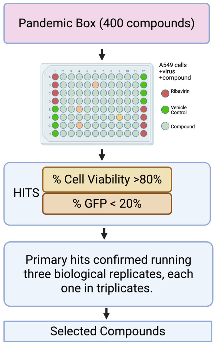 Figure 1
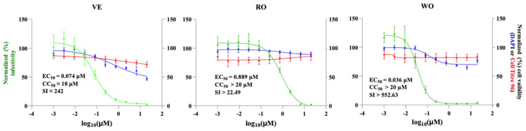 Figure 2
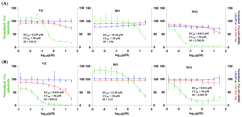 Figure 3
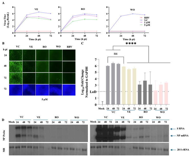 Figure 4
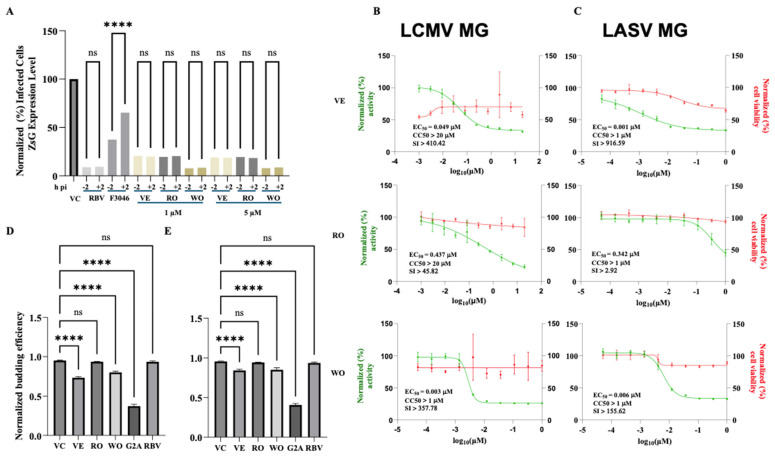 Figure 5
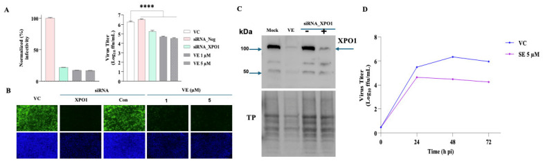 Figure 6
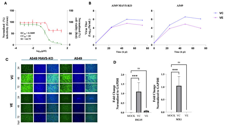 Figure 7
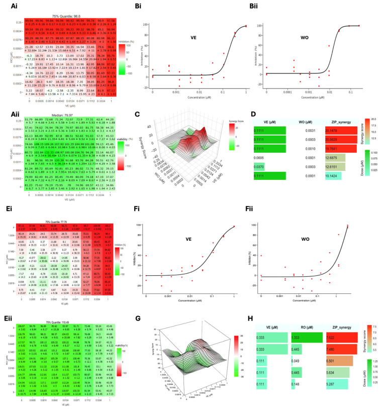 Figure 8
- —NIH/NIAID
Peer Reviews
No public reviews on file for this paper yet. If you reviewed it on a platform where reviews are public (OpenReview, ICLR, NeurIPS, ICML), you can paste yours below so the community can read it here.
Videos
No videos yet. Explain this paper in a talk, walkthrough, or lecture? Add one.
Taxonomy
TopicsViral Infections and Outbreaks Research · Mosquito-borne diseases and control · Intimate Partner and Family Violence
1. Introduction
Mammarenaviruses (MaAvs) cause persistent infections in their natural rodent reservoirs worldwide [1]. Human infections occur through mucosal exposure to aerosols or by direct contact of abraded skin with infectious materials [1]. Several MaAvs cause hemorrhagic fever (HF) in humans and represent important public health concerns in their endemic regions [1]. Lassa virus (LASV), endemic to Western Africa, is the etiologic agent of Lassa fever (LF), a HF disease associated with high morbidity and a case fatality rate as high as 69% among hospitalized confirmed LF cases [2,3,4,5]. LASV is estimated to infect >500,000 people annually [6], but limited diagnostic capabilities complicate an accurate assessment of LASV prevalence [7,8]. Recent studies indicate that LASV endemic regions are expanding [9,10,11], and increased traveling has led to the importation of LF cases into non-endemic metropolitan areas around the globe [12,13]. Notably, LASV ranks very high among zoonotic viruses with potential for spillover and spread in humans [14], and models based on projections of climate, land use, and population changes indicate that regions in Central and East Africa will become suitable for LASV over the next decades, resulting in a dramatic increase in the population at risk of LASV exposure [15]. Because of its impact on human health, pandemic potential, and lack of effective countermeasures, LF has been included in the World Health Organization (WHO) list of priority diseases for which there is an urgent need to develop therapeutics and vaccines [16]. Likewise, the MaAv Junin virus (JUNV) causes Argentine hemorrhagic fever with case fatality rates exceeding 15%, and several other New World MaAv cause VHFD throughout South America [17]. In addition, mounting evidence indicates that the worldwide-distributed MaAv lymphocytic choriomeningitis virus (LCMV) is a neglected human pathogen of clinical significance in congenital infections [18]. Notably, LCMV poses a serious threat to immunocompromised individuals [19], and although most LCMV infections in immunocompetent individuals are asymptomatic or result in mild symptoms [20], in some cases, LCMV-infected immunocompetent individuals can develop severe disease [21].
There are no US FDA-approved MaAv vaccines, and current anti-MaAv therapy is limited to an off-label use of ribavirin (RBV), whose efficacy remains controversial [22,23]. The broad-spectrum polymerase inhibitor favipiravir [24,25] and the cell entry inhibitors LHF-535 [26] and ARN-75039 [27,28] have shown promising results in preclinical models of LF, and LHF-535 is in phase I clinical trials. However, favipiravir has limited potency against LASV, and its short human half-life [29,30] are likely to compromise its efficacy. Likewise, the high degree of LASV genetic diversity [31,32] can compromise the use of cell entry inhibitors, as single mutations in the virus glycoprotein gene can confer cross-resistance to these cell entry inhibitors [33,34]. Accordingly, the development of broad-spectrum antivirals to combat human pathogenic MaAv represents a significant unmet biomedical need.
MaAv are enveloped viruses with a bisegmented, negative-strand RNA genome [1,35,36]. Each genome segment uses an ambisense coding strategy to express two viral proteins. The large (L) segment (ca. 7.3 kb) encodes the RING-finger matrix protein (Z) and the RNA-dependent RNA polymerase (RdRP) L protein. The small (S) segment (ca 3.4 kb) encodes the glycoprotein precursor (GPC) and nucleoprotein (NP). NP encapsulates the viral genome RNA to create the viral nucleocapsid (NC) that, together with the L protein, forms the viral ribonucleoprotein complex (vRNP) that directs replication and transcription of the viral genome [1,37]. GPC is co-translationally processed by the signal peptidase to form a stable signal peptide (SSP) and a GPC precursor, which is post-translationally processed by the S1P protease into GP1 and GP2. The mature tripartite GP complex, consisting of SSP, GP1, and GP2, incorporates into the viral envelope, and its ectodomain forms the spikes present at the virion surface and mediates virus cell entry via receptor-mediated endocytosis [37]. GP1 binds to cellular receptors, whereas GP2 mediates a pH-dependent fusion event between viral and cell membranes in the acidic environment of the late endosome [38]. Upon fusion, the vRNP is released into the cytosol, where it initiates viral transcription and replication [1]. MaAv assembly and budding is driven by the matrix protein Z [1].
Drug repurposing strategies can substantially decrease the time and resources necessary to advance a candidate antiviral drug into the clinic [39]. Furthermore, drug repurposing can yield valuable insights into viral biology by identifying novel pathways and host cell factors involved in various stages of the viral replication cycle, thereby revealing new antiviral targets [40,41,42,43].
Here, we document the results of the screening of the Pandemic Response Box (PRB) [44] for inhibitors of MaAv infection. The PRB was assembled by the Medicines for Malaria Venture, Geneva (MMV), and the Drugs for Neglected Diseases initiative (DNDi) and contains a collection of 400 compounds comprising 201 antibacterial, 153 antiviral, and 46 antifungal compounds, with several of these compounds demonstrating activity across multiple disease domains [44]. We identified three compounds listed in the PRB—Ro-24-7429 (RO), WO 2006118607 A2 (WO), and verdinexor (VE)—potent inhibitors of the activity of LCMV and LASV vRNP in cell-based minigenome assays and multiplication of LCMV in cultured cells.
RO is an antagonist of the HIV-1 transcriptional transactivator (Tat) protein that inhibits HIV-1 replication via interference with Tat-dependent initiation and elongation of transcription, resulting in reduced levels of viral RNA synthesis and protein production [45,46] but with no detectable antiviral effect [47]. RO inhibits RUNX family transcription factor 1, which has been associated with its antifibrotic and anti-inflammatory properties [48]. WO has been shown to have antiviral activity against the hepatitis C virus (HCV) via inhibition of dihydroorotate dehydrogenase (DHODH), a key enzyme in pyrimidine biosynthesis [49,50]. VE is a novel oral selective inhibitor of nuclear export (SINE) compound that targets exportin 1 (XPO1), the primary nuclear export regulator that shuttles proteins and RNAs from the nucleus to the cytoplasm [51]. VE has been shown to inhibit the multiplication of the influenza A virus (IAV) [52] and respiratory syncytial virus (RSV) [53] in cultured cells and has shown efficacy in animal models of IAV infection [52,54]. RO, WO, and VE target different, distinct host factors, rather than viral factors. Consistent with their distinct validated targets, VE and WO exhibit very strong synergistic activity when used in combination therapy. Host-directed antivirals (HDAs) represent a promising yet underexplored approach in contemporary antiviral research [55]. Unlike conventional direct-acting antivirals (DAAs) that target viral proteins and functions, HDAs disrupt host factors and cellular processes that viruses hijack for their replication and pathogenesis. Since members of a virus family share host dependencies, HDAs have the potential to act as broad-spectrum antivirals. In addition, HDAs pose a higher genetic barrier to the emergence of drug-resistant viral variants, which often compromise antiviral therapy [56]. Likewise, combination therapy of antivirals with synergistic effects can pose a high genetic barrier to the emergence of drug-resistant viral variants that often jeopardize monotherapy approaches and facilitate the use of reduced drug doses within the therapeutic range to alleviate side effects associated with high drug doses used in monotherapy.
2. Materials and Methods
2.1. Compound Source
Medicines for Malaria Venture, Geneva (MMV), and the Drugs for Neglected Diseases initiative, Geneva, Switzerland (DNDi), assembled the Pandemic Response Box (PRB), a collection of 400 compounds [44].
2.2. Cells and Viruses
HEK 293T (ATCC CRL-3216, Manassas, VA, USA), Vero E6 (ATCC CRL-1586, Manassas, VA, USA), and A549 (ATCC CCL-185, Manassas, VA, USA) cells were cultured in Dulbecco’s modified Eagle’s medium (DMEM; Thermo Fisher Scientific, Vacaville, CA, USA). The medium was supplemented with 10% heat-inactivated fetal bovine serum (FBS), 2 mM L-glutamine, and antibiotics (100 µg/mL streptomycin and 100 U/mL penicillin). Recombinant viruses rLCMV/GFP-P2A-NP [57] and the single-cycle infectious rARMΔGPC/ZsG-P2A-NP expressing the ZsGreen fluorescent protein [58] have been described. Using the same strategy described for the generation of rLCMV/GFP-P2A-NP, we generated rJUNV/GFP-P2A-NP based on the live-attenuated vaccine strain Candid 1 of JUNV.
2.3. Generation of rJUNV/GFP-P2A-NP
For the rescue of rJUNV/GFP-P2A-NP, based on the live-attenuated vaccine strain Candid 1 of JUNV, we used the same procedures and strategy described for the generation of rLCMV/GFP-P2A-NP [57]. Briefly, BHK-21 cells were seeded into 6-well plates at 7.0 × 10^5^/well 1 day prior to transfection to achieve an 80–90% confluence. Cells were transfected with 1.2 μg of pC-NP and 1.5 μg of pC-L (both NP and L corresponding to the Romero strain of JUNV), 1.2 μg of mouse pol-I S JUN GFP-P2A-NP, and 2.1 μg of the mouse pol-I L JUNV (total 6 μg/well) using Lipofectamine 3000 (LF3000) (Invitrogen, Carlsbad, CA, USA) (1.25 μL LF3000/μg of plasmid DNA) and P3000™ reagent (Invitrogen, Carlsbad, CA, USA) (1.75 μL P3000/μg of plasmid DNA) for more efficient transfection. At 72 h post-transfection, cell culture supernatants (CCSs) were collected, and virus titration was performed.
2.4. Virus Titration
Virus titers were determined by a focus-forming assay (FFA) [59] using Vero E6 cells. Briefly, cells were seeded in a 96-well (2.0 × 10^4^ cells/well) and 24 h later infected with 10-fold serial dilutions of the virus. At 20 h post-infection (hpi), the cells were fixed with 4% paraformaldehyde (PFA) in PBS, and infected cells were detected by epifluorescence based on their expression of GFP.
2.5. Cell Viability Assay
Cell viability was evaluated by measuring formazan produced from MTS [3-(4,5-dimethylthiazol-2-yl)-5-(3-carboxymethoxyphenyl)-2-(4-sulfophenyl)-2H-tetrazolium] through the activity of NADPH or NADH produced by living cells. Cells were seeded in a 96-well optical plate (3.0 × 10^4^ cells/well), and 20 h later, compounds (at designated concentrations, four replicates) or vehicle control (0.3% DMSO) were added, bringing the final volume to 100 µL. After 72 h treatment, the CellTiter 96 AQueous One Solution reagent (Promega, CAT#: G3580, Madison, WI, USA) was added to each well, and the plate was incubated for 15 min at 37 °C with 5% CO_2_. Absorbance was measured at 490 nm using a Biotek Cytation 5 plate reader (Agilent Technologies, Santa Clara, CA, USA). The results were normalized to the vehicle control, which was assigned a value of 100%. One Solution and media were then aspirated, and the cells were fixed with 4% paraformaldehyde and stained with DAPI.
2.6. Determination of Compounds’ EC50 and CC50 Values
Cells were seeded onto a 96-well optical plate (3.0 × 10^4^ cells/well), and 20 h later, cells were infected with rLCMV/GFP-P2A-NP at an MOI of 0.03 or rJUNV/GFP-P2A-NP at an MOI of 0.03. Infected cells were treated with three-fold serial dilutions of the indicated compound, starting at a concentration of 30 µM and using four replicates for each dilution. After 72 h treatment, cell viability was determined using the CellTiter 96 AQueous One Solution Cell Proliferation Assay to determine compound CC_50_ values. Compound EC_50_ values were determined by measuring GFP expression levels. After measuring GFP, cells were subjected to DAPI staining. GFP and DAPI were quantified by fluorescence using a Biotek Cytation 5 plate reader (Agilent Technologies, Santa Clara, CA, USA). This approach of determining compound CC_50_ values in infected cells was feasible because LCMV has a non-cytolytic multiplication. Mean values were normalized to an infected and vehicle (DMSO)-treated control, also set to 100%. The EC_50_ and CC_50_ values were calculated using GraphPad Prism v10 (GraphPad Software, San Diego, CA, USA). CC_50_ and EC50 values were used to determine compound selectivity index (SI) values (SI = CC_50_/EC_50_).
2.7. Virus Multi-Step Growth Kinetics
A549 cells were seeded (2.0 × 10^5^ cells/well, a 12-well plate) and the following day infected with rLCMV/GFP-P2A-NP at an MOI of 0.03. After a 90 min adsorption period, the inoculum was removed, and the cells were treated with VC (0.3% DMSO), specified compounds (1 µM and 5 µM), or RBV (100 µM) as a positive control. CCS were collected at 24, 48, and 72 hpi. At 72 hpi, cells were stained with Hoechst 33342 dye solution (Invitrogen, Thermo Fisher Scientific, Eugene, OR, USA). Live cell images were taken using a Keyence BZ-X710 all-in-one fluorescence microscope (Keyence corporation of America, Itasca, IL, USA)series. Cells were washed, and RNA was extracted using TRI Reagent (TR 118, Molecular Research Centre, Cincinnati, OH, USA). Virus titers in CCS were measured by FFA using Vero E6 cells (4 replicates), and the results were expressed as mean ± SD. Data were plotted using GraphPad Prism (GraphPad Software, San Diego, CA, USA).
2.8. RT-qPCR
RT-qPCR was conducted as described in [60]. RNA isolation was performed using TRI Reagent following the manufacturer’s guidelines. RNA was dissolved in RNA storage solution (Life Technologies, Carlsbad, CA, USA. AM 7000), and its concentration was measured with a NanoDrop™ 2000 spectrophotometer (ND-2000 Thermo Fisher Scientific™, Waltham, MA, USA). RNA (1 µg) was converted into cDNA using the SuperScript IV first-strand synthesis system (18091050, Life Technologies). For amplification of LCMV NP and the housekeeping gene GAPDH, Powerup SYBR (A25742, Life Technologies) was employed, utilizing the following primers: NP forward (F): 5′ CAGAAATGTTGATGCTGGACTGC-3′ and NP reverse (R): 5′-CAGACCTTGGCTTGCTTTACACAG-3′; GAPDH F: 5′-CATGAGAAGTATGACAACAGCC-3′ and GAPDH R: 5′-TGAGTCCTTCCACGATACC-3′; ISG15 F: 5′-CAGGACGACCTGTTCTGGC-3′ and ISG15 R: 5′-GATTCATGAACACGGTGCTCAGG-3′; and MX1 F: 5′-GCAGCTTCAGAAGGCCATGC-3′ and MX1 R: 5′-CCTTCAGGAACTTCCGCTTGTC-3′.
2.9. Time-of-Addition Assay
A549 cells were seeded (3.0 × 10^4^ cells/well) in a 96-well black-walled optical plate (353219, Falcon) and, after 16 h of culture, treated with VC (0.3% DMSO), indicated compounds at concentrations of 1 µM and 5 µM, and F3406 at 5 µM, with four replicates for each treatment condition. Treatments were administered either 2 h prior (−2 h) or 2 h after (+2 h) infection with the single-cycle infectious rARMΔGPC/ZsG-P2A-NP (MOI of 1.0), thereby obviating the need for NH_4_Cl treatment to avoid confounding effects from multiple rounds of infection. At 24 hpi, cells were fixed with 4% PFA, and ZsGreen expression levels were quantified using a Cytation 5 imaging reader.
2.10. Budding Assay
Budding assays were performed as described [61]. Briefly, HEK293T cells (1.75 × 10^5^ cells/well in a 12-well plate) were transfected with 0.5 μg of pC.LASV-Z-Gluc or pC.LASV-Z-G2A-GLuc (mutant control) or pCAGGS-Empty (pC-E) using Lipofectamine 3000 and P3000™. After 5 h of transfection, cells were washed three times and treated with VE, RO, WO (1 µM and 5 µM), RBV (100 µM), or vehicle control (VC). After 48 h of treatment, CCS containing virus-like particles (VLPs) was collected, and cell lysates were prepared. CCS samples were clarified from cell debris by centrifugation (13,000 rpm/4 °C/10 min), and aliquots (20 µL each) from CCS samples were added to 96-well black plates (VWR, West Chester, PA, USA), and 50 µL of Steady-Glo luciferase reagent (Promega) was added to each well. Cell lysates were prepared using 250 µL of lysis buffer (1% NP-40, 50 mM Tris-HCl (pH 8.0), 62.5 mM EDTA, 0.4% sodium deoxycholate). Lysates were clarified from cell debris by centrifugation (13,000 rpm/4 °C/10 min). GLuc activity in Z-containing VLP and whole-cell lysates (WCLs) was determined using the Steady-Glo luciferase assay system (Promega, Madison, WI, USA) according to the manufacturer’s protocol using a Berthold Centro LB 960 luminometer (Berthold Technologies, Oak Ridge, TN, USA). The GLuc activity in CCS and WCL served as a surrogate for Z protein levels. Z budding efficiency (in %) was determined by the ratio of VLP-associated GLuc levels (ZVLP) and total GLuc levels (ZVLP + ZWCL) times 100.
2.11. LCMV and LASV Cell-Based Minigenome (MG) Assays
The LCMV and LASV MG systems have previously been described [62,63]. HEK293T cells were seeded (0.75 × 10^6^ cells/well) onto a poly-L-lysine-treated 6-well plate 20 h prior to transfection with pCAGGS-T7 Cyt (0.625 μg) and the components (pT7MG-ZsGreen, pCAGGS-NP, and pCAGGS-L). Cells were transfected using Lipofectamine 3000™ (2.5 μL/2.4 μg DNA) and P3000™ (4 μL/2.4 μg DNA). After 2.5 h, the transfection medium was removed, and the cells were washed with DPBS. Cells were detached using 0.5 mL of Accutase, collected with 1 mL of fresh medium, and centrifuged at 1300 rpm at 6 °C for 5 min. The transfected cells were resuspended in an appropriate volume of fresh medium to achieve a cell density of 3.0 × 10^4^ cells per well in a 96-well plate. Subsequently, 10 μL (10×) of three-fold serial dilutions of the indicated compounds, starting at 20 µM (VE and RO) and 1 µM (WO), were prepared in four replicates and plated onto a 96-well plate. Then, 90 μL of the cell suspension (3.0 × 10^4^ cells/well) was added, bringing the final volume to 100 μL, and incubated for 72 h at 37 °C with 5% CO_2_. The cells were then fixed with 4% paraformaldehyde and stained with DAPI. EC50 values were determined by measuring GFP expression levels, while CC50 was evaluated by measuring DAPI. Both GFP and DAPI were quantified by fluorescence using a Biotek Cytation 5 plate reader (Agilent Technologies, Santa Clara, CA, USA). The mean values were normalized to the vehicle (DMSO)-treated control, which was set to 100%. The LASV L and NP sequences used in the MG assay were derived from the Josiah strain.
2.12. Epifluorescence
Images were collected using the Keyence BZ-X710 microscope. Images were transferred to a laptop for data processing purposes. Microsoft PowerPoint Version 16.103 (25110922) was used to assemble and arrange the images, with each one being imported separately and arranged in a cohesive manner within its respective composite. The canvas size was adjusted to ensure a harmonious layout.
2.13. siRNA-Mediated XPO1 Knockdown (KD)
ON-TARGETplus Human XPO1 (7514) siRNA—SMARTpool (Dharmacon Inc., Lafayette, CO, USA) with four target sequences—“AAGAAUGGCUCAAGAAGUA”, “GGACAAGAGUCGACACAAU”, “UAGAUAAUGUGGUGAAUUG”, and “CGAAAUGUCUCUC”—was used to KD XPO1 in A549 cells using non-targeting siRNA as a control. Briefly, A549 cells were seeded overnight in a 12-well plate at 2.0 × 10^5^/well and transfected with siRNA targeting XPO1 and a non-targeting siRNA control using DharmaFECT-1 reagent (Dharmacon). Then, 10 μL of 5 μM siRNA was diluted with 190 μL of Opti-MEM to give a concentration of 250 nM of siRNA. Next, 2 μL of DharmaFECT was diluted with 198 μL of Opti-MEM. Both were incubated for 5 min at room temperature (RT), mixed, and incubated for 20 min at RT. Media in each well was removed, 500 μL of the transfection media was added to each well plate with 2000 μL of antibiotic-free DMEM, giving a final concentration of 25 nM of siRNA, and incubated at 37 °C in 5% CO_2_ for 48 h.
2.14. Determining Virus Production in XPO1 Knockdown Cells
A549 cells were seeded at a density of 3.0 × 10^4^ cells per well in a 6-well black-walled optical plate and incubated at 37 °C in 5% CO_2_ for 20 h. Cells were transfected with 25 nM of siRNA targeting XPO1 or a non-targeting siRNA control and incubated at 37 °C in 5% CO_2_ for 48 h. Cells were then infected with rLCMV/GFP-P2A-NP at an MOI of 0.03. After a 90 min adsorption period, the inoculum was removed, and cells were treated with VC (0.5% DMSO), VE (1 µM and 5 µM), or RBV (100 µM). CCS were collected at 48 hpi, cells were washed, fixed with 4% PFA, and stained with DAPI. Production of infectious particles was determined by measuring virus titers in CCS by FFA using Vero E6 cells (4 replicates), and the results were expressed as mean ± SD. Data were plotted using GraphPad Prism (GraphPad Software, San Diego, CA, USA). Virus infectivity was determined based on GFP expression levels quantified by fluorescence using a Biotek Cytation 5 plate reader (Agilent Technologies, Santa Clara, CA, USA). Mean values were normalized to an infected and vehicle (DMSO)-treated control, also set to 100%, and plotted using GraphPad Prism v10 (GraphPad Software, San Diego, CA, USA).
2.15. Determining Synergistic Activities
A549 cells were seeded onto a 96-well optical plate at a density of 3.0 × 10^4^ cells per well. After 20 h, the cells were infected with rLCMV/GFP-P2A-NP MOI of 0.03. At 90 min post-infection, a concentration matrix of compounds, prepared in three-fold serial dilutions at 2×, was added in various combinations 1:1, bringing the total concentration to 1× according to a predetermined synergy matrix (an 8-by-8 matrix for all possible 64 combinations of two drug combinations). After 72 h post-treatment, GFP expression levels were determined. After measuring GFP, cells were subjected to DAPI staining. GFP and DAPI were quantified by fluorescence using a Biotek Cytation 5 plate reader (Agilent Technologies, Santa Clara, CA, USA). Mean values were normalized to an infected and vehicle (DMSO)-treated control, also set to 100%. Normalized values derived from matrix data were subjected to analysis utilizing the SynergyFinder+ software version 07.09.2024-R-3.10.3 (https://synergyfinder.org/ accessed 21 October 2025) [64]. Our dose–response assay yields sigmoidal (logistic) curves, and because the combined drugs act through distinct mechanisms of action, synergy is most appropriately assessed using the zero interaction potency (ZIP) model rather than HSA, Bliss, or Loewe [65].
In SynergyFinder+, average ZIP scores >10 are generally considered biologically meaningful synergy, scores between −10 and +10 indicate additive or non-interacting effects, and scores < −10 indicate antagonism, consistent with prior implementations and published usage of the ZIP model.
3. Results
3.1. Screening of the Pandemic Response Box (PRB) for Inhibitors of LCMV Multiplication
To identify potential anti-MaAv compounds within the PRB, we implemented a high-throughput screening (HTS) based on a cell-based infection assay using the rLCMV/GFP-P2A-NP, hereafter called rLCMV/GFP, that allowed us to use the GFP signal as an accurate surrogate of virus multiplication [65]. To identify primary hits, we infected (MOI = 0.03) A549 cells with rLCMV/GFP and treated them with each compound at a single dose (5 µM). At 72 hpi, we determined cell viability using the CellTiter 96 AQueous One Solution Cell Proliferation Assay. After absorbance values were recorded, we fixed cells using 4% PFA, and GFP expression levels were quantified. Both formazan absorbance and GFP signal were determined using a Synergy H4 Hybrid Multi-Mode microplate reader (BioTek Instruments, Winooski, VT, USA). As controls, we used treatment with the validated MaAv inhibitor RBV (100 µM) and vehicle control (VC). Cell viability and GFP expression levels were normalized to those of vehicle-treated (VC) controls. Primary hits (n = 22) were selected based on causing ≥50% reduction in infectivity (GFP signal) and ≤20% cytotoxicity (DAPI signal). Selected primary hits were confirmed in biological replicates, using each compound at a single concentration (5 µM) in triplicate (Figure 1). We identified Ro-24-7429 (RO), WO 2006118607 A2 (WO), and verdinexor (VE) as the top three hits with a potent inhibitory effect on LCMV multiplication (≥80% reduction in infectivity and ≤20% reduction in cell viability) (Supplementary Table S1).
3.2. Dose-Dependent Effect of VE, RO, and WO on LCMV Multiplication in Cultured Cells
To evaluate the magnitude of the antiviral activity of the selected hits, we determined their dose-dependent effect on rLCMV/GFP multiplication in A549 cells (Figure 2). Cells were infected with rLCMV/GFP (MOI = 0.03) and treated with three-fold serial dilutions of each compound. At 72 hpi, we determined cell viability using the CellTiter 96 AQueous One Solution Cell Proliferation Assay. After absorbance values were recorded, we fixed cells using 4% PFA and stained them with DAPI. GFP and DAPI staining signals, as well as formazan absorbance, were measured using the Cytation 5 Cell Imaging Multi-Mode Reader. Normalized cell viability values and GFP expression levels were used to determine compounds’ CC_50_ and EC_50_ values using GraphPad Prism software v10. The three selected hits, VE, RO, and WO, exhibited potent anti-LCMV activity with EC_50_ values (µM) of 0.074, 0.889, and 0.036, respectively, and selectivity index (SI) values of >242 (VE), >22.49 (RO), and >552.6 (WO) (Figure 2).
3.3. Efficacy of VE, RO, and WO on JUNV Multiplication in Cultured Cells
To assess the broad-spectrum anti-MaAv of VE, RO, and WO, we assessed their efficacy against the LCMV genetically distantly related New World MaAv JUNV. To overcome the need for BSL4 containment required for the use of live forms of pathogenic strains of JUNV, we used rJUN GFP-P2A-NP, hereafter called rJUNV/GFP, based on the live-attenuated vaccine strain Candid#1 of JUNV, which can be safely used in BSL2. We infected (MOI = 0.5) Vero E6 (Figure 3A) and A549 (Figure 3B) cells with rJUNV/GFP and treated them with a three-fold serial dilution of each compound. At 72 hpi, we determined cell viability using the CellTiter 96 AQueous One Solution Cell Proliferation Assay. After absorbance values were recorded, we fixed cells using 4% PFA and stained them with DAPI. GFP and DAPI staining signals, as well as formazan absorbance, were measured using the Cytation 5 Cell Imaging Multi-Mode Reader. Normalized cell viability values and GFP expression levels were used to determine compounds’ CC_50_ and EC_50_ values using GraphPad Prism software v10. VE and WO exhibited potent antiviral activity against JUNV in both Vero E6 (Figure 3A) and A549 (Figure 3B) cells, whereas RO was less efficient in inhibiting the multiplication of JUNV in both Vero E6 and A549 cells (Figure 3).
3.4. Effects of VE, RO, and WO on LCMV Multi-Step Growth Kinetics
To examine the effects of VE, RO, and WO on the production of infectious viral progeny over time and virus cell propagation, we determined their effects on LCMV multi-step growth kinetics in A549 cells. We infected (MOI = 0.03) A549 cells with rLCMV/GFP and treated them with the indicated concentrations of each compound, VC, or RBV (100 µM). At the indicated hpi, CCSs were collected and titers of infectious virus determined by FFA (Figure 4A). Cells were stained with Hoechst in FluoroBrite DMEM, and epifluorescence images of live cells were taken from each sample (Figure 4B) using a Keyence BZ-X710 microscope. After collection of the images, total cellular RNA was isolated from each sample, and levels of viral RNA synthesis were determined by Northern blotting (NB) (Figure 4D) and RT-qPCR (Figure 4C). Treatment with VE at 1 µM or 5 µM caused about a 2-log reduction in virus peak titers (Figure 4A). RO exhibited reduced efficacy at 1 µM compared to 5 µM (Figure 4A). Treatment with WO at either 5 or 1 µM resulted in the absence of detectable levels of infectious virus in CCS at all hpi tested (Figure 4A). The observed reduction in production of LCMV infectious progeny in CCS of infected cells treated with each of the compounds correlated with restricted viral cell propagation (Figure 4B) and viral RNA synthesis, as quantified by RT-qPCR (Figure 4C) or NB (Figure 4D). VE, RO, and WO inhibited to a similar degree both virus replication and transcription as determined by NB results (Figure 4D).
3.5. Effects of VE, RO, and WO on Different Steps of LCMV Life Cycle
To gain insights about the mechanisms whereby VE, RO, and WO exert their anti-MaAv activity, we investigated their effects on various stages of the LCMV life cycle. We first assessed whether the compounds affected virus cell entry. For this, we performed a time-of-addition experiment using the single-cycle infectious rLCMV∆GPC/ZsG to prevent the confounding effects associated with multiple rounds of infection cycles in the absence of NH_4_Cl treatment. We treated A549 cells (3.0 × 10^4^ cells/well, 96-well plate) with each compound (1 µM or 5 µM), the LCMV cell entry inhibitor F3406 (5 µM), or the LCMV multiplication inhibitor RBV (100 µM) starting 2 h prior to (−2) or 2 h post (+2) infection with rARMΔGPC/ZsG-P2A-NP at an MOI of 1. At 48 hpi, we determined ZsG expression levels and normalized them to those of VC-treated cells, which were assigned a value of 100%. The results from the time-of-addition experiment showed that VE, RO, and WO exerted a similar inhibitory effect on ZsG expression when administered 2 h prior to or 2 h following infection (Figure 5A), indicating that all three targeted a post-cell entry step of the LCMV life cycle.
We next examined the effect of the compounds on the activity of the vRNP using both LCMV and LASV cell-based MG systems that accurately recapitulate all the steps of viral RNA synthesis, where the MG-directed reporter gene expression serves as an accurate surrogate of vRNP activity. We transfected HEK293T cells with the components of the LCMV (Figure 5B) or LASV (Figure 5C) MG system. At 2.5 h post-transfection, single-cell suspensions were prepared and seeded onto poly-L-lysine-coated 96-well plates (3.0 × 10^4^ cells/well) with media containing different concentrations (four replicates per concentration) of each compound. At 72 h post-transfection, cells were fixed with 4% PFA and stained with DAPI. ZsG expression levels and DAPI staining were quantified using the Cytation 5 plate reader and used to determine levels of MG activity and cell viability (total nuclei), respectively. VE and WO potently inhibited the activity of both LCMV and LASV MGs, whereas RO was significantly less efficient in inhibiting either LCMV or LASV MG activities (Figure 5B,C).
To assess the effect of VE and RO on the budding process directed by the matrix Z protein [66], we used a previously established cell-based budding assay wherein the activity of the Gaussia luciferase (GLuc) reporter gene serves as an indicator of Z budding activity [61]. We transfected HEK293T cells with a plasmid encoding LCMV Z-GLuc and subsequently treated them with VE, RO, WO (5 µM and 1 µM), or VC. As controls, we used the budding-deficient mutant G2A of the Z [67] and treatment with RBV (100 µM). At 48 h post-transfection, we measured levels of GLuc activity associated with virus-like particles (VLPs) present in CCSs and intracellular Z protein in whole-cell lysates (WCLs). Z budding efficiency was determined by the ratio of VLP-associated GLuc levels (CCSGLuc) and total GLuc levels (CCSGLuc + WCLGLuc) × 100. VE and WO, but not RO, had a significant inhibitory effect on LCMV Z budding activity at 5 µM (Figure 5D) and 1 µM (Figure 5E).
3.6. Effect of XPO1 siRNA-Mediated Knockdown (KD) on Production of LCMV Infectious Progeny
VE is a selective inhibitor of the nuclear export protein XPO1 and has been shown to exhibit antiviral activity against IAV [54,68], whose replication and gene transcription take place in the nucleus, and RSV [52,53], whose M protein appears to have a nuclear function required for RSV replication. In contrast, there is no evidence that the MaAv life cycle involves a nuclear phase [69], raising the intriguing question of why VE targeting of XPO1 results in MaAv inhibition. To further investigate the role of XPO1 on LCMV infection, we assessed the effect of siRNA-mediated knockdown (KD) of XPO1 on LCMV multiplication. For this, we transfected A549 cells with the ON-TARGETplus Human XPO1 (7514) siRNA—SMARTpool (Dharmacon) and a non-targeting siRNA as a control. At 48 h post-transfection, we infected cells with rLCMV/GFP (MOI of 0.03). Non-transfected cells were treated with VE (1 µM or 5 µM) or VC. At 48 hpi, CCSs were collected and titers of infectious virus determined by FFA (Figure 6A). Cells were fixed with 4% PFA and stained with DAPI, and representative images of each sample were collected using a Keyence BZ-X710 microscope (Figure 6B). Compared to the non-targeting siRNA-transfected cells, XPO1 KD in A549 cells significantly reduced the production of infectious virus (Figure 6A), which correlated with a reduction in virus cell propagation (Figure 6B). To confirm the KD of XPO1 in A549 cells by VE and TARGETplus Human XPO1 siRNA, we treated A549 cells with VE (5 µM), mock-treated them, or transfected them with siRNA targeting XPO1 and a non-targeting siRNA control. At 48 h post-treatment or transfection, we prepared cell lysates, and levels of XPO1 protein were determined by Western blot (Figure 6C). To further validate that the anti-LCMV activity of VE was via targeting XPO1, we examined the effect of the VE analog Selinexor (SE) on LCMV multi-step growth kinetics in A549 cells. SE exhibited a potent inhibitory effect on LCMV multiplication (Figure 6D).
3.7. Assessment of the Contribution of the Interferon (IFN) Response to VE Anti-MaAv Activity
Treatment with VE and the related second generation of SINE Eltanexor has been shown to increase expression of type I interferon (T1IFN) and other innate immune-related genes in Kaposi’s sarcoma-associated herpesvirus (KSHV) and human cytomegalovirus (HCMV) infections [70,71], whereas treatment with VE and SE has been shown to decrease levels of type II IFN and other proinflammatory cytokines in IAV and SARS-CoV-2 infections [68,72]. To assess the contribution of the T1IFN response to the anti-MaAv of VE, we evaluated the dose–response effect of VE on LCMV multiplication in A549 cells deficient in mitochondrial antiviral-signaling protein (MAVS) required for induction of IFNb in LCMV-infected cells [73]. VE exerted a strong dose-dependent inhibitory effect on LCMV multiplication in MAVS-KO A549 cells (Figure 7A), which correlated with reduced production of infectious progeny (Figure 7B) and restricted virus propagation in the infected cell monolayer (Figure 7C). These findings indicated that the T1IFN response did not significantly contribute to the anti-LCMV activity of VE. Unexpectedly, we found that in contrast to the findings cited above with KSHV- and HCMV-infected cells, under our experimental conditions, treatment with VE resulted in reduced expression levels of the interferon-stimulated genes ISG15 and MX1 (Figure 7D).
3.8. Assessment of the Synergistic MaAv Antiviral Activity of VE in Combination with WO and RO
To investigate the synergistic antiviral activity of VE with WO and RO against MaAv, we designed an 8 × 8 matrix to test 8 serial dilutions of two compounds in their 64 possible combinations for their ability to inhibit the multiplication of LCMV. To assess synergy, we selected the zero interaction potency (ZIP) synergy model [74]. VE showed strong synergistic antiviral activity in combination with WO (Figure 8A), with the highest ZIP synergy score of 20 observed at concentrations of 0.11 μM (VE) and 0.0003 μM (WO). Monotherapy treatment with VE and WO at these concentrations resulted in a moderate antiviral effect of VE (22.64% inhibition) and non-detectable activity for WO. However, when combined, VE + WO exhibited 50.05% inhibition. Cell viability with individual compound treatment at these concentrations was 69.57% (VE) and 81.24% (WO), whereas combination therapy resulted in 72.72% cell viability. The combination treatment of VE and WO also showed five other combinations with ZIP score values > 10. The combination therapy of VE and RO only exhibited very modest synergistic antiviral activity, with the highest ZIP synergy score of 7.6 (Figure 8B) observed at concentrations of 0.333 μM (VE) and 1.333 μM (RO). The individual treatments of VE and RO at these concentrations displayed moderate antiviral effects with percent inhibition values of 77.62% and 40.14%, respectively. However, when combined, 96.24% inhibition was exhibited. Cell viability with individual treatment at the same concentration was 53.41% and 100%, respectively, while cell viability when in combination was 54.3%. The combination treatment of VE and RO also showed four other combinations with ZIP score values > 5.
4. Discussion
The lack of US FDA-approved vaccines or antivirals to prevent and treat diseases caused by human pathogenic MaAv represents an important unmet medical need. Current treatment of infections by LASV and other human pathogenic MaAv is limited to an off-label use of RBV, whose efficacy remains controversial [22,23,76]. Significant efforts are being directed towards the discovery and development of antiviral drugs against LASV and other hemorrhagic fever-causing MaAv, which has resulted in the identification of several promising candidates [40]. However, expanding the landscape of MaAv candidate antivirals will facilitate overcoming some current challenges, including limited potency and the development of resistance. Drug repurposing strategies can accelerate the progression of a candidate antiviral drug into clinical trials, thereby reducing the labor- and resource-intensive efforts associated with the preclinical optimization of newly discovered hits in traditional drug-discovery approaches [39,77]. The open-source PRB is a collection of 400 compounds assembled based on publicly available information regarding chemotypes currently in the discovery and early development stages, which have demonstrated beneficial activities [44].
Compounds from the PRB have been identified as potential antivirals against different viruses, including enterovirus A71 [78], KSHV [79], SARS-CoV-2 [80,81], Ebola virus [82], Chikungunya virus [83], and the Zika virus [84]. Here, we have presented our results from the screening of the PRB for compounds with anti-MaAv activity. We used a cell-based infection assay with features amenable to HTS [62] to screen the PBR for inhibitors of LCMV multiplication. We identified 22 primary hits. Validation and reassessment assays identified RO, WO, and VE as the top candidate inhibitors of LCMV multiplication in cultured cells. RO (Ro-24-7429) was originally developed as an inhibitor of the HIV-1 Tat protein [45,46] but with no detectable antiviral effect in patients with HIV [47,85]. Subsequently, RO was found to inhibit runt-related transcription factor 1 (RUNX1) protein [48] and to ameliorate lung fibrosis and inflammation in a mouse model of bleomycin-induced pulmonary fibrosis [48]. Additionally, RO was shown to reduce the expression of angiotensin-converting enzyme 2 (ACE-2) and furin, which are host proteins essential for SARS-CoV-2 infection, supporting the repurposing of RO for the treatment of SARS-CoV-2 infection [48]. We found that RO exhibited strong antiviral activity against LCMV (EC50 = 0.89 µM and SI > 22). RO did not affect viral cell entry or Z-mediated budding processes but rather inhibited RNA synthesis mediated by vRNP (Figure 4D and Figure 5). However, unexpectedly, RO showed significantly lower activity against JUNV (EC50 = 15.34 µM and SI = 1.96). At the present time, we do not have an explanation for this finding, which may question the use of RO as a broad-spectrum MaAv antiviral. WO, a tetrahydrocarbazole amide, has been shown to exhibit broad-spectrum antiviral activity [86], which has been linked to its inhibitory effect on dihydroorotate dehydrogenase (DHODH) [87], a key enzyme in the biosynthesis of pyrimidines [49,50]. WO showed potent antiviral activities against LCMV (EC_50_ = 0.04 µM and SI > 552.63) and JUNV (EC_50_ = 0.01 µM and SI > 2.8) via targeting the activity of the vRNP, a result consistent with our published findings showing that DHODH inhibitors, including brequinar [66], vidofludimos calcium, and related analogs [88], have potent anti-MaAv activity.
VE, an oral selective inhibitor of nuclear export (SINE) compound that targets the XPO1/CRM1 exportin [51], has been shown to inhibit the replication of IAV [52] and RSV [53] in cultured cells. Notably, VE has demonstrated efficacy in animal models of IAV infection [52,54]. VE inhibits IAV multiplication by blocking XPO1-mediated nuclear export of vRNP [52], whereas inhibition of RSV by VE has been linked to the nuclear accumulation of RSV M protein due to VE targeting of XPO1 [53]. VE has also been shown to have antiviral activity against several DNA viruses via nuclear retention of viral proteins and also promoting the production of type I interferon [71,89]. MaAv’s life cycle is restricted to the cytoplasm of infected cells [69]. Hence, it was unexpected to find that VE exhibited a potent antiviral effect against MaAv, with EC_50_ values of 0.07 µM and 0.24 µM against LCMV and JUNV, respectively, and corresponding SI values of 242 and 124.31. We found that siRNA-mediated KD of XPO1 phenocopied the effect of VE in LCVM-infected cells, characterized by XPO1 reduced expression levels that correlated with inhibition of production of LCMV infectious progeny (Figure 6), supporting that VE exerted its anti-MaAv activity via targeting XPO1. Increased expression of T1IFN has been implicated in the inhibition of KSHV and HCMV infection by treatment with SINE compounds [71]. However, we found that VE exhibited an anti-LCMV activity of similar magnitude in WT and MAVS-KO A549 cells, indicating that the host cell T1IFN response did not contribute significantly to the antiviral effect of VE on LCMV. Moreover, contrary to the reported VE-induced expression of ISGs in KSHV- and HCMV-infected cells [70,71], we observed a decreased expression of ISG15 and MX1 ISGs upon VE treatment (Figure 7). Nevertheless, we cannot rule out the possibility that MAVS-independent signaling pathways might have contributed to VE anti-MaAv activity. Recent evidence suggests that XPO1 has activities beyond its role in nuclear export, including epigenomic regulation [90], which could influence the host cell response to infection. Examining the effect of VE and other SINE compounds on the global cellular gene expression profile may provide insights into the molecular bases of VE anti-mammarenavirus activity. However, we consider these studies to be beyond the scope of the present work.
HDAs represent a promising yet underexplored approach in contemporary antiviral research [91,92]. Unlike conventional direct-acting antivirals (DAAs) that target viral proteins and functions, HDAs disrupt host factors and cellular processes that viruses hijack for their replication and pathogenesis. Since members of a virus family share host dependencies, HDAs have the potential to act as broad-spectrum antivirals. In addition, HDAs pose a higher genetic barrier to the emergence of drug-resistant viral variants, which often compromise antiviral therapy [55,56]. The current fast pace of identification and characterization of virus–host interactions has accelerated the discovery of many novel potential host druggable targets for the development of HDAs and has facilitated drug repurposing strategies for drugs with established safety profiles, thus reducing longer development timelines and regulatory barriers associated with the development of novel drugs [39]. HDA-based antiviral strategies face the challenge of risks associated with toxicity and unintended consequences of disruption of cell physiological processes. However, therapies against acute viral infections, such as HF diseases caused by MaAv, require short-term treatment, which increases the likelihood of identifying effective therapeutic regimens with acceptable safety profiles.
The success of combination therapy is illustrated by the current therapeutic approaches to treat HIV and HCV infections involving combinations of two to four antivirals targeting different steps of the virus life cycle [93,94,95,96]. This has the advantage of fostering a synergistic antiviral effect [97,98], which facilitates reducing drug doses within the therapeutic range and alleviating side effects associated with high drug doses used in monotherapy, as well as posing a higher genetic barrier to the emergence of drug-resistant viral variants that often jeopardize monotherapy [99,100]. To investigate the synergistic efficacy of VE with RO and WO, we used the ZIP synergy model because it is well-suited for drug combination therapy studies in which each compound dose–response follows a sigmoidal behavior, and the combined compounds act through distinct mechanisms of action. ZIP defines non-interaction as the absence of changes in both potency (EC_50_) and efficacy upon combination and evaluates deviations across the entire dose–response surface, rather than at individual dose pairs. This makes ZIP particularly appropriate for antiviral combination matrices, where interactions frequently manifest as shifts in dose–response potency rather than simple additive or maximal effects. We considered alternative synergy models, but they were less suitable for this experimental design. The Highest Single Agent (HSA) model evaluates each dose pair independently by comparing the combination effect to the most active single agent at the same concentrations; however, it does not account for dose–response behavior or potency shifts and therefore provides limited interpretability for matrix-based antiviral assays. The Bliss independence model assumes probabilistic independence of drug effects but similarly operates on observed effect levels without explicitly incorporating fitted dose–response relationships, making it less sensitive to interaction effects driven by changes in drug potency. Conversely, the Loewe additivity model requires the assumption of similar or interchangeable mechanisms of action, which is not applicable to the mechanistically distinct antiviral compounds tested here. The ZIP synergy model integrates Bliss-type independence with Loewe-style dose–response modeling, allowing assessment of non-interaction under the assumption of unchanged potency while accommodating mechanistically distinct drugs. ZIP synergy scores are centered around zero, with positive values indicating synergy and negative values indicating antagonism. In SynergyFinder+, average ZIP scores > 10 are generally considered biologically meaningful synergy, scores between −10 and +10 indicate additive or non-interacting effects, and scores < −10 indicate antagonism. We found that VE exhibited strong synergistic antiviral activity in combination with WO (Figure 8A), with a ZIP synergy score of 20. In contrast, the combination therapy of VE and RO only exhibited very modest synergistic antiviral activity with a ZIP synergy score of 7.6 (Figure 8B). Future studies should examine whether VE exhibits synergistic antiviral activity in combination with inhibitors of LASV cell entry that have been advanced to clinical trials [26], which could facilitate the development of combination therapies of HDAs and DAAs. It should be noted that research aimed at developing VE and related XPO1-targeting SINE compounds as a host-directed antiviral against MaAv could be facilitated by insights gathered from cancer research, including preclinical and clinical data from the oncology pipeline for XPO1 inhibitors [101].
The reference list from the paper itself. Each links out to its DOI / PubMed record.
- 1Howley P.M. Knipe D.M. Whelan S. Arenaviridae: The Viruses and Their Replication Fields Virology: Emerging Viruses 7th ed. Knipe D.M. Whelan S.P.J. Lippincott Williams & Wilkins Philadelphia, PA, USA 2021 Volume 1978-1-9751-1254-7
- 2Smith D.R.M. Turner J. Fahr P. Attfield L.A. Bessell P.R. Donnelly C.A. Gibb R. Jones K.E. Redding D.W. Asogun D. Health and Economic Impacts of Lassa Vaccination Campaigns in West Africa Nat. Med.2024303568357710.1038/s 41591-024-03232-y 39198710 PMC 11645265 · doi ↗ · pubmed ↗
- 3Garry R.F. Lassa Fever—The Road Ahead Nat. Rev. Microbiol.202321879610.1038/s 41579-022-00789-836097163 PMC 9466315 · doi ↗ · pubmed ↗
- 4Fichet-Calvet E. Rogers D.J. Risk Maps of Lassa Fever in West Africa P Lo S Neglected Trop. Dis.20093 e 38810.1371/journal.pntd.0000388 PMC 264476419255625 · doi ↗ · pubmed ↗
- 5Grant D.S. Samuels R.J. Garry R.F. Schieffelin J.S. Lassa Fever Natural History and Clinical Management Lassa Fever: Epidemiology, Immunology, Diagnostics, and Therapeutics Springer Cham, Switzerland 2023 Volume 44016519210.1007/82_2023_26337106159 · doi ↗ · pubmed ↗
- 6Basinski A.J. Fichet-Calvet E. Sjodin A.R. Varrelman T.J. Remien C.H. Layman N.C. Bird B.H. Wolking D.J. Monagin C. Ghersi B.M. Bridging the Gap: Using Reservoir Ecology and Human Serosurveys to Estimate Lassa Virus Spillover in West Africa P Lo S Comput. Biol.202117 e 100881110.1371/journal.pcbi.100881133657095 PMC 7959400 · doi ↗ · pubmed ↗
- 7Hastie K.M. Melnik L.I. Cross R.W. Klitting R.M. Andersen K.G. Saphire E.O. Garry R.F. The Arenaviridae Family: Knowledge Gaps, Animal Models, Countermeasures, and Prototype Pathogens J. Infect. Dis.2023228 S 359S 37510.1093/infdis/jiac 26637849403 PMC 10582522 · doi ↗ · pubmed ↗
- 8Moore K.A. Ostrowsky J.T. Mehr A.J. Johnson R.A. Ulrich A.K. Moua N.M. Fay P.C. Hart P.J. Golding J.P. Benassi V. Lassa Fever Research Priorities: Towards Effective Medical Countermeasures by the End of the Decade Lancet Infect. Dis.202424 e 696e 70610.1016/S 1473-3099(24)00229-938964363 · doi ↗ · pubmed ↗