Divalent HIV-1 gp120 Immunogen Exhibits Selective Avidity for Broadly Neutralizing Antibody VRC01 Precursors
Ryan Bailey, Kalista Kahoekapu, Albert To, Ludwig I. Mayerlen, Helmut Kae, Gabriel Manninen, Brien K. Haun, John M. Berestecky, Cecilia Shikuma, Axel T. Lehrer, Iain S. MacPherson

TL;DR
Researchers designed a rigid HIV immunogen that binds strongly to a key antibody, which could help in developing better vaccines.
Contribution
The study introduces a novel rigid divalent immunogen with selective avidity for VRC01-class antibodies.
Findings
The rigid immunogen shows higher affinity for VRC01 antibodies compared to a flexible control.
The immunogen binds divalently to VRC01 and monovalently to non-CD4bs antibodies.
The design may have broad applications for immune focusing in vaccine development.
Abstract
Background: A major goal for the vaccine field is the elicitation of broadly neutralizing antibodies (bnAbs) against pathogens that exhibit extensive antigenic diversity. Methods: In this study, we designed a rigid divalent immunogen for high avidity binding to the bnAb, VRC01, which targets the CD4 binding site (CD4bs) of the HIV spike protein. This was accomplished by covalently linking two HIV-1 gp120 antigens to a complementary antibody and crosslinking the light chains. Binding kinetics were analyzed using a novel gel shift assay and surface plasmon resonance. Results: The rigid divalent immunogen exhibits a higher affinity for VRC01-class antibodies compared to a flexible control, likely due to antigen pre-organization limiting the entropic penalty for divalent binding. Crucially, this immunogen exhibited divalent binding to VRC01 and monovalent binding to a non-CD4bs Ab, A32—a…
Genes, proteins, chemicals, diseases, species, mutations and cell lines named across the full text — each resolved to its canonical identifier and authoritative record.
Click any figure to enlarge with its caption.
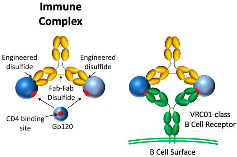 Figure 1
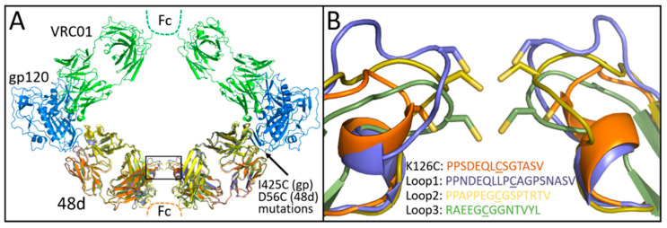 Figure 2
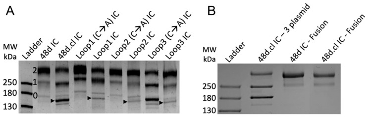 Figure 3
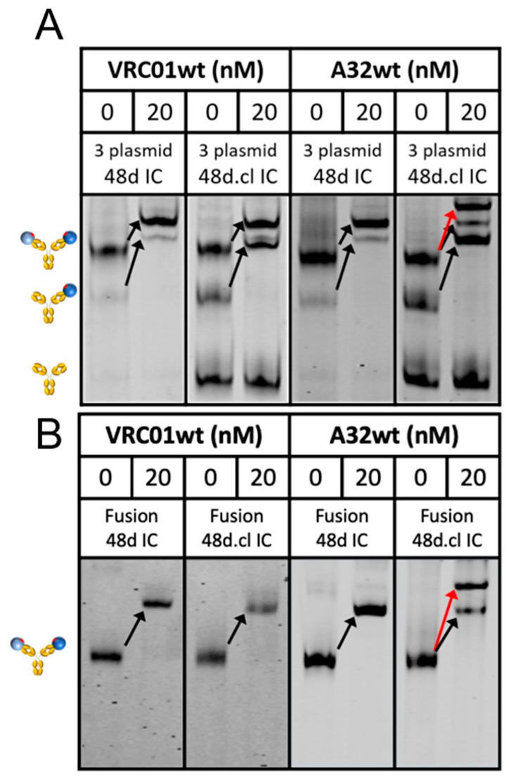 Figure 4
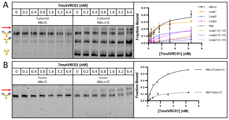 Figure 5
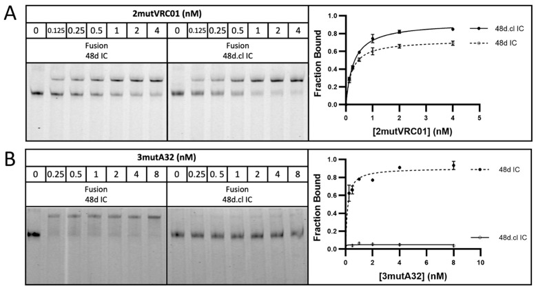 Figure 6
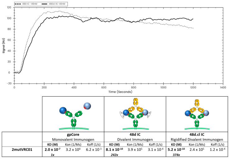 Figure 7
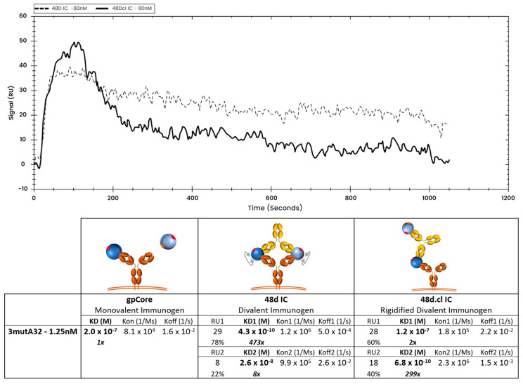 Figure 8
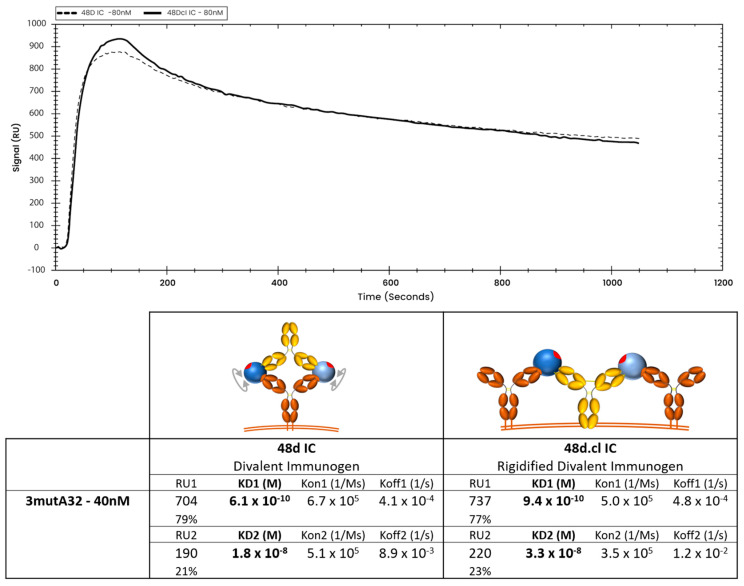 Figure 9
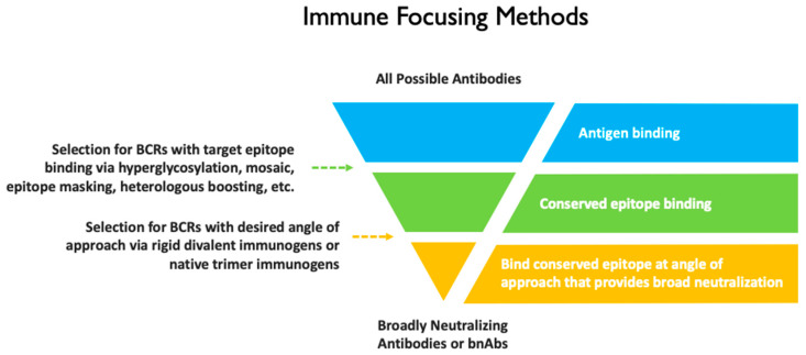 Figure 10
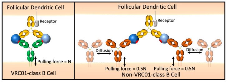 Figure 11
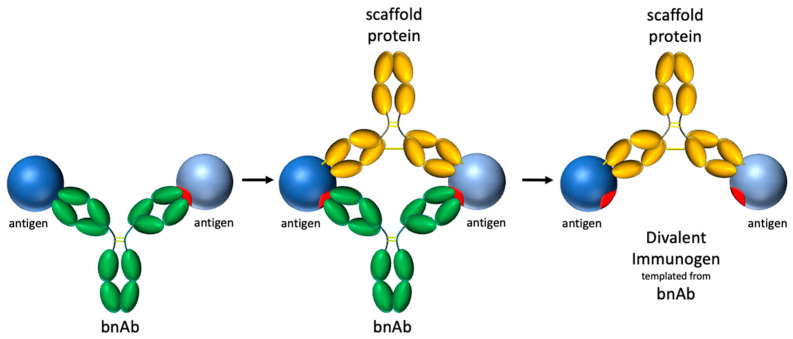 Figure 12
- —National Institute of Allergy and Infectious Diseases of the National Institutes of Health
- —National Heart, Lung and Blood Institute of the National Institutes of Health
- —Hawaii Center for AIDS
- —National Institute of General Medical Sciences of the National Institutes of Health
- —Department of Navy
- —Office of Naval Research
Peer Reviews
No public reviews on file for this paper yet. If you reviewed it on a platform where reviews are public (OpenReview, ICLR, NeurIPS, ICML), you can paste yours below so the community can read it here.
Videos
No videos yet. Explain this paper in a talk, walkthrough, or lecture? Add one.
Taxonomy
TopicsHIV Research and Treatment · Immunotherapy and Immune Responses · vaccines and immunoinformatics approaches
1. Introduction
Human Immunodeficiency Virus (HIV) remains one of the most prevalent and deadly infectious diseases in the world, with 40.8 million people globally living with HIV at the end of 2024, and 630,000 deaths in that year [1]. For the same year, there were 1.3 million new infections, a statistic that can be directly addressed with the deployment of an effective vaccine. Despite a decades-long effort, only a single large-scale vaccine study in 2009 showed slight (30%) efficacy against HIV in Thailand [2], and attempts to build upon this study have failed, resulting in discontinuation of the additional clinical trial (reviewed in [3,4]).
The biggest barrier to an effective vaccine is the vast antigenic diversity in the gp120 spike protein, driven by the rapid mutation rate of the virus [5,6,7]. However, many chronically infected patients are able to generate broadly neutralizing antibodies (bnAbs) that target conserved sites on the trimeric HIV spike and this has buoyed confidence that a vaccine is indeed possible [8,9,10]. VRC01, isolated from a chronically infected patient in 2009, is a prototypical bnAb that targets the conserved CD4bs of gp120 [11]. Multiple VRC01-lineage bnAbs originating from a VH1-2*2 and 5 amino acid CDRL3 have been isolated from numerous other patients [8,12,13,14]. VRC01 has recently been shown in a clinical trial to be protective against susceptible viruses, albeit with a higher stringency than anticipated (IC80 < 1 µg/mL in an in vitro neutralization assay representing approximately 30% of the circulating viruses in the clinical trial setting) [15]. Thus, the clinical trial results highlight the need for vaccines eliciting multiple potent bnAb classes, each targeting a conserved site for protection, of which VRC01-class bnAbs can play an important role.
Germline antibodies for multiple bnAbs paradoxically have no measurable affinity for wild-type gp120 [11,16,17]. Therefore, a major goal in the HIV vaccine field is to develop immunogens able to activate germline B cells and drive the evolution of their B cell receptors (BCRs) toward the bnAb affinity and breadth [8,17,18,19,20,21]. eOD-GT8 is a germline targeting immunogen recently shown to effectively activate VRC01 germline B cells in 97% of participants in a clinical trial [22,23,24] and GT1.1 is a trimeric gp120 (gp140) germline targeting immunogen shown to stimulate early maturation of the VRC01-class germline in a clinical trial [25]. In multiple lineage-based vaccination studies, VRC01-like antibodies were produced in mice that had been primed with eOD-GT8 60mer and boosted with gp120 and gp140 antigens [18,26,27]. Elicited antibodies were effective against N276 glycan-deficient viruses and had some neutralization breadth against N276 glycan-containing viruses [18,26,27]. Lineage-based approaches have been developed for other anti-HIV bnAbs. For example, germline targeting immunogens have been developed for specific VRC01-class bnAb CH31 [28], IOMA-class bnAbs, which also target the CD4bs with a similar angle of approach to VRC01 [29], PGT121, a broadly neutralizing antibody that targets the conserved V3 “glycan supersite” [17] and bnAbs targeting the V2 Apex [30].
All germline-targeting vaccination strategies aim to focus the immune response to rare B cell lineages and effectively maintain large populations of these cells in germinal centers. To this end, it will be useful to develop broadly applicable methods for bolstering the competitive fitness of specific B cells. Here, we propose immune-focusing contingent on a BCR’s target epitope and angle of approach. Following the initial binding of an antigen by one Fab of an IgG, the binding of a second antigen in close proximity by the second Fab can form a much more stable divalent complex. Simultaneous engagement of both Fabs of an antibody has been studied extensively and shown to increase the overall avidity 50- to 1500-fold [31,32,33,34]. We hypothesize that divalent binding is an ideal mechanism for imparting a “selective avidity” advantage to specific B cell populations to compete for antigen against non-bnAb-lineage B cells that bind to other epitopes or bind to the same epitope at non-neutralizing angles of approach. This strategy may effectively complement current lineage-based HIV vaccines and may also support novel lineages with similar binding properties. We have chosen VRC01 as our model system and have designed divalent constructs for precise, rigid positioning of two copies of gp120 such that only a select few BCRs, including VRC01-class BCRs, can bind with geometry that allows divalent binding. The divalent immunogens exhibit a higher affinity for VRC01 germline revertants than a flexible control, likely due to reduction in the entropic penalty associated with divalent binding. Experiments testing binding with VRC01 and a non-CD4bs IgG antibody, A32, to our divalent gp120 design demonstrate selective avidity. Pending future in vivo studies, this immunogen design concept may serve as a blueprint for the construction of immune-focusing vaccines that present an epitope for high avidity binding only with a specific angle of approach.
2. Materials and Methods
2.1. Plasmids and DNA Synthesis
Original plasmids for VRC01 and gp120 were obtained from the AIDS Reagent program. Plasmids for gpCore and other clones were constructed from gene fragments (Twist Biosciences, San Francisco, CA, USA) or primers (Integrated DNA Technologies, Coralville, IA, USA) and using megaprimer-based whole-plasmid synthesis, protocol described in [35]. Sequences for all proteins used in this study are included in the Supplementary Materials.
2.2. Protein Expression and Purification
Proteins were expressed by transient transfection of expression plasmids into ExpiCHO cells (Thermo Fisher, Waltham, MA, USA). Standard or max titer protocols were followed and protein harvest took place after 8–14 days of expression. Following expression in ExpiCHO cells, the ICs and antibody proteins were purified using Protein A Magnetic Sepharose Xtra Beads (Cytiva, Marlborough, MA, USA). Proteins were aliquoted and stored at −80 °C.
2.3. Site-Specific Covalent Aptamer Conjugation to 48d ICs
ICs were site-specifically labeled at the Fc with a fluorophore-linked covalent DNA aptamer sequence (ACG AGC GCG GAA CCG [3]GC C[3]G GCA CAG ACA AAC GAA CAC CAC AAG AGC CAT GGC CAT ATC AAG AAT CTA CT where [3] is ethynyldeoxyuridine) (BaseClick GMBH, Neuried, Germany). Briefly, aptamer (8 µM), 2.5 mM CuSO_4_ and 2.5 mM Tris-hydroxypropyltriazolylmethylamine (THPTA), 25 mM MES pH 6.0 and 5 mM MgSO_4_ in a 25 µL total volume were combined in a capless 0.5 mL microcentrifuge tube and placed along with a tube containing 15 µL of 25 mM hydroxysulfosuccinimidyl-4-azidobenzoate (sulfo-HSAB, G-Biosciences, St. Louis, MO, USA) and a last tube containing 20 µL of 25 mM sodium ascorbate in a 25 mL two-necked flask. Argon gas was flowed into the flask and out through a rubber septum/16 gauge needle for 30 min at low flow rate. Then, while maintaining a low argon flow rate into the flask, the exhaust septum/needle were removed, 8 µL sulfo-HSAB and 1.25 µL of sodium ascorbate were transferred to the tube containing the DNA followed by pipette mixing. The reaction was allowed to proceed for 30 min, after which it was stopped by the addition of 2 µL 100 mM THPTA and two Sephadex G-50 spin column buffer exchanges into 25 mM MES/5 mM MgSO_4_ buffer and one spin column buffer exchange into 25 mM HEPES pH 7.2, 150 mM NaCl and 2 mM MgSO_4_. The modified aptamer was then incubated with the immune complex (5–10 µg) followed by CuSO_4_ to a final concentration of 10 µM.
2.4. Gel Shift Assay
Covalent aptamer-modified ICs (0.1–1 nM) were mixed with varying concentrations of Ab for 2 h followed by addition of fluorophore-labeled imager strand (IRDye700-AGT AGA TTC TTG ATA TGG CCA TGG CTC TTG) and 12.5% Ficoll. The samples were loaded into 6% acrylamide/0.5X TBE gels and run at 250 V for 35 min. Gels were imaged on a Li-Cor Odyssey laser imager. Fluorescence measurements were made using Li-Cor Odyssey software (Image Studio 4.0) from which binding fractions were calculated.
2.5. Surface Plasmon Resonance Binding Analysis
SPR binding analysis was performed using OpenSPR-XT (Nicoya, Kitchener, ON, CA). The following immobilization strategy was used: Biotin SPR sensors (Nicoya), a layer of 400 nM streptavidin (New England Biolabs, Ipswich, MA, USA), a layer of 1 µM biotinylated Strep-Tag (WSHPQFEK) peptide (Genscript, Piscataway, NJ, USA), a layer of 200 nM Strep-tactin XT (IBA Lifesciences, Göttingen, Germany), then immobilization of our C terminus Strep-tagged ligand antibody, either VRC01-class Ab or A32 class Ab, at various concentrations between 1 nM and 80 nM. A schematic of this binding immobilization strategy is shown in Figure S1. This multi-step immobilization strategy was developed as a means of maximizing both (a) the vertical orientation of the immobilized Ab, so that both Fabs of the Ab would be available for divalent binding, and (b) the stability of the immobilized ligand. The analyte consisted of our divalent immunogens with gpCore or monovalent gpCore at various concentrations for the purpose of establishing monovalent affinities. Bmax values, summarized in Table S1, represent the maximum binding response observed under a given SPR experimental condition, reflecting the apparent binding capacity of the immobilized ligand. Bmax is an experimental parameter that depends on the ligand immobilization level, ligand orientation and accessibility, and therefore varies with antibody loading concentration on the SPR sensor. Fit model selection was guided by expected binding stoichiometry, antibody and immunogen geometry, ligand density and sensorgram behavior. Reported kinetic parameters should be interpreted as apparent kinetic values reflecting dominant binding modes under the specified experimental conditions, rather than absolute intrinsic constants. Graphing of the sensorgram was completed using TraceDrawer software (version 1.10.1).
2.6. Data Analysis
Statistical analysis and gel shift assay results were prepared using GraphPad Prism 9 software.
3. Results
3.1. Design of an Immune Focusing Rigid Divalent Immunogen
We imagined an immune focusing immunogen having two copies of an antigen fixed in space such that divalent binding is limited to B cell receptors with a specific binding footprint and angle of approach (Figure 1). To this end, we designed a covalent immune complex (IC) using a complementary anti-gp120 Ab, 48d, which was selected after an exhaustive search in the Protein Data Bank. 48d binds at the coreceptor binding site of core gp120 (gpCore), less than 1 nm from the VRC01 footprint [36,37]. Binding by VRC01 Fabs to the two copies of gpCore would result in an Ab paratope-paratope distance of ~14 nm. The rationale is that Fab-Fab geometry of an IgG antibody has a strong impact on the avidity conveyed by divalent binding, especially for antibodies with low monovalent affinity. Under these circumstances, an antigenic spacing of 13–16 nm permitted the highest avidity [33,34]. gpCore (of strain BG505) was chosen as our gp120 antigen because it removes potentially distracting variable loops V1/V2 and V3.
Covalent linkage between 48d Fab and gp120 was initially accomplished by D56C and I423C substitutions to 48d heavy chain and gp120 core, respectively, for disulfide bonding as predicted by the program Disulfide by Design [38]. For Fab-Fab crosslinking, we identified Lys126 in the light chain of 48d as a potential site for cysteine substitution and disulfide formation by modeling 48d and VRC01 Fabs simultaneously bound to 2 copies of gpCore (Figure 2A). Modeling of the initial design suggested that the resulting construct might constrain divalently bound VRC01 via a narrower than optimal Fab-Fab distance. Therefore, we sought to identify additional designs that relax the ring complex. Specifically, the light chain loop containing Lys126 was remodeled using the programs ColabPaint [39] based on [40] as well as ProteinMPNN [41] and AlphaFold2 [42] using ColabFold (version 1.5.5) [43]. Starting with models initially generated by ColabPaint, we performed iterative residue optimization with ProteinMPNN followed by structure prediction by AlphaFold2 using ColabFold. After modeling many preliminary designs, the top three designs were selected for testing on the basis of (1) the geometry of the construct and (2) the predicted confidence in successful folding as measured by pLDDT in AlphaFold2. AlphaFold2 predictions were used qualitatively to assess foldability and overall structural plausibility of loop-modified constructs; experimental characterization was used to validate functional integrity. The sequences and AlphaFold2 predicted structures for these three designs–named Loop1, Loop2 and Loop3—that are shown in Figure 2B.
We initially expressed these designs by co-transfecting 3 plasmids encoding the 48d heavy chain, 48d light chain and gp120 into ExpiCHO cells (sequences in Supplementary Materials). In this format, covalent binding via disulfide formation between the gpCore and 48d happens post-translationally and, due to incomplete saturation of gp120 on 48d, results in three species of immune complexes–zero, singly, and doubly gpCore-bound IC—with the doubly bound IC being our desired species. The immune complexes were purified using Protein A chromatography and analyzed by SDS-PAGE and Coomassie staining (Figure 3A). Differentially gp120-occupied species are identified with the 2, 1 and 0 in Figure 3A. Comparison of the banding pattern between non-Fab-Fab-crosslinked and Fab-Fab-crosslinked designs indicated that crosslinking efficiency might be less than 100%. Arrows in Figure 3A represent the band believed to be zero gp120-bound, Fab-Fab crosslinked ICs, with the non-crosslinked IC running slower in the gel, and we suspected that the singly- and doubly gp120 bound ICs had similar crosslinking efficiencies despite the lack of clear separation in their bands. We estimate 48d.cl IC, incorporating the K126C substitution, to have the highest crosslinking efficiency, approximately 80–95%, while our three computational loop designs to have a lower crosslinking efficiency of approximately 40–60%. These crosslinking efficiency estimates are corroborated by a gel shift assay (Section 3.3). The presence of non-crosslinked species in our samples and the lack of a clear method to effectively purify them away from the crosslinked species presented a challenge to our characterization of their binding. For this reason, we focused most of our surface plasmon resonance (SPR) binding assays on 48d and 48d.cl ICs due to the relatively high purity of these products.
3.2. Design of gp120 + 48d Heavy Chain Fusion Proteins
In order to eliminate unwanted species of immune complex containing zero or one gpCore, we designed a gpCore + 48d heavy chain fusion protein by circularly permuting gpCore (creating a new N and C terminus) and linking the new gpCore peptide to 48d.heavy via a 3x(GGGS) linker, similar to the approach used in [44]. Transfection with two plasmids, one encoding the gpCore + 48d heavy chain fusion and one encoding the 48d light chain, was performed and the complexes were purified via protein A chromatography and analyzed via SDS-PAGE (Figure 3B). The banding pattern suggests a majority of protein encoding the complete fusion construct, with a faster migrating minor contaminant. The fusion protein approach represents an improved expression format for the divalent immunogens, given the simpler transfection protocol and stoichiometric gp120 occupancy of the heavy chain.
3.3. Engineered Divalent Immunogens Bind to VRC01 with 1:1 Stoichiometry and to a Non-CD4bs Ab with 1:2 Stoichiometry
We sought to characterize the binding of our constructs to VRC01 as well as a non-CD4bs Ab, A32, which recognizes a conformational epitope involving the C1 and C4 gp120 region [45]. In contrast to VRC01, A32 is predicted to be unable to bind divalently to a Fab-Fab-crosslinked divalent immunogen to form a ring structure and therefore should bind with 2:1 stoichiometry. Accordingly, non-crosslinked Fabs in control ICs should be able to rotate to accommodate 1:1 binding. To test this, we developed a novel assay that utilizes a covalent DNA aptamer irreversibly bound to the Fc of 48d [46]. During electrophoresis, the negatively charged aptamer pulls the immune complex through a polyacrylamide gel. A fluorophore-tagged oligonucleotide hybridized to the covalent aptamer enables sensitive detection with an imaging system. Incubation with wtVRC01 caused a complete shift in singly and doubly gp120-bound ICs, regardless of Fab-Fab crosslinking, which was expected due to the high monovalent affinity of ~5 nM (Figure 4). In contrast, incubation of 48d.cl with wtA32 shows a different binding pattern, where an extra shifted band consistent with 2:1 binding (red arrow) is observed, but only in the crosslinked sample. The single-shifted band agrees with the presence of non-Fab-Fab crosslinked minor species in the 48d.cl sample. Together, the VRC01 and A32 gel shift data strongly suggest different binding stoichiometries for each Ab, in agreement with our intended design for selective avidity. We ran a similar assay with Loops 1-3, and the shift data agree with our estimated Fab crosslinking efficiencies from the SDS-PAGE data (Figure S2). We also asked whether our constructs would bind other VRC01-class antibodies, such as N6, with 2:1 or 1:1 stoichiometry. The results with N6 demonstrated a single shift for all of our constructs, indicating 1:1 divalent binding (Figure S3), which is consistent with an antibody binding divalently to the Fab-Fab crosslinked immunogen.
3.4. The Divalent Immunogen Binds mutVRC01 Antibodies with Higher Affinity Compared to a Flexible Control
In order to test the ability of the divalent gp120 immune complexes to bind to VRC01 precursors, we selected two VRC01 variants containing germline reversion mutations from [47]—seven point mutations (7mutVRC01), reported to have a monovalent affinity for gp120 of 152 nM, and a two-alanine insertion (2mutVRC01), reported to have a monovalent affinity for gp120 of 320 nM. Using SPR, we experimentally determined the 7mutVRC01 and 2mutVRC01 to have a monovalent affinity for gpCore of approximately 7 µM and 200 nM, respectively. The difference between the previously reported affinities and our experimentally determined affinities could be due to differences in the gp120 constructs used (wtGP120 from HXB2 vs. gpCore from BG505 in this study). We also observed signs of instability in 7mutVRC01 (loss of binding upon −80 °C storage) and as a result it frequently exhibited variance in binding characteristics, although binding trends within an assay (with Fab-Fab-crosslinked and non-crosslinked control run in parallel) were consistent from batch to batch and 7mutVRC01 protein was frozen in aliquots to ensure the same freeze–thaw exposure in all experiments. In order to compare binding by a non-CD4bs bnAb of similar monovalent affinity, we created a mutant of A32 with reduced affinity by incorporating R100A in the heavy chain and Y30F and Y32F in the light chain. The resulting Ab, 3mutA32, was determined to have an affinity for gpCore of approximately 200 nM—nearly identical to our 2mutVRC01 mutant. A summary of the antibodies used for testing binding of the divalent immunogens is shown in Table 1.
Incubation with 7mutVRC01 resulted in a stronger shifted band for doubly gp120-bound, Fab-Fab crosslinked versions of 48d.cl IC and Loops1-3 ICs compared with their non-crosslinked or cysteine → alanine variants (resulting in an inability to crosslink Fabs), strongly suggesting that crosslinking reduces the entropic penalty for divalent binding (Figure 5). A summary of the gel shift assay results for all eight constructs with 7mutVRC01 is shown in Figure 5A, and the gel shift assay for the 48d IC and 48d.cl IC fusion constructs is shown in Figure 5B. The incubation of 48d and 48d.cl ICs with 2mutVRC01 (K_D_ = 200 nM) suggests that while the overall affinities are higher than for 7mutVRC01 (estimated K_D_ = 7 µM), the affinity difference between Fab-Fab-crosslinked and non-Fab-Fab-crosslinked species is significantly diminished (Figure 6A). For 2mutVRC01, we consistently observed crosslinked IC to have a higher estimated maximum fraction bound (B_max_), despite the similar apparent affinities between species. This may indicate the ability of the crosslinked IC to bind to more glycosylation variants of gpCore, which are known to exist in any given preparation of HIV gp120 [49]. It should be noted that in theory, the Bmax should be near 1 for all species in Figure 5 and Figure 6; although, in reality the gel shift may not perfectly reflect the theoretical B_max_ because the nature of the assay favors tight binding interactions that survive the gel migration.
3.5. The Divalent Immunogen Binds a VRC01 Revertant with Significantly Higher Avidity than an A32 Variant of Similar Monovalent Affinity
In an ideal vaccination scenario, the divalent immunogen would selectively bind to low-monovalent affinity VRC01 BCRs with higher avidity than either a non-CD4bs BCR or a CD4bs BCR with non-neutralizing angle of approach with similar monovalent affinities. In agreement, incubation with 3mutA32 weakly bound to 48d.cl IC, with the shifted species approaching the fraction of non-crosslinked IC apparent from the reaction with wtA32 in Figure 4 (Figure 6). As expected, 3mutA32 bound much more strongly to non-Fab-Fab-crosslinked control IC, due to Fab rotation and divalent binding. These data strongly support the idea that the rigid divalent immunogen could selectively target VRC01-class B cells for high avidity binding. A summary of the gel shift assay results for 48d and 48d.cl ICs with 3mutA32 is shown in Figure 6.
3.6. SPR Analysis Supports the Interpretation of Reduced Dissociation Rates (Koff) and Equilibrium Dissociation Constants (KD) of the Crosslinked Divalent Immunogen Binding to 2mutVRC01
To achieve the most vertical presentation of antibodies on a biotin sensor, we developed a novel immobilization strategy, starting with streptavidin, which is expected to bind to two biotins, leaving two surface-exposed, unoccupied biotin-binding pockets. We then layered on a biotinylated Strep-Tag peptide, which is expected to occupy the remaining binding pockets (Figure S1). A variant of streptavidin having a high affinity for the Strep-Tag (Strep-Tactin XT™) was then layered onto the sensor, and finally, we bound our C terminal Strep-tagged antibody, either VRC01 or A32, at concentrations between 1 nM and 80 nM. This multi-step immobilization strategy maximizes both (a) the vertical orientation of the immobilized Ab so that both Fabs of the Ab would be available for divalent binding, and (b) the stability of the immobilized ligand. Using this immobilization strategy, we characterized binding of 48d.cl IC and control 48d IC (Figure 7 and Figure S5A) to 2mutVRC01 by SPR. We consistently observe slower dissociation rates and lower equilibrium constants for the crosslinked 48d.cl IC when binding with 2mutVRC01 (Figure 7), suggesting higher avidity resulting from a lower entropic penalty for divalent binding. We also immobilized 7mutVRC01 but were unable to consistently observe a signal. This may be due to the instability in the variant that we also observed in gel shift assays.
3.7. SPR Data Suggests Monovalent Binding to 3mutA32 by the Crosslinked Immunogen
At low ligand density (loading 1.25 nM 3mutA32 on the SPR chip), crosslinked 48d.cl IC exhibited a rapid initial dissociation consistent with mostly monovalent binding followed by tighter binding species consistent with divalent binding (Figure 8 and Figure S5A). In contrast, non-Fab-Fab-crosslinked control IC showed tight binding and a slow dissociation consistent with mostly divalent binding (Figure 8 and Figure S5B). A logical interpretation of this is that there is a minor population of 48d.cl that exhibits divalent binding, despite poor divalent binding as observed in the gel shift assay. This component is most easily explained by (1) the portion of the 48d.cl IC which is not crosslinked, estimated to be 5–20%, and which is also visible in the gel shift assay, and (2) divalent binding to two adjacent immobilized 3mutA32 antibodies on the SPR sensor that were close enough for the divalent immunogen to bind both antibodies simultaneously. In agreement with the latter, increasing the density of immobilized 3mutA32 ligand (loading with 40 nM Ab), results in nearly all the 48d.cl IC binding strongly (Figure 9). Reducing A32 ligand density resulted in lower divalent binding behavior but also lower SPR signal and greater noise. We found that the lower bound 3mutA32 ligand density for reliable signal was accomplished by loading with ~1 nM antibody.
3.8. The Divalent Immunogen, When Combined with Germline-Targeting Mutations to gp120, Binds to Monomeric VRC01 IgM Unmutated Common Ancestor, Suggesting It Could Function as a Prime
While we initially envisioned the fixed divalent design as a boosting immunogen, we wondered whether the divalent immunogen could also function as a priming immunogen. We thus sought to characterize the binding of the immunogen with monomeric VRC01 IgM unmutated common ancestor (UCA) [50,51]. Given that gpCore, even when presented on a highly multimerized nanoparticle, has been shown to not bind VRC01 IgM UCA [16,52], we designed three germline-targeting gp120s, named GTi, GTii and GTiii, and characterized their binding, along with the original gpCore immunogen, with monomeric VRC01 IgM UCA via gel shift assays.
GTi mutations included N276D, N386D and N462D to remove three glycans surrounding the CD4 binding site (N197, also implicated in steric restriction of VRC01 to the CD4bs, is already absent in our gpCore due to removal of V1/V2 loop region). GTii mutations included the GTi glycan deletions plus T278R and G471S, paralleling a germline targeting trimer GT1 [53]. GTiii contained 12 mutations and is based on the eOD-GT6 immunogen [24].
Consistent with previous studies, no measurable binding was observed between the gpCore divalent immunogen and monomeric VRC01 IgM UCA (Figure S6A). In contrast, all three GT-based immunogens (including the minimally mutated GTi) did bind VRC01 IgM UCA (Figure S6B–D). Based on our gel shift assays, we estimate that GTi binds VRC01 IgM UCA with a K_D_ of ~500 nM for the wild-type (WT) version and ~800–1000 nM for the Fab-Fab-crosslinked version (Figure S6B). Similarly, GTii binds with a K_D_ of ~30 nM (WT) and ~60 nM (crosslinked) (Figure S6C), while GTiii binds with a K_D_ of ~16 nM (WT) and ~25 nM (crosslinked) (Figure S6D). The lower avidity of the Fab-Fab-crosslinked immunogen compared to WT likely reflects suboptimal geometry for IgM, which is less flexible than IgG and is predicted to have difficulty accommodating the narrow 48d Fab-Fab angle modeled for the 48d.cl IC (Figure 2). Furthermore, introducing VRC01 “knockout” mutations (D279K/D368V) into the GT-based immunogens abrogated binding to VRC01 IgM UCA, confirming the specificity of the interaction. These results suggest that a minimally mutated gp120-based divalent immunogen could potentially function as a prime in immunization regimens.
4. Discussion
In this study, we engineered rigid divalent immunogens that bind both Fab regions of an IgG1 isotype BCR simultaneously with the goal of leveraging avidity to enhance selectivity for BCRs with a specific target epitope and angle of approach. Binding assays demonstrated “selective avidity”: high-avidity divalent binding to the VRC01-class Ab and low avidity monovalent binding to a non-CD4bs Ab. The divalent immunogens also displayed increased affinity for VRC01-class antibodies compared to their non-Fab-Fab-crosslinked controls, a likely consequence of gp120 antigen pre-organization minimizing the entropic cost of divalent binding, as reflected by reduced dissociation rates (K_off_) and equilibrium dissociation constants (K_D_), particularly at lower monovalent affinities.
This study represents a new approach to immune focusing. It differs from most other immune focusing approaches in that in addition to selecting for BCRs that bind the target epitope, it also imposes an angle of approach requirement (Figure 10). In contrast to a previous HIV-1 Env–antibody complex aiming to focus responses by masking variable epitopes [54], our design rigidly and precisely positions two antigens for selective divalent binding, thereby directly targeting VRC01-class B cell receptors. Importantly, the selective avidity design accommodates existing immune focusing methods such as simultaneous incorporation of distant gp120s [55,56] and hyperglycosylation [57,58].
Our divalent immunogens were designed with the goal of selectively promoting IgG^+^ VRC01-class B cell proliferation in the germinal center (GC), to be administered as a boost to eOD-GT8 60mer, which is a multivalent immunogen effective in activating VRC01-class naïve B cells in healthy volunteers [23]. Features of GC B cells are likely to be more conducive to selective activation by our divalent immunogens than for naïve B cells. We expect entropic penalty minimization to enhance BCR engagement by VRC01-class GC B cells because adjacent BCRs of a non-VRC01-class B cell must undergo negentropic ordering for 2:1 (BCR:IC) binding (Figure 11). GC B cells have a 5- to 10-fold lower BCR density on GC B cells [59], which may place an even greater emphasis on individual BCR binding and promote greater binding selectivity. Whereas naïve B cells are readily activated by BCR crosslinking induced by multivalent antigens, GC B cells are largely insensitive to soluble multivalent antigen and BCR signaling is re-wired, resulting in an intrinsic affinity threshold of GC B cells at least 100-fold higher than that of naïve B cells [60,61]. Instead, GC B cells signal in response to membrane-anchored antigen, likely a result of mechanosensing of pulling forces applied by the B cell to extract antigen from follicular dendritic cells [60]. When controlled for T cell help, BCR signaling in response to high affinity binding results in greater positive selection signals than for low affinity binding [62], suggesting that stronger pulling forces resulting from high affinity BCR-antigen interactions promote positive selection signals. We reason that ring dimer formation will increase the pulling force experienced by the BCR compared to 2:1 (BCR:IC) binding (Figure 11), resulting in significantly stronger positive selection signals that will complement entropic penalty-driven binding selectivity.
Naïve B cells express IgM and IgD BCRs, which have different hinge regions and flexibilities compared to IgG1 [63], and this will affect the geometry and avidity of the 1:1 ring-dimer complex with our IC designs. IgD has a long (30 residue) upper hinge and this is expected to convey low avidity for a divalent interaction due to a large entropic penalty, particularly at low monovalent affinity. In contrast, IgM has a Cμ2 domain instead of a hinge region. Intuitively, this would suggest a more constrained range of independent Fab motion compared to IgG1, in agreement with a recent cryo-EM study [64]. However, a TEM study of the ability to form ring dimer complexes [65] and a study of divalent IgM binding using DNA nanotechnology and SPR [33] suggest a large range of motion in monomeric IgM. Our gel shift assays suggest at least some degree of independent Fab motion in IgM, enabling ring dimer formation with our IC design. Since high avidity ring dimer formation with IgM is indeed possible, we hypothesize that multimerization with the minimal number of divalent immunogens required for robust activation of naïve B cells may maximize the immune focusing effect when administered as a priming immunogen.
The concept of selective avidity extends beyond the targeting of HIV bnAb germline precursors to other potential applications across the vaccine field. Divalent immunogens may have utility both in recruiting and supporting known bnAb germlines as well as novel germlines that are otherwise outcompeted by immunodominant responses elicited by conventional vaccines. Recent work by Dvorscek et al. demonstrates how immune complexes can elicit strong activation and proliferation of B cells with complementary BCRs [66], suggesting that precisely engineered antigen–antibody complexes-such as those in our selective avidity design-could be broadly applicable for enhancing immune responses. A high-level design process for developing a divalent immunogen targeting a bnAb of interest is suggested in Figure 12. In the example below, and in our design, a complementary antibody was used. A potential advantage of using an immune complex design is that the antibody portion can be humanized to be minimally immunogenic. However, other proteins, including de novo designs created using new protein design tools [67,68], could be used to rigidly support two antigens for specific divalent engagement of targeted BCRs.
5. Conclusions
Rigid divalent gp120 immunogens were designed to convey “selective avidity”—divalent binding to VRC01-class Abs and monovalent binding to non-CD4bs Abs or CD4bs Abs—with a different angle of approach. We have demonstrated this effect with a VRC01 and a non-VRC01-class Ab, A32. Selective avidity may enhance the survival of VRC01 lineage B cells during vaccination, and in vivo studies are planned to clarify the broader applicability of this immune focusing strategy.
6. Patents
I.M., R.B. and A.T.L. are inventors on a patent application (PCT/US23/82376) filed by the University of Hawaii that covers divalent immunogen designs as described in this work.
The reference list from the paper itself. Each links out to its DOI / PubMed record.
- 1World Health Organization Hiv Data and Statistics World Health Organization Geneva, Switzerland 2025 Available online: https://www.who.int/teams/global-hiv-hepatitis-and-stis-programmes/hiv/strategic-information/hiv-data-and-statistics(accessed on 6 December 2025)
- 2Rerks-Ngarm S. Pitisuttithum P. Nitayaphan S. Kaewkungwal J. Chiu J. Paris R. Premsri N. Namwat C. de Souza M. Adams E. Vaccination with Alvac and Aidsvax to Prevent Hiv-1 Infection in Thailand N. Engl. J. Med.20093612209222010.1056/NEJ Moa 090849219843557 · doi ↗ · pubmed ↗
- 3Adepoju V.A. Udah D.C. Onyezue O.I. Adnani Q.E.S. Jamil S. Bin Ali M.N. Navigating the Complexities of Hiv Vaccine Development: Lessons from the Mosaico Trial and Next-Generation Development Strategies Vaccines 20251327410.3390/vaccines 1303027440266130 PMC 11946271 · doi ↗ · pubmed ↗
- 4Desrosiers R.C. The Failure of Aids Vaccine Efficacy Trials: Where to Go from Here J. Virol.202397 e 002112310.1128/jvi.00211-2336916947 PMC 10062124 · doi ↗ · pubmed ↗
- 5Alizon S. Fraser C. Within-Host and between-Host Evolutionary Rates across the Hiv-1 Genome Retrovirology 2013104910.1186/1742-4690-10-4923639104 PMC 3685529 · doi ↗ · pubmed ↗
- 6Mansky L.M. Retrovirus Mutation Rates and Their Role in Genetic Variation J. Gen. Virol.1998791337134510.1099/0022-1317-79-6-13379634073 · doi ↗ · pubmed ↗
- 7Menendez-Arias L. Mutation Rates and Intrinsic Fidelity of Retroviral Reverse Transcriptases Viruses 200911137116510.3390/v 103113721994586 PMC 3185545 · doi ↗ · pubmed ↗
- 8Kwong P.D. Mascola J.R. Hiv-1 Vaccines Based on Antibody Identification, B Cell Ontogeny, and Epitope Structure Immunity 20184885587110.1016/j.immuni.2018.04.02929768174 · doi ↗ · pubmed ↗