On the Importance of Correct Snake Identification. Comment on Chippaux et al. Snakebites in Cameroon by Species Whose Effects Are Poorly Described. Trop. Med. Infect. Dis. 2024, 9, 300
Wolfgang Wüster, David A. Warrell, David J. Williams

Abstract
Genes, proteins, chemicals, diseases, species, mutations and cell lines named across the full text — each resolved to its canonical identifier and authoritative record.
Click any figure to enlarge with its caption.
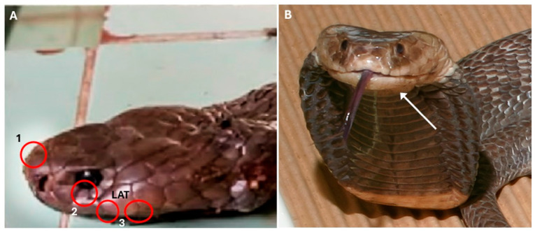 Figure 1
Peer Reviews
No public reviews on file for this paper yet. If you reviewed it on a platform where reviews are public (OpenReview, ICLR, NeurIPS, ICML), you can paste yours below so the community can read it here.
Videos
No videos yet. Explain this paper in a talk, walkthrough, or lecture? Add one.
Taxonomy
TopicsVenomous Animal Envenomation and Studies · Rabies epidemiology and control · Beetle Biology and Toxicology Studies
One of the major obstacles to improving the management of snakebite envenoming is the lack of accurate identification of species responsible for clinical cases, which prevent the improvement of definitions of species-specific syndromes. Understanding which snakes are biting people in a given region of the world, and what the clinical consequences of these cases are, is a cornerstone to the formulation of strategies to reduce the burden of snakebite. Yet, in many parts of the world, this knowledge remains elusive. Nowhere is this problem more acute than in the forested zones of Central and Western Africa, where detailed epidemiological studies remain conspicuous by their absence, the key species involved in severe envenoming remain largely unknown, and their syndromes poorly understood and ill-defined. It is thus with considerable excitement that we read the recent paper by Chippaux et al. [1], promising a description of the syndromes of envenoming by several rarely documented snake species in Cameroon.
However, we were troubled to find that upon comparing clinical notes and photographs, there were multiple misidentifications of specimens to which cases were attributed. Coupled with clinical incongruities, these inaccuracies cast considerable doubt on the value of the paper.
The following are clear misidentifications of the snakes shown in the photographs:
- Figure 7 [1]: the snake shown is unquestionably a Naja nigricollis, not a N. haje. An enhanced version of the Figure is shown in our Figure 1A. Its identity is evident from the clearly visible second (lower) preocular, the contact between the 3rd supralabial and the eye (Figure 1A—the scale contacting the eye is clearly contiguous with the 3rd supralabial, although there may be a small surface irregularity conveying an impression of partial separation), and the small penultimate supralabial, all characters that distinguish African spitting cobras (subgenus Afronaja) from the Naja haje group (subgenus Uraeus) [2,3,4]. The temporal and supralabial scalation is more challenging to discern. Superficial examination gives the impression of a single anterior temporal, bordered below by a high supralabial that is also in broad contact with the postoculars. However, closer inspection suggests that the lower supralabial edges are curled inwards towards the mouth, and that what looks like a high supralabial is in fact a lower temporal, bordered below by the typical low 4th and 5th supralabials of Afronaja spp. that are, in this photo, largely hidden by the overhanging temporal region (Figure 1A). A corner of the low 4th supralabial is just about visible between the posterior edge of the third supralabial, the anterior edge of the lower anterior temporal and the edge of the mouth. Additionally, the illustrated specimen has approximately square internasals in broad contact with each other and a low rostral scale, whereas in N. haje, the rostral is high and separates the anterior inner edges of the internasals from each other. This identification is also supported by the clinical syndrome of envenoming that consisted of massive swelling of the entire bitten arm with large blisters on the dorsum of the hand, in the absence of any neurotoxic signs. The patient died 30 h after the bite, possibly from hypovolaemic shock as there is no mention of fluid replacement being given. This clinical picture is consistent with N. nigricollis but not with N. haje envenoming, which entails significant neurotoxicity that can sometimes be accompanied by local swelling and minor blistering, but not cytotoxicity without neurotoxic signs [5,6].
In summary, 5 out of 12 snakes (42%) shown in the photos in this paper are unambiguously misidentified, a ratio worsened by the fact that several of the remaining photos do not show the level of detail required for a species-level identification, either in the PDF file of the article or in the HTML version on the journal website. If this high rate of misidentification, which compares unfavourably with the correct identification rates achieved by crowdsourcing snake identifications from photos [17], were to be extrapolated to the other snake identifications in the article, it would cast grave doubts on the reliability of the entire paper. Importantly, the lack of locality information for individual cases impedes the verification of records where the snake cannot be confidently identified, and represents a wasted opportunity to provide more geospatially and herpetologically detailed epidemiological information.
The issues highlighted here point to the wider problems of misidentifications in the clinical snakebite literature [18]. Photos of snakes responsible for bites, taken either at the bedside or by the victim or their companions at the time of the accident, represent valuable vouchers allowing for the verification of identifications in studies such as this, and have allowed for the reidentification of snakes in other studies, in some cases leading to retractions (e.g., [10], where the snake responsible was a Trimeresurus purpureomaculatus rather than Rhabdophis subminiatus). However, even when misidentifications can later be corrected, the damage may have been done. For instance, because of the paper discussed here, the literature now contains a false record of a fatal cytotoxic envenomation by Naja haje, potentially misleading future work.
As always, the publication of inaccurate scientific information suggests a failure to exercise due diligence by both the authors and the editorial review process of the journal. To maintain the integrity of the scientific record, we recommend that editors of scientific journals should send all manuscripts involving snake identifications to qualified herpetologists for review, even when one or several authors are themselves widely acknowledged snake and snakebite experts, as is the case here. Also, since multiple misidentifications in the figures shown in manuscripts erode confidence in the identification of bite cases where the snake is not depicted, we suggest that studies of this nature should always provide, in a supplementary online appendix, all photos used for the identification of snakes responsible for bites, at full original resolution and with locality details, all unfortunately absent in the paper discussed here. Ideally, the photos should show the upper and lower sides of the head and neck, both sides of the head (in case of anomalies), and the entirety of the ventral and dorsal surface of the snake. Deposition of photos in natural history museums and online databases such as iNaturalist or HerpMapper will add further to their value. Where possible under local circumstances, the collection of tissue samples for DNA analysis and the deposition of specimens in natural history collections will allow for further verification of the material and thus future-proof the output of epidemiological studies. These suggestions will increase both the verifiability of the data and add valuable information on the distribution of medically important species and on the epidemiology of snakebite, and thereby prevent erroneous information from entering and being disseminated through the scientific literature.
The reference list from the paper itself. Each links out to its DOI / PubMed record.
- 1Chippaux J.-P. Madec Y. Amta P. Ntone R. Noël G. Clauteaux P. Boum Y.II Nkwescheu A.S. Taieb F. Snakebites in Cameroon by Species Whose Effects Are Poorly Described Trop. Med. Infect. Dis.2024930010.3390/tropicalmed 912030039728827 PMC 11679889 · doi ↗ · pubmed ↗
- 2Wallach V. Wüster W. Broadley D.G. In Praise of Subgenera: Taxonomic Status of Cobras of the Genus Naja Laurenti (Serpentes: Elapidae)Zootaxa 20092236263610.11646/zootaxa.2236.1.2 · doi ↗
- 3Chippaux J.-P. Jackson K. Snakes of Central and Western Africa Johns Hopkins University Press Baltimore, MD, USA 2019
- 4Trape J.-F. Guide des Serpents d’Afrique Occidentale, Centrale et d’Afrique du Nord IRD Editions Marseille, France 2023
- 5Warrell D.A. Greenwood B.M. Davidson N.M. Ormerod L.D. Prentice C.R.M. Necrosis, Haemorrhage and Complement Depletion Following Bites by the Spitting Cobra (Naja nigricollis)QJM Int. J. Med.19764512210.1093/oxfordjournals.qjmed.a 067448943796 · doi ↗ · pubmed ↗
- 6Warrell D.A. Barnes H.J. Piburn M.F. Neurotoxic Effects of Bites by the Egyptian Cobra (Naja Haje) in Nigeria Trans. R. Soc. Trop. Med. Hyg.197670787910.1016/0035-9203(76)90012-21265823 · doi ↗ · pubmed ↗
- 7Wüster W. Crookes S. Ineich I. ManéY. Pook C.E. Trape J.-F. Broadley D.G. The Phylogeny of Cobras Inferred from Mitochondrial DNA Sequences: Evolution of Venom Spitting and the Phylogeography of the African Spitting Cobras (Serpentes: Elapidae: Naja nigricollis Complex)Mol. Phylogenet. Evol.20074543745310.1016/j.ympev.2007.07.02117870616 · doi ↗ · pubmed ↗
- 8Spawls S. Branch B. The Dangerous Snakes of Africa Bloomsbury London, UK 2020