Functional Dissection of Leishmania major Membrane Components in Resistance to Cholesterol-Dependent Cytolysins
Chaitanya S. Haram, Sebastian J. Salinas, Coleman Wilson, Salma Waheed Sheikh, Kai Zhang, Peter A. Keyel

TL;DR
This study explores how Leishmania major resists cholesterol-dependent cytolysins, revealing that sterols and glycans play key roles in toxin binding and pore formation.
Contribution
The study identifies distinct mechanisms in Leishmania major compared to mammals for resisting cholesterol-dependent cytolysins.
Findings
Loss of GP63 enhances perfringolysin O toxicity but not streptolysin O.
Plasmenylethanolamine and lipophosphoglycan have minimal roles in CDC binding.
Sterols protect cells from CDCs but do not reduce toxin binding.
Abstract
Bacteria use cholesterol-dependent cytolysins (CDCs) to damage eukaryotes. While well-studied in mammals, the mechanisms by which CDCs bind to and kill protozoans remain unclear. CDCs bind to the human pathogen Leishmania major but only kill in the absence of sphingolipids. The contribution of other leishmanial membrane components to CDC binding and cytotoxicity remains unknown. Here, we used genetic knockouts and inhibitors to determine the contribution of key membrane components to CDC binding and killing in L. major. We analyzed toxin binding and killing using flow cytometry and Western blotting. Loss of the virulence factor GP63 enhanced toxicity of perfringolysin O but not streptolysin O. Plasmenylethanolamine and lipophosphoglycan had minimal contributions to CDC binding and cytotoxicity. Removal of sterols protected cells from CDCs yet failed to reduce binding. We used CDCs…
Genes, proteins, chemicals, diseases, species, mutations and cell lines named across the full text — each resolved to its canonical identifier and authoritative record.
Click any figure to enlarge with its caption.
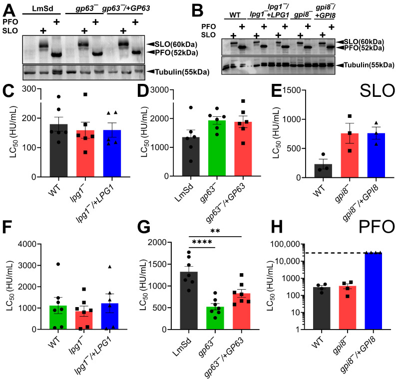 Figure 1
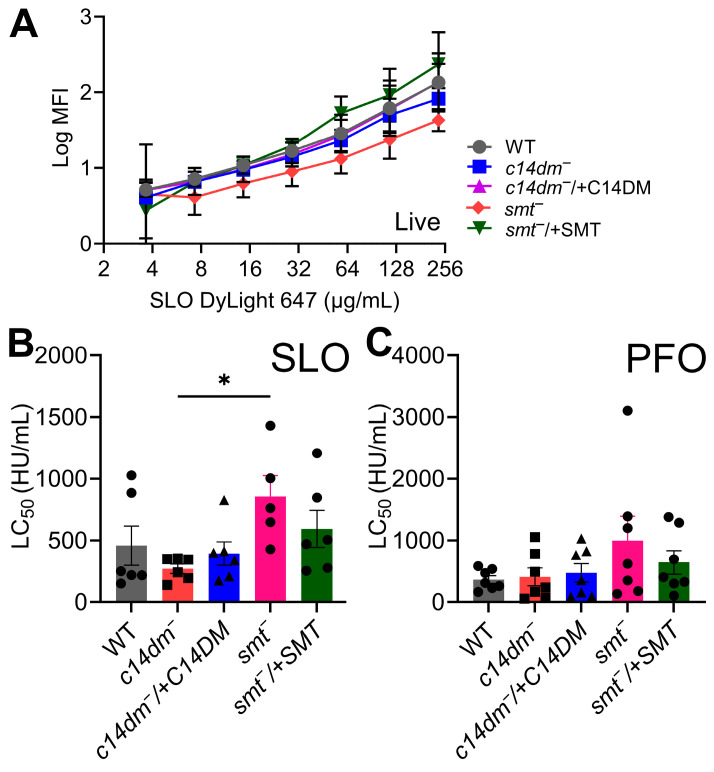 Figure 2
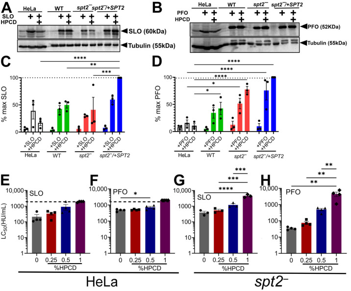 Figure 3
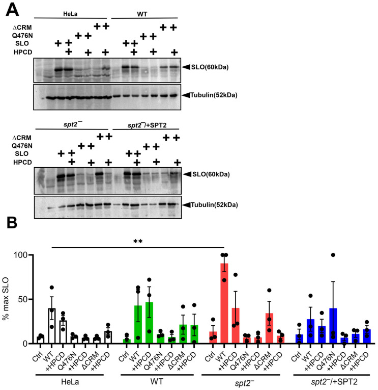 Figure 4
- —National Institute of Allergy and Infectious Diseases of the National Institutes of Health
Peer Reviews
No public reviews on file for this paper yet. If you reviewed it on a platform where reviews are public (OpenReview, ICLR, NeurIPS, ICML), you can paste yours below so the community can read it here.
Videos
No videos yet. Explain this paper in a talk, walkthrough, or lecture? Add one.
Taxonomy
TopicsResearch on Leishmaniasis Studies · Lipid Membrane Structure and Behavior · Sphingolipid Metabolism and Signaling
1. Introduction
One neglected tropical disease with a global impact is leishmaniasis. Leishmaniasis afflicts over two million people, with 70,000 deaths annually, and is characterized by disfiguring skin or mucocutaneous lesions and lethal visceral phenotypes [1]. Leishmaniasis is caused by deeply branching protozoan parasites in the genus Leishmania, including L. major. L. major cycles between mammalian hosts and sandfly vectors. In both life stages, L. major competes with bacteria and other microorganisms, though the competitive mechanisms remain poorly defined.
One aspect of interspecies competition is the secretion of and resistance to pore-forming toxins. Pore-forming toxins comprise the largest family of bacterial toxins [2], but their ability to destroy non-mammalian cells is poorly described. The best-studied pore-forming toxins are cholesterol-dependent cytolysins (CDCs). CDCs are a family of β-barrel, pore-forming toxins that are secreted as monomers and oligomerize to form 30 nm pores [3]. The archetypal CDCs, streptolysin O (SLO) and perfringolysin O (PFO), from Streptococcus pyogenes and Clostridium perfringens, respectively, bind to cholesterol in the mammalian cell membrane via cholesterol-recognition motifs (CRMs) and then use cholesterol for pore formation [4]. Differences in the cholesterol-binding region enable SLO to damage the membrane faster and with less sensitivity to the rest of the lipid environment compared to PFO, which takes longer to form pores and has greater sensitivity to the surrounding environment [5,6,7]. The cholesterol-binding domain of PFO is frequently used as a cholesterol sensor to determine the amounts of accessible cholesterol in mammalian membranes [8]. However, the binding of SLO and PFO to mammalian cell membranes might also involve glycans during initial monomer binding [9,10]. Thus, CDCs are well-characterized in mammalian systems.
In contrast to mammalian systems, the mechanisms by which protozoans resist pore-forming toxins are poorly understood. While some protozoans prevent toxin binding, we previously showed that L. major is bound by CDCs [11]. However, the loss of sphingolipids is necessary for killing [11]. We chose L. major as a system to understand how protozoans deal with pore-forming toxins because L. major must compete with bacteria that secrete pore-forming toxins in the sandfly [12], it is a pathogen with human clinical significance [1], and it is a genetically tractable system. Knockouts and complemented strains exist for many enzymes that synthesize key membrane components, including phospholipids, sphingolipids, ergosterol, and GPI-anchored molecules [13,14,15,16,17,18]. Our prior work showed that inositol phosphorylceramide (IPC), the major sphingolipid in L. major, prevents CDC-mediated cytotoxicity, despite CDC binding [11]. The contributions of other membrane components, such as phospholipids, glycoconjugates, and GPI-anchored proteins, to CDC resistance remain unresolved.
In this study, we systematically examined the role of other L. major plasma membrane constituents—including GPI-anchored proteins, phospholipids, and sterols—in resistance to CDCs. Using genetically engineered L. major knockout strains and cytotoxicity assays with SLO and PFO, we assessed the contribution of specific membrane factors to toxin susceptibility. We found that the virulence factor lipophosphoglycan (LPG) and plasmalogen phospholipid plasmenylethanolamine (PME) are dispensable for protection. The GPI-anchored metalloproteinase GP63 conferred resistance to PFO but not to SLO. Moreover, depletion of ergosterol protected cells from CDC-mediated killing, despite continued toxin binding. This binding is likely mediated by glycans because SLO variants deficient in sterol recognition still bound to Leishmania membranes, while SLO deficient in glycan binding had worse recognition. These data suggest that in Leishmania spp., CDCs may use sterols for pore formation instead of binding. Collectively, our findings indicate that Leishmania employ distinct, non-mammalian strategies to defend against CDCs, offering potential insights into novel therapeutic targets.
2. Results
2.1. GPI-Anchored Proteins Protect L. Major from PFO
To determine the contribution of L. major plasma membrane components other than sphingolipids to CDC binding and cytotoxicity, we first examined the abundant glycosylated virulence factors GP63 and LPG. Glycans may contribute to toxin binding in mammalian cells [9,10], and our prior work with spt2^−^ promastigotes suggested glycans contribute to toxin binding in L. major [11]. To test the contribution of GP63 and LPG, we used promastigotes deficient in one of three genes: gp63, which encodes the GPI-anchored metalloprotease GP63 [18]; lpg1, which encodes a galacto-furanosyltransferase pivotal for synthesis of the LPG glycan core [17]; and gpi8, which encodes the transamidase that links GPI anchors to proteins [19].
Since the gpi8^−^ did not exist in L. major, we created this mutant and complemented it via CRISPR (Supplementary Figure S1). To confirm the successful generation of the gpi8^−^ mutant in L. major, promastigotes were screened for drug resistance and verified by PCR analysis. PCR amplification using gpi8^−^-specific open reading frame (ORF) primers showed no detectable band in the gpi8^−^ mutant, whereas amplification with selection marker-specific primers produced the expected fragments, confirming complete replacement of the gpi8 alleles (Supplementary Figure S1A). In contrast, the wild-type strain had the intact gpi8 ORF and no signal for the drug maker. To verify the genetic knockout by complementation, the gpi8 ORF was episomally expressed in the gpi8^−^ background. The presence of gpi8 in the complemented line (gpi8^−^/+GPI8) was confirmed by PCR analysis (Supplementary Figure S1A). GPI8 is required for the synthesis of all GPI-anchored proteins. Consistent with loss of gpi8, the gpi8^−^ mutant lost GP63 expression, whereas wild-type and gpi8^−^/+GPI8 promastigotes maintained expression (Supplementary Figure S1B). Conversely, LPG was significantly elevated in the gpi8^−^ mutant compared to wild-type and gpi8^−^/+GPI8 promastigotes (Supplementary Figure S1B). These results validated the generation of the gpi8^−^ mutant line and genetic complementation of gpi8^−^/+GPI8, providing a reliable system to test the role of GPI-anchored proteins in L. major.
We then tested the binding and cytotoxicity of CDCs in gp63^−^, lpg1^−^, or gpi8^−^ promastigotes and their wild-type counterparts and complemented strains. The gp63^−^ promastigotes were generated in L. major strain Seidman and were defective in GP63 synthesis [18], while the other strains were generated in strain LV39. Since our prior work on spt2^−^ promastigotes [11] showed increased cytotoxicity despite no alterations to binding, we tested both binding and cytotoxicity of SLO and PFO. In the absence of the SPT2 inhibitor myriocin, SLO and PFO failed to kill any of the promastigotes (Supplementary Figure S2), consistent with the protection that sphingolipids afford to L. major against CDCs [11]. When gp63^−^, lpg1^−^, or gpi8^−^ promastigotes were treated with myriocin, CDC binding was similar across all groups (Figure 1A,B). These data indicate that CDCs can bind to L. major in the absence of glycans. We then compared killing using dose–response curves and calculating the LC_50_ by logistic modeling. Lower LC_50_ values represent increased killing. Myriocin-treated lpg1^−^ promastigotes showed equal levels of killing by SLO and PFO compared to the myriocin-treated wild-type (LV39) and the complemented lpg1^−^/+LPG1 cells (Figure 1C,F, Supplementary Figure S2A,B). Loss of GP63 trended to show a decrease in death by SLO (high LC_50_), which was not reversed in the complemented strain (Figure 1D, Supplementary Figure S2C). In contrast, loss of GP63 enhanced cytotoxicity against PFO, which was partially reversed in the complemented gp63^−^/+GP63 cells (Figure 1G, Supplementary Figure S2D). The myriocin-treated gpi8^−^ and gpi8−/+GPI8 promastigotes largely phenocopied the gp63^−^ and gp63^−^/+GP63 cells when challenged by SLO, including the failure of gpi8^−^/+GPI8 cells to reverse the decreased killing (Figure 1E, Supplementary Figure S2E). Surprisingly, myriocin-treated gpi8^−^ cells, which lack GP63, among other GPI-anchored proteins, were no more sensitive to PFO than wild-type cells (Figure 1H, Supplementary Figure S2F). However, overexpression of GPI8 in these cells protected them from PFO, even at the excessive dose of 64,000 HU/mL (Figure 1H, Supplementary Figure S2F). Overall, we conclude that LPG fails to contribute to resistance to CDCs, while GP63 may protect L. major from PFO.
2.2. Plasmenylethanolamine Is Unnecessary to Resist CDCs
Kinetoplastids like L. major contain both standard phospholipids and plasmalogens like plasmenylethanolamine (PME). Plasmalogens contain an ether linkage at the sn1 and an ester linkage at the sn2 position of the phospholipid. PME comprises 80–90% of the ethanolamine-containing phospholipids in L. major promastigotes [16]. Genetic deletion of ethanolamine phosphate cytidylyltransferase (ept) eliminates plasmenylethanolamine from L. major, while phosphatidylethanolamine (PE) levels remain unperturbed due to production of PE from phosphatidylcholine (PC) [16]. PC is essential in L. major promastigotes and loss of its biosynthesis is lethal [20]. We tested the contribution of PME to toxin resistance by treating wild-type, ept^−^, and ept^−^/+EPT promastigotes with myriocin and then measured binding and cytotoxicity. Myriocin-pretreated ept^−^ promastigotes bound SLO to a similar level compared to both wild-type and ept^−^/+EPT promastigotes (Supplementary Figure S3). We then challenged these cells with SLO or PFO. Without myriocin pretreatment, all genotypes resisted both SLO and PFO (Supplementary Figure S3). With myriocin pretreatment, ept^−^ had equal sensitivity to both SLO and PFO compared to both wild-type and ept^−^/+EPT promastigotes (Supplementary Figure S3). Based on these data, we conclude that PME is dispensable for the binding and cytotoxicity of SLO and PFO.
2.3. Cholestane-Based Sterols Reduce Binding and Cytotoxicity in L. major
We next tested the other major membrane component used by CDCs for binding and cytotoxicity: sterols. We used mutants lacking the key ergosterol synthesis genes c14 demethylase (c14dm) and sterol 24-C methyltransferase (smt). Instead of ergosterol, these mutants have elevated levels of 14-methylated ergosterol or cholestane-based sterols, respectively [15,21]. The c14dm^−^ promastigotes are more sensitive to membrane stress [15], including detergents like Triton-X-100 [11,22], even without myriocin. We tested the binding of SLO to these cells. While SLO bound to c14dm^−^ promastigotes like wild-type, SLO bound less to the smt^−^ promastigotes (Figure 2A). This reduced binding was rescued in the smt^−^/+SMT cells (Figure 2A). We then tested the cytotoxicity of CDCs in these cell types. Consistent with our prior work [11], these cells were resistant to CDCs without myriocin treatment (Supplementary Figure S4). In contrast to the increased detergent sensitivity, myriocin-treated c14dm^−^ promastigotes showed equivalent sensitivity to both CDCs compared to wild-type and complemented strains (Figure 2B,C, Supplementary Figure S4A,B). Consistent with the reduced binding, there was a trend towards resistance of the smt^−^ cells to both CDCs that was reversed in the smt^−^/+SMT cells (Figure 2B,C, Supplementary Figure S4C,D). Based on these data, we conclude that CDCs do not bind well to cholestane-based sterols in the membrane, and this may reduce the cytotoxicity of CDCs in L. major.
2.4. Ergosterol Is Needed for CDC Cytotoxicity but Not Binding in Promastigotes
We further explored sterol dependence using chemical approaches to probe the contribution of sterols. We previously showed that spt2^−^ cells are more sensitive than wild-type cells to Triton-X-100 [11]. We extended this work by testing their sensitivity to the sterol-binding detergent saponin. The spt2^−^ promastigotes were more sensitive to saponin than wild-type or spt2^−^/+SPT (Supplementary Figure S5A).
We next used a less invasive method to remove membrane sterols: short-term treatment with 2-hydroxypropyl-β-cyclodextrin (HPCD). In mammalian cells, short-term treatment removes accessible cholesterol from the plasma membrane without depleting intracellular stores [8,23]. We treated HeLa cells as a control and wild-type, spt2^−^, and spt2^−^/+SPT promastigotes with HPCD, then challenged them with SLO or PFO, and measured binding and cytotoxicity. For binding assays, we used the non-hemolytic, poorly oligomeric “monomer-locked” variants of SLO and PFO to reduce complications from cytotoxicity and oligomerization [11]. HPCD pretreatment decreased toxin binding to HeLa cells but failed to reduce binding to L. major promastigotes (Figure 3). Indeed, quantitation of the blots revealed a slight trend towards increased binding of SLO and PFO after HPCD treatment in L. major promastigotes (Figure 3). Overall, limiting ergosterol in the plasma membrane using HPCD failed to reduce CDC binding in L. major.
We next tested cytotoxicity in HPCD-treated cells. We observed a HPCD-dose-dependent increase in resistance to SLO and PFO of spt2^−^ cells (Figure 3, Supplementary Figure S5B,C), which was consistent with an HPCD-dose-dependent increase in resistance to SLO and PFO in HeLa cells (Figure 3, Supplementary Figure S5D,E). We did not observe toxicity due to HPCD in L. major. We conclude that sterols are needed for cytotoxicity but not for the binding of CDCs to L. major.
2.5. SLO Uses Glycan-Binding Residues to Bind L. major Promastigotes
Since CDCs bound to L. major independent of sterol removal, we tested the contribution of the SLO sterol- and glycan-binding sites to L. major binding. SLO sterol binding in mammalian cells can be abrogated by elimination of the cholesterol-recognition motif (SLO ΔCRM) [4], while glycan binding is lost by mutating Q476N in domain 4 of SLO (SLO Q476N) [10]. We previously showed that neither of these mutant toxins kill L. major [11], but we did not test binding. We measured the binding of wild-type SLO, SLO Q476N, and SLO ΔCRM to HeLa cells, wild-type, spt2^−^, and spt2^−^/+SPT promastigotes with and without HPCD. Consistent with previous results [10], both SLO Q476N and ΔCRM had minimal binding to HeLa cells independent of HPCD (Figure 4). In L. major, both SLO Q476N and ΔCRM had reduced binding independent of genotype, with SLO ΔCRM trending towards greater binding (Figure 4). The weak binding from SLO Q476N showed a trend to further reduction after HPCD treatment in WT and spt2^−^/+SPT (Figure 4). Together, we interpret these results to indicate that the glycan-binding site is important for binding to L. major, with sterol engagement more important for toxicity.
3. Discussion
In this study, we tested the contribution of leishmanial plasma membrane components to protection from CDCs. We found that GPI-anchored proteins may contribute to protection against PFO, while PME and LPG failed to limit CDC cytotoxicity. Cholestane-based sterols reduced CDC binding. Removal of ergosterol from the plasma membrane limited CDC cytotoxicity without reducing binding. Consistent with this finding, SLO lacking the cholesterol-recognition motif could bind, but SLO deficient in glycan binding failed to bind. We interpret these data to suggest that single-celled protozoans like Leishmania use distinct defense strategies compared to mammalian cells.
Kinetoplastids rely on the packing of GPI-anchored proteins and glycans to protect them from environmental and immune stressors. We found that in L. major, loss of GP63 sensitized cells to PFO, while a global loss of GPI-anchored proteins failed to alter cellular sensitivity. However, overexpression of GPI8 made the cells resistant to PFO. Since binding was not altered, it is possible that GP63 interferes with oligomerization or pore formation, while the other GPI-anchored proteins might better support oligomerization. At high concentrations (i.e., gpi8^−^/+GPI8), the GPI-anchored proteins might saturate binding and act in a dominant negative fashion, similarly to how overexpression of key components in the Endosomal Sorting Complex Required for Transport (ESCRT) can interfere with ESCRT function. Another possibility is that strain-specific differences in glycosylation, GP63, and membrane composition between the LV39 and Seidman strains could account for discrepancies between the gp63^−^ and gpi8^−^ promastigotes. Alternatively, GP63 localizes to the detergent-resistant membranes enriched in sterols, so the tight packing of GP63 could limit PFO cytotoxicity [24]. PFO has stricter membrane-binding requirements than SLO in both mammals and L. major [5,11], which may account for why SLO is not affected by GP63 but PFO is. While PFO and SLO are structurally quite similar, small differences exist, including in the lipid-binding interface [5,6,7,11] and in binding residues for glycans [9,10], which could account for the differences observed. LPG and PME localization to the non-detergent-resistant membrane fraction [16,25] could account for the failure of these species to protect cells from CDC cytotoxicity. Glycan preference could also account for differences in cytotoxicity. In mammalian systems, SLO prefers galactose-rich glycans, while PFO prefers N-acetylneuraminic acid in glycans [9,10]. In L. major, LPG is rich in galactose residues interspersed with mannose residues, while GP63 is mannose-rich [26]. Thus, these results provide new insight into the L. major membrane environment that CDCs, especially PFO, require for cytotoxicity.
The key lipid for CDC activity is sterol. In mammalian cells, SLO and PFO use cholesterol both for binding and pore formation [2,4], while other CDCs rely on the GPI-anchored protein CD59 for binding and cholesterol for pore formation [27]. We found that the accumulation of cholestane-based sterols in the smt^−^ mutant [14] reduced both the binding and cytotoxicity of SLO in L. major. In contrast, methylation on C24 made no difference to CDC cytotoxicity. This is surprising because c14dm^−^ promastigotes are generally more sensitive to membrane stress [15]. Whether these differences are due to lipid packing or direct engagement of the CDC with the sterol remain to be determined.
In contrast to other systems, where cholesterol can be sufficient for CDC binding and cytotoxicity [2], we found that ergosterol is only needed for cytotoxicity, not binding in L. major promastigotes. These findings are consistent with previous reports in mammalian cells that SLO and PFO can bind to glycans and glycosphingolipids in addition to cholesterol, while the process of membrane insertion depends on cholesterol [9,10]. We used toxins defective in cholesterol or glycan binding in the presence of HPCD to tease apart the relative contributions of both sites. While both mutants had reduced binding compared to wild-type, they bound better to promastigotes than to HeLa cells. This suggests the CDCs may use the CRM to recognize membrane components other than ergosterol in the leishmanial membrane. This is consistent with the failure of HPCD to eliminate binding of the glycan-binding mutant. Overall, we find that ergosterol is needed for CDC-mediated killing, but the CDCs use both glycan-binding and cholesterol-recognition regions to bind to multiple cryptic sites on the promastigote membrane.
Our findings provide new insights into the competition between L. major and bacteria. Promastigotes compete with bacteria in the sandfly midgut. While IPC is the major protective determinant against CDCs in L. major promastigotes, the toxins still bind. The recent discovery of CDC-like toxins in Elizabethkingia anopheles and other bacteria [28] suggests that bacterial pore-forming toxins are used for more than human pathogenesis. While it is possible that toxin binding to L. major is incidental to their major function against other organisms, our work raises the possibility that these toxins could have secondary roles opsonizing L. major. Notably, SLO is capable of translocating the lethal NAD+ glycohydrolase (Spn) across mammalian membranes via its N-terminal domain [29]. We did not test Spn in L. major, but it could be a back-up killing mechanism that bacteria use to compete. Clostridium perfringens α-toxin has sphingomyelinase activity [30], which suggests PFO could be lethal if α-toxin cleaves IPC. Future work is needed to determine how CDC binding alters the balance between L. major and the sandfly gut microbiota.
While this study provides a comprehensive analysis of key Leishmania major plasma membrane components involved in defense against pore-forming toxins, it had some limitations. We measured toxin binding via Western blot, which could also detect internalized toxins. We did not measure the HPCD extraction of ergosterol directly. Amastigotes have different membrane compositions compared to promastigotes, especially sterols and glycans. We did not test amastigotes because L. major amastigotes cannot be grown axenically. Purification from macrophages would make killing and binding assays challenging to interpret. Some experiments were performed with three independent biological replicates, which could be low given variation. We only investigated one phospholipid, PME. The functional roles of other abundant phospholipids, such as PC and PE, remain unexplored because their genetic knockout is lethal. Differences in our findings between gpi8− and gp63^−^ promastigotes suggest that the minor GPI-anchored molecules contribute to binding and cytotoxicity. However, identifying which of these components contribute is beyond the scope of our study. Due to this, we were unable to pinpoint the specific glycans in L. major promastigotes responsible for CDC binding. We did not challenge L. major with sandfly gut microbiota. The present work provides a foundation for future studies to address these gaps and better understand membrane integrity and repair in L. major.
4. Materials and Methods
4.1. Reagents
All reagents were from Thermo Fisher Scientific (Waltham, MA, USA), unless otherwise noted. Cysteine-less His-tagged PFO (PFO WT) in pET22 was a generous gift from Rodney Tweten (University of Oklahoma Health Sciences Center, Oklahoma City, OK, USA). Cysteine-less codon-optimized SLO (SLO WT) in pBAD-gIII, monomer-locked (G298V/G299V) PFO, monomer-locked (G398V/G399V) SLO, SLO S101C, SLO Q476N, and SLO ΔCRM (T564A/L565A) were previously described [11]. 6D11 anti-SLO monoclonal antibody (mAb) was from Fisher (catalog NBP105126). WIC79.3 anti-LPG mAb was from Stephen Beverley (Washington University School of Medicine, St Louis, MO, USA) [31]. Anti-GP63 monoclonal antibody m235 was from Robert McMaster (The University of British Columbia, Vancouver, BC, Canada) [32]. Mouse anti-tubulin antisera were from Fisher. Cross-adsorbed HRP-conjugated anti-mouse IgG or anti-rabbit IgG was from Jackson Immunoresearch (West Grove, PA, USA).
4.2. Recombinant Toxins
Toxins were induced and purified from Escherichia coli BL21 GOLD cells as previously described [33,34]. Toxins were induced with 0.2% arabinose (SLO) or 0.2 mM IPTG (PFO) for 3 h at room temperature and purified using Nickel-NTA beads. Protein concentration was determined by Bradford assay and hemolytic activity was determined as previously described using human red blood cells (Zen Bio, Research Triangle Park, NC, USA) [34]. One hemolytic unit is defined as the amount of toxin required to lyse 50% of a 2% human red blood cell solution in 30 min at 37 °C in 2 mM CaCl_2_, 10 mM HEPES, pH 7.4, and 0.3% BSA in PBS. Hemolytic units were used to control for differences in toxin activity. The specific activities of monomer-locked toxins, SLO Q476N, and SLO ΔCRM were <100 HU/mg. They were used at a mass equivalent to wild-type SLO and PFO. Multiple toxin preparations were used to control for variation by preparation (Table 1). Where indicated, wild-type or monomer-locked toxins were conjugated to Cy5 using NHS chemistry as described [33]. Alternatively, SLO S101C, which retains wild-type activity [33], was conjugated to AlexaFluor647 or DyLight 647 using maleimide chemistry as described [33].
4.3. Leishmania Strains and Culture
Leishmania major strains and mutants were previously characterized (Table 2). LV39 clone 5 (Rho/SU/59/P) was used as the wild-type strain for all experiments except when the gp63^−^ L. major was used. For these experiments, the L. major strain NIH S (MHOM/SN/74/Seidman) clone A2 was used as the wild-type strain [18]. L. major promastigotes were cultured at 27 °C in 1X M199 medium (pH 7.4) with 10% fetal bovine serum and additional supplements. The gpi8^−^ mutant was generated using the CRISPR-Cas9 system as described [35]. The endogenous gpi8 gene was disrupted by replacing gpi8 with a drug-resistance marker through homologous recombination. The resulting gpi8^−^ was selected based on resistance to blasticidin (BSD) and hygromycin (HYG). The replacement of gpi8 alleles was confirmed by PCR using ORF primers and drug-specific primers to confirm integration. For genetic complementation, gpi8 was cloned into pXG1a. The construct was then transfected into gpi8^−^ promastigotes to generate the gpi8^−^/+GPI8 complemented strain.
Promastigotes were cultured at 27 °C in M199 medium (Gibco, Waltham, MA, USA) with 0.182% NaHCO_3_, 40 mM HEPES, pH 7.4, 0.1 M adenine, 1 µg/mL biotin, 5 µg/mL hemin, 2 µg/mL biopterin, and 10% heat-inactivated fetal bovine serum, pH 7.4. Episomal complemented cells were maintained in the same medium with the addition of 10 µg/mL neomycin (G418) except for experimental passages. Culture density and cell viability were determined by hemocytometer counting and flow cytometry after propidium iodide (PI) staining at a final concentration of 20 µg/mL. Log phase promastigotes were replicative parasites at 2.0–8.0 × 10^6^ cells/mL.
4.4. HeLa Cell Culture
HeLa cells (ATCC (Manassas, VA, USA) CCL-2) were maintained at 37 °C, 5% CO_2_ in DMEM (Corning, Corning, NY, USA) supplemented with 10% fetal bovine serum (R&D Biosystems, Minneapolis, MN, USA) and 1× L-glutamine (D10). They were negative for mycoplasma by microscopy.
4.5. Myriocin Treatment of L. major
Log phase cells were seeded at 1.0 × 10^5^ cells/mL in complete medium and either treated with 10 µM myriocin dissolved in 1× DMSO (experimental) or an equivalent volume of diluent 1× DMSO (control). Cells were cultured and allowed to reach log phase in 48 h before harvesting and processing cells for experiments.
4.6. Leishmania Processing
Cells were processed as described [11] and either resuspended in serum-free 1× M199 to a final concentration of 1.0 × 10^7^ cells/mL for Western blot or in serum-free Tyrode’s buffer (137 mM NaCl, 2.7 mM KCl, 1.8 mM CaCl_2_, 0.49 mM MgCl_2_, 0.42 mM NaH_2_PO_4_, 11.9 mM NaHCO_3_, and 11.1 mM dextrose, pH 7.5) to a final concentration of 1.0 × 10^6^ cells/mL for cytotoxicity. For cytotoxicity, 1.0 × 10^5^ cells were plated per well in a V-bottom 96-well plate or per Marsh tube.
4.7. Binding Assay via Western Blot
L. major promastigotes were resuspended to a final cell concentration of 1.0 × 10^7^ cells in serum-free 1× M199 supplemented with 2 mM CaCl_2_. Where indicated, L. major promastigotes were pretreated with 0%, 0.25%, 0.5%, or 1% HPCD for 30 min at 27 °C before toxin challenge. Cells were challenged with 5 μg of SLO WT, PFO WT, SLO ML, PFO ML, SLO ΔCRM, and SLO Q476N for 30 min at 4 °C. After toxin challenge, cells were centrifuged for 10 min at 3000 RPM (Rotor SX 47_50_) at room temperature (25 °C). Cells were washed once with 1× PBS and centrifuged at 3000 RPM for 10 min at room temperature (25 °C). Cell pellets were washboarded and then resuspended in 1× SDS sample buffer (with freshly added 2-mercaptoethanol) and heated at 95 °C for 10 min. SDS-PAGE and Western blots were performed as previously described [36]. Cell lysates were resolved on 10% acrylamide gels and transferred to nitrocellulose at 110 V for 90 min. Blots were blocked with 5% bovine serum albumin (BSA) in 1× Tris-buffered saline (15.2 mM NaCl, Tris base 46.2 mM, and 150 mM NaCl) at 4 °C for 2 h; probed with primary antibodies overnight in 1% BSA in 1× TBST at 4 °C; washed three times with 1× TBST for 10 min each; incubated with HRP-conjugated secondary antibodies in 1% BSA in 1× TBST for 1 h; washed with 1× TBST three times for 10 min each; developed with enhanced chemiluminescence reagent (1.25 mM luminol (Sigma, Lavonia, MI, USA) 0.01% H_2_O_2_ (Walmart, Fayetteville, AR, USA), 0.2 mM p-coumaric acid (Sigma), 0.1 mM Tris-HCl, pH 8.4); and imaged on an iBlot (Fisher). Antibody dilutions were anti-SLO 1:2000; anti-LPG 1:1000; anti-GP63 1:1000; tubulin 1:1000; anti-mouse HRP 1:10,000; and anti-rabbit HRP 1:10,000.
4.8. Protein Expression Levels of GPI-Anchored Proteins
Total protein lysates were prepared from promastigotes of LV39WT, LV39Cas9, gpi8^−^, or gpi8^−^/+GPI8 promastigotes. Parasites were harvested at late-log phase, washed in PBS, and lysed in SDS-containing buffer by boiling at 95 °C for 10 min. Equal amounts of protein were resolved using SDS-PAGE gels and transferred to PVDF membranes. Membranes were blocked with 5% BSA in TBST and incubated with anti-GP63 (1:1000), anti-LPG WIC79.3 (1:1000), and anti-tubulin (1:1000) primary antibodies. HRP-conjugated anti-mouse antibodies were used for detection, and signals were visualized by chemiluminescence. Parallel blots were stained with Ponceau S staining solution to verify equal protein loading across samples.
4.9. Flow Cytometry Cytotoxicity Assay
Killing assays were performed as described [36]. Promastigotes were resuspended at 1 × 10^6^ cells/mL in Tyrode’s buffer supplemented with 2 mM CaCl_2_ and 20 μg/mL PI. HeLa cells were resuspended at 1 × 10^6^ cells/mL in RPMI media supplemented with 2 mM CaCl_2_ and 20 μg/mLPI. When indicated, cells were treated with 0.25%, 0.5%, or 1% HPCD at 27 °C for 30 min. Cells were challenged with toxins for 30 min at 37 °C and analyzed using an Attune flow cytometer. Specific lysis and LC_50_ were determined as described [36]. For fluorescence binding assays, Cy5, Alexa Fluor 647, and DyLight 647 conjugated toxins were used. The median fluorescence intensity (MFI) was quantitated and background-subtracted using cells receiving no fluorescent toxin.
4.10. Statistics
Prism 10.6.1 (Graphpad, San Diego, CA, USA), Sigmaplot 11.0 (Systat Software Inc., San Jose, CA, USA), and Excel (Version 2211) were used for statistical analysis. Data are represented as mean ± SEM. The LC_50_ for toxins was calculated by logistic modeling [36]. Logistic modeling was chosen because cytotoxicity is near zero at low concentrations and rapidly saturates at higher concentrations according to a sigmoidal curve [36]. This is best modeled with a logistic equation, as measured by high R^2^ values when assessing model fit. Statistical significance was determined by one-way ANOVA with Tukey post-testing, one-way ANOVA (Brown–Forsythe method) with Dunnett T3 post-testing, or Kruskal–Wallis, as appropriate. p < 0.05 was considered statistically significant. Graphs were generated in GraphPad and organized in Photoshop (Creative Cloud 2022, Adobe, San Jose, CA, USA).
The reference list from the paper itself. Each links out to its DOI / PubMed record.
- 1Telleria E.L. Martins-da-Silva A. Tempone A.J. Traub-Cseko Y.M. Leishmania, microbiota and sand fly immunity Parasitology 20181451336135310.1017/S 003118201800101429921334 PMC 6137379 · doi ↗ · pubmed ↗
- 2Tweten R.K. Cholesterol-dependent cytolysins, a family of versatile pore-forming toxins Infect. Immun.2005736199620910.1128/IAI.73.10.6199-6209.200516177291 PMC 1230961 · doi ↗ · pubmed ↗
- 3Shatursky O. Heuck A.P. Shepard L.A. Rossjohn J. Parker M.W. Johnson A.E. Tweten R.K. The mechanism of membrane insertion for a cholesterol-dependent cytolysin: A novel paradigm for pore-forming toxins Cell 19999929329910.1016/S 0092-8674(00)81660-810555145 · doi ↗ · pubmed ↗
- 4Farrand A.J. La Chapelle S. Hotze E.M. Johnson A.E. Tweten R.K. Only two amino acids are essential for cytolytic toxin recognition of cholesterol at the membrane surface Proc. Natl. Acad. Sci. USA 20101074341434610.1073/pnas.091158110720145114 PMC 2840085 · doi ↗ · pubmed ↗
- 5Farrand A.J. Hotze E.M. Sato T.K. Wade K.R. Wimley W.C. Johnson A.E. Tweten R.K. The Cholesterol-dependent Cytolysin Membrane-binding Interface Discriminates Lipid Environments of Cholesterol to Support beta-Barrel Pore Insertion J. Biol. Chem.2015290177331774410.1074/jbc.M 115.65676926032415 PMC 4505022 · doi ↗ · pubmed ↗
- 6Ray S. Thapa R. Keyel P.A. Multiple Parameters Beyond Lipid Binding Affinity Drive Cytotoxicity of Cholesterol-Dependent Cytolysins Toxins 201811110.3390/toxins 1101000130577571 PMC 6356533 · doi ↗ · pubmed ↗
- 7Johnson B.B. Moe P.C. Wang D. Rossi K. Trigatti B.L. Heuck A.P. Modifications in perfringolysin O domain 4 alter the cholesterol concentration threshold required for binding Biochemistry 2012513373338210.1021/bi 300313222482748 · doi ↗ · pubmed ↗
- 8Das A. Brown M.S. Anderson D.D. Goldstein J.L. Radhakrishnan A. Three pools of plasma membrane cholesterol and their relation to cholesterol homeostasise Life 20143 e 0288210.7554/e Life.0288224920391 PMC 4086274 · doi ↗ · pubmed ↗