Magnetically Sculpted Microfluidics for Continuous-Flow Fractionation of Cell Populations by EpCAM Expression Level
Zhenwei Liang, Xiaolei Guo, Xuanhe Zhang, Yiqing Chen, Chuan Du, Yuan Ma, Jiadao Wang

TL;DR
This paper introduces a microfluidic system that uses magnetic fields to separate cells based on their surface protein expression levels, enabling precise cell fractionation for research and assays.
Contribution
The novel contribution is a magnetic-field design strategy using soft magnetic strips to enable expression-level-dependent cell sorting in continuous-flow microfluidics.
Findings
The system successfully partitions cells into four EpCAM-related subgroups (high, medium, low, near-negative).
The sorted fractions maintain high cell recovery (>90%) and viability (98.2 ± 1.3%).
The magnetic interface design is optimized using a COMSOL–MATLAB framework and a force-equivalent metric.
Abstract
Continuous-flow separation of magnetically labeled cells according to surface-marker expression levels is increasingly needed to study phenotypic heterogeneity and support downstream assays. Here, we present a microfluidic platform that uses spatially engineered soft magnetic strips (SMS) to sculpt lateral magnetic deflection fields for quantitative, label-guided cell fractionation. Under a uniform bias field, the SMS generates controllable magnetic gradients within the microchannel, producing distinct lateral velocities among EpCAM-labeled tumor cells that carry different Dynabead loads, which indirectly report membrane protein expression. Multi-outlet collection converts these “race-based” trajectory differences into discrete expression-level-resolved fractions. A COMSOL–MATLAB framework and a force-equivalent metric |(H·∇)H| are used to optimize key structural parameters of the…
Genes, proteins, chemicals, diseases, species, mutations and cell lines named across the full text — each resolved to its canonical identifier and authoritative record.
Click any figure to enlarge with its caption.
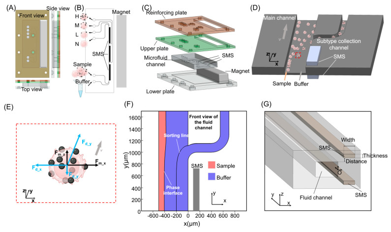 Figure 1
 Figure 2
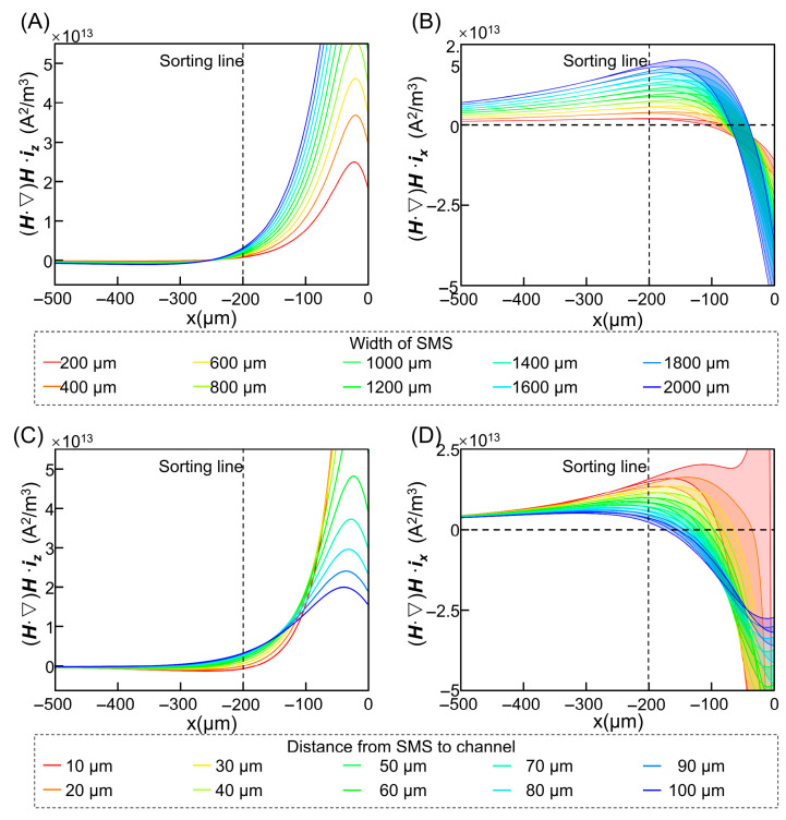 Figure 3
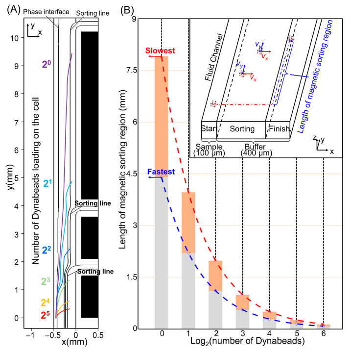 Figure 4
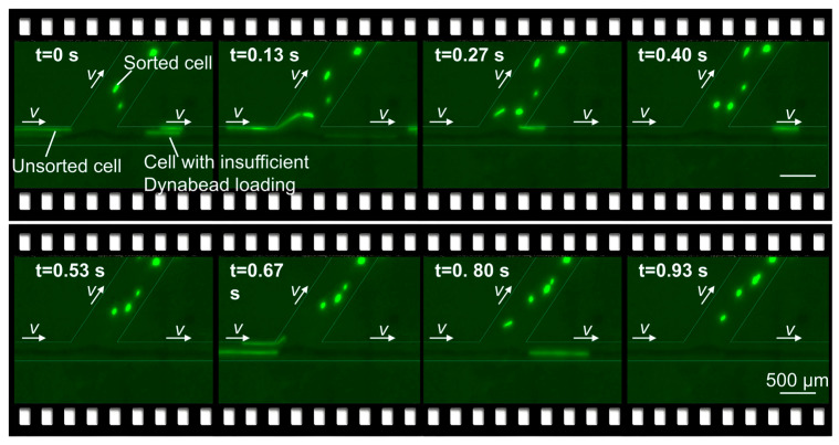 Figure 5
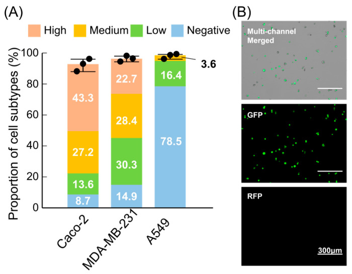 Figure 6
Peer Reviews
No public reviews on file for this paper yet. If you reviewed it on a platform where reviews are public (OpenReview, ICLR, NeurIPS, ICML), you can paste yours below so the community can read it here.
Videos
No videos yet. Explain this paper in a talk, walkthrough, or lecture? Add one.
Taxonomy
TopicsMicrofluidic and Bio-sensing Technologies · 3D Printing in Biomedical Research · Cancer Cells and Metastasis