Expression of miR-210-3p as a Prognostic Marker for Development of Diabetic Neuropathy
Savelia G. Yordanova, Diana Nikolova, Zdravko Kamenov, Vera Karamfilova, Traykov Lachezar, Yavor Assyov, Tsvetan Gatev, Radka Kaneva, Olga Belcheva, Darina Kachakova, Veronika Petkova, Yavor Zhelev, Antoaneta Trifonova Gateva

TL;DR
This study explores how miR-210-3p gene expression relates to diabetic neuropathy severity and suggests it could help diagnose and monitor the condition.
Contribution
The study identifies miR-210-3p as a potential prognostic marker for diabetic neuropathy when combined with corneal confocal microscopy.
Findings
Lower ΔCt values (higher miR-210-3p expression) were found in patients with combined neuropathy.
miR-210-3p expression was inversely correlated with neuropathy severity but positively with diabetes duration.
Corneal nerve parameters were significantly reduced in diabetic neuropathy patients.
Abstract
Background/Objectives: Diabetic neuropathy (DN) is one of the most common complications of type 2 diabetes mellitus (T2DM), involving complex metabolic, vascular, and epigenetic mechanisms. MicroRNA-210-3p (miR-210-3p), a hypoxia-responsive molecule, has been implicated in various diabetic complications, but its role in DN is not well defined. This study aimed to investigate the relationship between miR-210-3p expression, measured as delta Ct (ΔCt), and the presence and type of diabetic neuropathy, as well as correlations with corneal nerve parameters assessed by corneal confocal microscopy (CCM). Methods: Eighty patients with T2DM were stratified into four groups: no neuropathy, autonomic neuropathy, peripheral neuropathy, and combined neuropathy. Expression of miR-210-3p was quantified using RT-qPCR, and CCM was used to measure corneal nerve fiber density (CNFD), length (CNFL), and…
Genes, proteins, chemicals, diseases, species, mutations and cell lines named across the full text — each resolved to its canonical identifier and authoritative record.
Click any figure to enlarge with its caption.
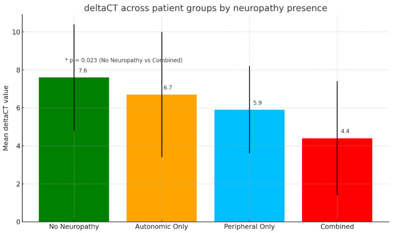 Figure 1
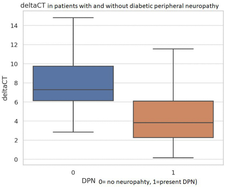 Figure 2
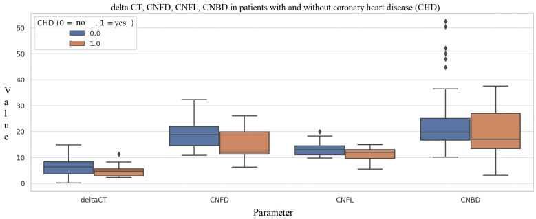 Figure 3
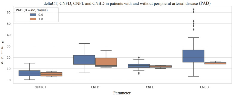 Figure 4
Peer Reviews
No public reviews on file for this paper yet. If you reviewed it on a platform where reviews are public (OpenReview, ICLR, NeurIPS, ICML), you can paste yours below so the community can read it here.
Videos
No videos yet. Explain this paper in a talk, walkthrough, or lecture? Add one.
Taxonomy
TopicsOcular Surface and Contact Lens · Corneal Surgery and Treatments · Connexins and lens biology