Contrast-Enhanced Mammography and Deep Learning-Derived Malignancy Scoring in Breast Cancer Molecular Subtype Assessment
Antonia O. Ferenčaba, Dora Galić, Gordana Ivanac, Kristina Kralik, Martina Smolić, Justinija Steiner, Ivo Pedišić, Kristina Bojanic

TL;DR
This study explores how contrast-enhanced mammography and AI can help identify breast cancer subtypes by analyzing tumor features and malignancy scores.
Contribution
The study introduces the use of deep learning-derived malignancy scores in contrast-enhanced mammography for breast cancer molecular subtype assessment.
Findings
Luminal tumors were more often spiculated with heterogeneous enhancement, while HER2-positive/triple-negative tumors were round with homogeneous enhancement.
Deep learning scores showed higher median values for malignant lesions compared to benign ones, with a significant difference observed.
AI scores varied across subtypes but differences were not statistically significant.
Abstract
Background and Objectives: Contrast-enhanced mammography (CEM) provides both morphological and functional information and may reflect breast cancer biology similarly to Magnetic Resonance Imaging (MRI). Materials and Methods: This single-center retrospective study included 399 women with Breast Imaging Reporting and Data System (BI-RADS) category 0 screening mammograms who subsequently underwent CEM. A total of 76 malignant lesions (68 invasive cancers, 8 ductal carcinoma in situ (DCIS)) with complete imaging and pathology data were analyzed. Invasive cancers were classified into luminal A, luminal B, luminal B/Human Epidermal Growth Factor Receptor 2 (HER2)-positive, HER2-enriched, and triple-negative, and grouped as luminal (Group 1) versus HER2-positive/triple-negative (Group 2). Results: Luminal subtypes predominated (47 of 68, 69%), while 21 of 68 (31%) were HER2-positive or…
Genes, proteins, chemicals, diseases, species, mutations and cell lines named across the full text — each resolved to its canonical identifier and authoritative record.
Click any figure to enlarge with its caption.
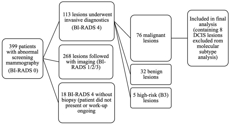 Figure 1
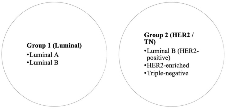 Figure 2
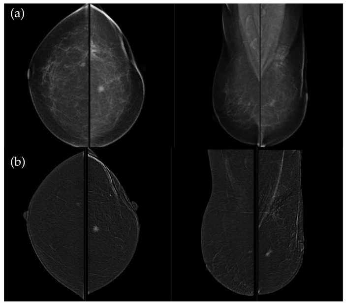 Figure 3
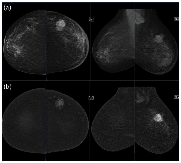 Figure 4
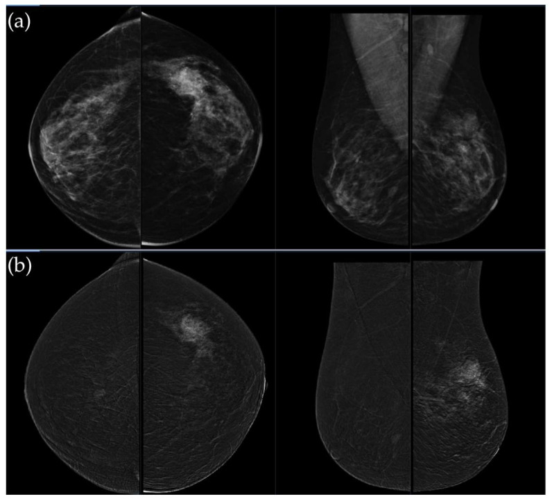 Figure 5
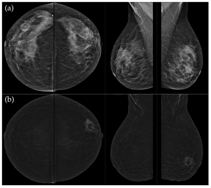 Figure 6
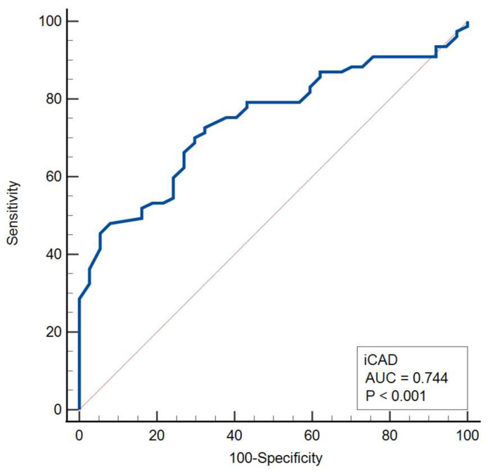 Figure 7
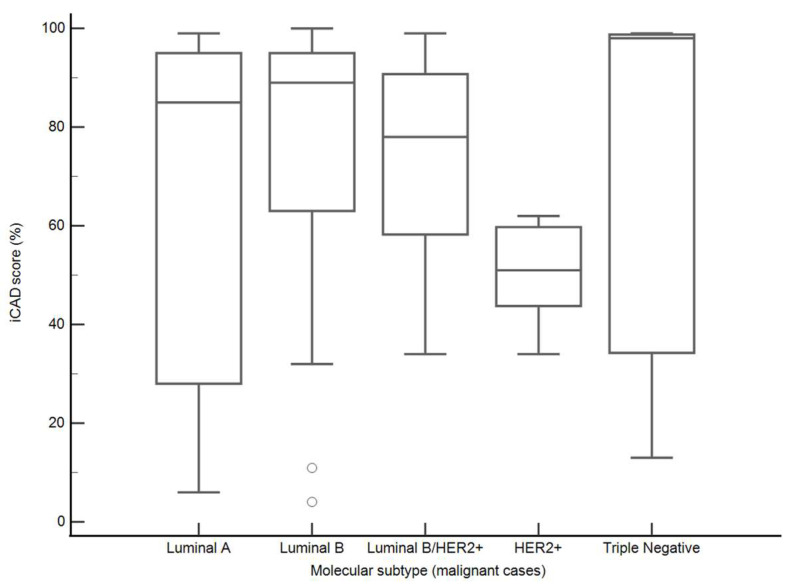 Figure 8
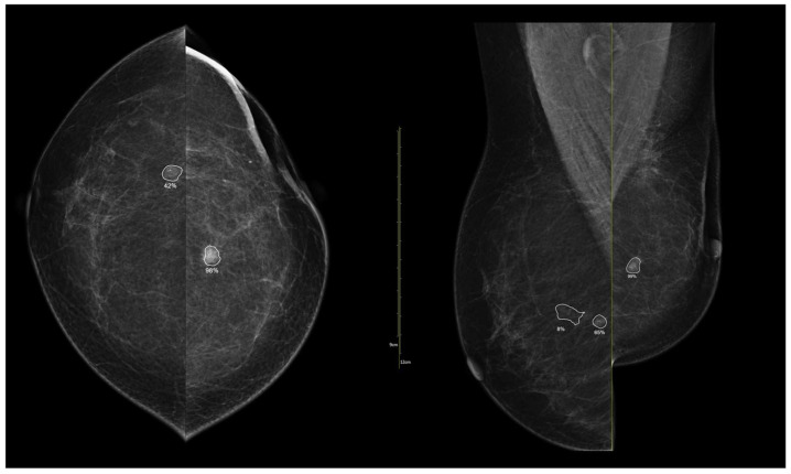 Figure 9
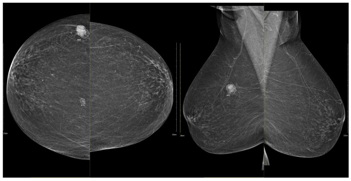 Figure 10
Peer Reviews
No public reviews on file for this paper yet. If you reviewed it on a platform where reviews are public (OpenReview, ICLR, NeurIPS, ICML), you can paste yours below so the community can read it here.
Videos
No videos yet. Explain this paper in a talk, walkthrough, or lecture? Add one.
Taxonomy
TopicsBreast Cancer Treatment Studies · MRI in cancer diagnosis · Digital Radiography and Breast Imaging