Evaluation of the Effectiveness of MicroMega Remover, ProTaper Universal Retreatment, Reciproc, and Hedstrom Files in the Retreatment of Curved Root Canals Obturated with Different Techniques: A Micro-Computed Tomography Study
Pınar Hava Dursun, Fatma Semra Sevimay, Arda Buyuksungur, Berkan Celikten

TL;DR
This study compares different dental tools for removing filling material from curved root canals, finding that some tools work faster without damaging the canal structure.
Contribution
This is one of the first studies to evaluate the MicroMega Remover system in curved canals with different filling techniques.
Findings
MM Remover and PTUR systems had significantly shorter retreatment times compared to the CWC–Hedstrom group.
No significant differences were found among the systems in terms of filling material removal or apical transportation.
All systems preserved canal anatomy within acceptable limits, but none achieved complete filling material removal.
Abstract
Background and Objectives: The anatomically demanding structure of curved root canals increases the technical difficulty of retreatment procedures. This study aimed to evaluate the retreatment efficacy of various rotary and reciprocating instruments in curved root canals obturated with cold and warm techniques regarding root canal filling material removal, apical transportation, and retreatment time. Materials and Methods: Sixty-four curved mesial root canals of mandibular molars with Vertucci type IV morphology were prepared using the ProTaper Gold system and obturated with AH Plus sealer using either the single-cone (SC) (n = 32) or continuous wave vertical compaction (CWC) (n = 32) technique. Each group was divided into four subgroups (n = 8) and retreated using MicroMega Remover (MM Remover), ProTaper Universal Retreatment (PTUR), Reciproc (Rec), and Hedstrom file systems.…
Genes, proteins, chemicals, diseases, species, mutations and cell lines named across the full text — each resolved to its canonical identifier and authoritative record.
Click any figure to enlarge with its caption.
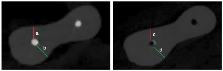 Figure 1
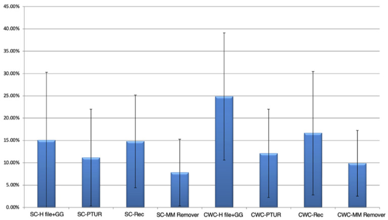 Figure 2
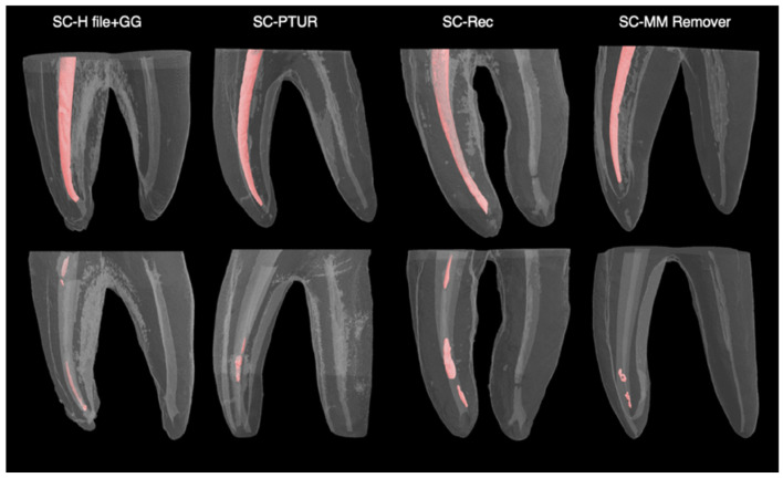 Figure 3
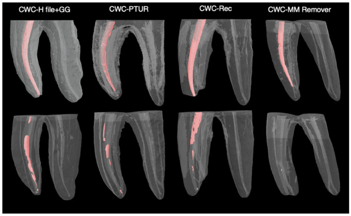 Figure 4
- —Ankara University Scientific Research Projects Coordination Unit
Peer Reviews
No public reviews on file for this paper yet. If you reviewed it on a platform where reviews are public (OpenReview, ICLR, NeurIPS, ICML), you can paste yours below so the community can read it here.
Videos
No videos yet. Explain this paper in a talk, walkthrough, or lecture? Add one.
Taxonomy
TopicsEndodontics and Root Canal Treatments · Dental Radiography and Imaging · Sinusitis and nasal conditions
1. Introduction
Root canal (RC) therapy aims to eradicate intraradicular infection, allowing periapical healing and preserving tooth function [1]. Despite high success rates of endodontic therapy (86–98%) [2], failures still occur for various reasons [3]. In such situations, nonsurgical retreatment is commonly considered the preferred initial management approach [1]. An essential phase of nonsurgical retreatment involves removing the previous RC filling material, as remnants can harbor microorganisms and impair the success of disinfection and re-obturation [4]. However, the literature indicates that no technique can completely remove filling materials from the RC system [5,6,7], and achieving complete cleanliness remains a clinical challenge.
Hedstrom hand files (Dentsply, Ballaigues, Switzerland) remain widely used in endodontic retreatment procedures due to their sharp cutting-edge design, which facilitates effective engagement and removal of RC filling materials [8]. Their retreatment performance has been assessed in several previous studies [9,10,11]. Nickel–Titanium (NiTi) file systems are favored for flexibility, high fracture resistance, efficiency, and ease of use [12]. The ProTaper Universal Retreatment (PTUR) system (Dentsply, Ballaigues, Switzerland), a multi-file NiTi instrument specifically developed for retreatment procedures, is commonly used in both curved and straight RCs [6,13,14]. The Reciproc (Rec) system (VDW, Munich, Germany), a single-file reciprocating instrument, has also been shown to effectively remove filling materials during retreatment while maintaining procedural efficiency [5,6,7,15]. The MicroMega Remover (MM Remover) (Micro-Mega, Besancon, France) is a newly developed single-file retreatment instrument manufactured using heat-treated C-wire technology and featuring a variable triple-helix cross-sectional design [16]. According to the manufacturer, the file is designed to mechanically remove gutta-percha without the use of solvents and, owing to its non-cutting tip, aims to minimize procedure-related risks while preserving the RC anatomy [17]. It is recommended for use at a rotational speed of 400–800 rpm with a torque setting of 2.5 Ncm. A recent study reported that the MM Remover demonstrated higher cyclic fatigue resistance than PTUR [16]. However, comprehensive studies assessing the retreatment performance of the MM Remover remain limited.
In recent years, warm obturation techniques have gained widespread acceptance as they provide a more homogeneous and well-adapted gutta-percha mass within complex RC anatomies. Nevertheless, these techniques may increase the difficulty of filling removal during retreatment, especially in severely curved RCs, where risks such as apical transportation, ledge formation, or perforation are elevated [7,18]. Therefore, the selection of appropriate file systems during retreatment is crucial to achieve optimal clinical outcomes.
Tomography-based imaging modalities are widely used in endodontic research due to their ability to provide three-dimensional (3D) visualization of RC systems [19,20]. Micro-computed tomography (micro-CT) provides non-invasive, high-resolution, and reproducible evaluations [7,21]. Owing to these advantages, it has been extensively employed to investigate RC morphology before and after instrumentation, including the assessment of dentin volume alterations [22]. Moreover, micro-CT has been used to evaluate debris removal following RC irrigation, residual filling material, and the shaping ability of endodontic instruments [19,23].
Although various micro-CT studies have evaluated different retreatment systems, comprehensive investigations assessing the performance of newly developed instruments are still required. In this context, the design features of the MM Remover, including a heat-treated C-wire alloy and a variable triple-helix cross-sectional geometry, may influence both the efficiency of filling material removal and apical transportation, particularly in curved RC anatomies, which are clinically challenging during nonsurgical retreatment. Such comparative data obtained under varying experimental conditions are essential for clarifying the clinical applicability of this system. According to the current literature, there is no available micro-CT study comparing the performance of the recently introduced MM Remover with established rotary and reciprocating file systems under different obturation techniques. Therefore, this in vitro study evaluated the effectiveness of four different instruments in terms of RC filling material removal and apical transportation in curved RCs obturated with different techniques using micro-CT. The instruments evaluated included PTUR, Rec, Hedstrom files, and the MM Remover. Additionally, the time required for RC filling material removal was compared.
2. Materials and Methods
2.1. Ethical Approval and Sample Size Calculation
Ethical approval for this research was obtained from the Ethics Committee of Ankara University Faculty of Dentistry (Decision No. 10/06, dated 19 June 2023). The study protocol was conducted in accordance with the Declaration of Helsinki. Before the teeth were extracted and collected, all participants were fully informed and provided written informed consent. Sample size estimation was conducted using G*Power software (version 3.1.9.7; Dusseldorf, Germany). Based on residual filling material as the primary outcome, an effect size of 0.6, a statistical power of 80%, and a type I error rate of 5% were applied, indicating that a minimum of six specimens per subgroup were required. Adequate power for apical transportation was verified by post-hoc analysis. Accordingly, eight specimens were included per group.
Sample Preparation, Inclusion, and Exclusion Criteria
The mesial roots of fully developed permanent mandibular molars extracted for periodontal reasons were included; teeth with restorations, previous RC treatment, fractures, cracks, or resorptions were excluded. Root surfaces were cleaned with periodontal curettes, and the teeth were stored in 0.1% thymol solution. Root curvature was assessed radiographically, and, according to Schneider’s classification [24], teeth with severe mesial curvature (25–40°) were selected. In total, 64 mesial RCs with Vertucci type IV configuration were included [25]. To achieve standardized root lengths of 16 ± 1 mm, the crowns were sectioned and access cavities were prepared with a diamond fissure bur.
2.2. Root Canal Preparation and Obturation
Apical patency of mesiobuccal and mesiolingual RCs was verified with a #10 K-file (Dentsply Maillefer, Ballaigues, Switzerland). The working length (WL) was determined at 1 mm coronal to the apical foramen. RCs were prepared to F2 (25/0.06) with the ProTaper Gold (Dentsply Sirona, Ballaigues, Switzerland), driven by an endodontic motor (AI Motor T-mode Yoshi Terauchi Edition; Woodpecker, Guilin, China). Throughout instrumentation, 2.5% sodium hypochlorite (NaOCl) (Microvem, Istanbul, Turkey) was used as the irrigant. The smear layer was removed by sequential irrigation with 17% ethylenediaminetetraacetic acid (EDTA) (Promida, Eskisehir, Turkey), 2.5% NaOCl, and distilled water (2 mL each). Finally, the RCs were dried with sterile paper points.
Samples were randomized to single-cone (SC; Group A) (n = 32/group) or continuous wave vertical compaction (CWC; Group B) (n = 32/group). AH Plus sealer (Dentsply Maillefer, Ballaigues, Switzerland) with F2 gutta-percha was used in both groups. In the SC group, gutta-percha cones were cut at the canal orifice and condensed with a plugger. In the CWC group, the F2 master cone was cut with the Elements Free down-pack device at 200 °C (Kerr Endodontics, Orange, CA, USA), leaving five mm apically, and compacted with a size 1 hand plugger (Machtou plugger; VDW, Munich, Germany). Backfilling at 100 °C was performed in 3–4 mm increments, and gutta-percha was condensed with pluggers (sizes 2–4) until obturation was complete. Coronal access cavities were temporarily sealed with Cavit-G (3M ESPE, Seefeld, Germany). Obturation quality was verified radiographically in buccolingual and mesiodistal projections. Samples were stored at 37 °C in 100% relative humidity for 14 days to allow sealer setting.
2.3. Root Canal Retreatment
The obturated samples were randomly allocated to four retreatment subgroups.
Group A1, B1 (H file + GG): The coronal third was enlarged with GG drills #3 and #2 (Mani, INC., Tochigi, Japan) at 1500 rpm. The procedure continued using Hedstrom files (#30 to #20) in peripheral push-pull movements along the canal walls. After reaching the WL with a #15 K-file (Dentsply Maillefer, Ballaigues, Switzerland), the apical enlargement to size 25 was performed.
Group A2, B2 (PTUR): The retreatment procedure was carried out with a rotational speed of 500 rpm and a torque set at 2 Ncm. D1 (30/0.09), D2 (25/0.08), and D3 (20/0.07) were used sequentially in the coronal, middle and apical thirds, respectively, for gutta-percha removal.
Group A3, B3 (Rec): The R25 file (25/0.08) was used on the WL with three consecutive reciprocating pecking motions, then withdrawn for debris removal.
Group A4, B4 (MM Remover): This single-file system (30/0.07) was used at a speed of 500 rpm and a torque of 2.5 Ncm. The file was operated with a gentle brushing motion without applying apical pressure. When resistance was encountered, the file was withdrawn from the RC for debris removal.
All retreatment procedures, except for those performed with hand files, were conducted using the AI Motor T-mode Yoshi Terauchi Edition (Woodpecker), following the manufacturer’s protocol. Each rotary file was used only once per RC. After each removal, irrigation was performed using 2.5% NaOCl (2 mL). Retreatment was considered complete when no filling material remained on the instrument surface and the RC walls were free of detectable obturation material [7,21]. To standardize the procedure, the WL was reached five times in each canal [15]. Retreatment time was recorded from first instrument use, excluding irrigation and instrument-change time. Subsequently, the RCs were irrigated sequentially with 17% EDTA, 2.5% NaOCl, and distilled water, each in a volume of 2 mL. The RCs were dried with sterile paper points, and the coronal access cavities were sealed temporarily with Cavit-G. All retreatment procedures were carried out by the same clinician to ensure consistency.
2.4. Micro-CT Scanning, Reconstruction and Analysis
All specimens were scanned with a SkyScan 1275 micro-CT device (Bruker MicroCT, Kontich, Belgium) following both obturation and retreatment procedures. Scanning was performed at 80 kVp and 125 µA with a 360° rotation, a 0.2° rotational step, a pixel size of 15.0 μm, and a 1 mm aluminum filter. On average, 1800 cross-sectional slices were obtained per sample. 3D reconstruction was carried out using NRecon software (version 1.7.4.2, Bruker MicroCT, Kontich, Belgium) with predefined parameters based on a previous study [26]. Reconstructed cross-sections were transferred to CTAn software (version 1.23.0.2, Bruker MicroCT, Kontich, Belgium) for quantitative analysis, morphometric measurements, and 3D visualization. RC filling material was isolated and quantified through segmentation of volumes of interest following both obturation and retreatment. All volumetric measurements were expressed in cubic micrometers (μm^3^). The residual filling material volume for each RC was calculated as a percentage using the formula: (A/B) × 100, where A is the residual filling material volume and B is the initial obturation volume. Apical transportation was analyzed using DataViewer (version 1.5.6.2, Bruker Micro-CT, Kontich, Belgium). Axial sections located three mm coronal to the apical foramen were selected from post-obturation and post-retreatment images (Figure 1). Apical transportation (μm) was calculated using the published formula [7]. An apical transportation value of ‘0’ indicates no apical transportation; positive values indicate mesial transportation of the RC, whereas negative values indicate distal transportation. All images were analyzed and measured twice by the same observer with over 10 years of experience in micro-CT evaluation, with a one-month interval between assessments, to evaluate intra-observer repeatability using the intraclass correlation coefficient (ICC).
2.5. Statistical Analysis
Data normality was evaluated with the Shapiro–Wilk test. The Kruskal–Wallis H test was applied to compare the groups, since the data for residual filling material percentage and retreatment time were not distributed normally. When a statistically significant difference was detected among more than two groups, pairwise comparisons were performed using the Bonferroni-corrected Mann–Whitney U test. A One-Way ANOVA was conducted to compare the data for apical transportation, as it exhibited a normal distribution. Intra-observer repeatability was evaluated using the ICC, according to commonly accepted thresholds [27]. Analyses were performed in IBM SPSS Statistics (version 11.5, SPSS Inc., Chicago, IL, USA), and p < 0.05 was accepted as statistically significant.
3. Results
The mean retreatment times (in seconds) obtained for each file system used during the retreatment procedure are presented in Table 1. Statistical analysis revealed a significant difference among the groups (p < 0.001). Group B1 (CWC–H file + GG) exhibited a significantly longer mean retreatment time than that of Group A2 (SC–PTUR) (p = 0.044), Group A4 (SC–MM Remover) (p < 0.001), Group B2 (CWC–PTUR) (p = 0.001), and Group B4 (CWC–MM Remover) (p = 0.005) (p < 0.05).
The mean percentages of residual RC filling material for each group are presented in Table 2 and Figure 2. The lowest mean percentage of residual filling material was observed in Group A4 (SC–MM Remover), while the highest was recorded in Group B1 (CWC–H file + GG). However, Kruskal–Wallis H test revealed no statistically significant differences among the groups regarding the residual filling material (%) (p > 0.05). Micro-CT images of the groups obturated using the SC (Figure 3) and CWC techniques (Figure 4), following RC obturation and retreatment, are also presented.
The apical transportation values (µm) of the experimental groups are shown in Table 3, with negative values observed in Groups A1 (SC–H file + GG), A2 (SC–PTUR), A3 (SC–Rec), and B3 (CWC–Rec), whereas positive values were recorded in Groups A4 (SC–MM Remover), B1 (CWC–H file + GG), B2 (CWC–PTUR), and B4 (CWC–MM Remover). The highest mean apical transportation in the distal direction was observed in Group B3 (−108.57 µm), while the highest in the mesial direction was found in Group B2 (+154.29 µm). However, according to the statistical evaluation, none of the differences among the groups were statistically significant (p > 0.05). Intra-observer repeatability demonstrated good to excellent reliability, with ICCs ranging from 0.87 to 0.90 across the evaluated measurements.
4. Discussion
When RC treatment fails, nonsurgical retreatment is often indicated. Instrument performance plays a crucial role in filling material removal during retreatment procedures, especially in curved RCs [28]. Previous studies have shown that none of the existing techniques can completely eliminate filling materials from the RC system [6,7,29]. This circumstance has led to a continuous search for an ideal technique or material.
Mandibular molars often present mesial roots with complex morphology and frequent curvatures [30,31]. Owing to their challenging anatomy, these teeth were selected for inclusion in the present study.
Currently, the CWC technique is widely preferred because it produces a more homogeneous volume of gutta-percha and better adaptation to RC curvatures [32,33]. However, warm obturation techniques may make the removal of RC filling materials more challenging when retreatment is required [34]. Despite various studies on retreatment efficacy of different instruments in RCs filled with warm or cold techniques, comparative data for curved RCs comparing warm versus cold obturation remain limited.
Various methods have been used to assess residual filling materials and apical transportation following retreatment, including two-dimensional (2D) radiographic examination [9,14,35], image analyzer software [36,37], and 3D tomographic techniques such as cone-beam computed tomography (CBCT) [20,38] and micro-CT [7,15]. Micro-CT offers higher resolution images with minimal artifacts compared to other imaging techniques [39,40]. These advantages enable accurate 3D visualization and quantitative assessment of RC morphology, dentinal removal and residual filling material following retreatment [10]. Therefore, it was selected as the imaging technique in the present study.
In this study, the retreatment effectiveness of PTUR, Rec, Hedstrom files, and the newly introduced MM Remover was evaluated with respect to residual filling material volume, apical transportation, and retreatment time in curved RCs. In this context, the evaluated instruments exhibit variability in metallurgical properties and design characteristics with respect to cross-sectional geometry, alloy composition, tip configuration, and taper design. The Rec system features an S-shaped cross-sectional design [7] and is manufactured using M-wire alloy [34]. By comparison, PTUR presents a triangular cross-sectional design [20], while the MM Remover is produced with a C-wire alloy and incorporates a variable triple-helix cross-sectional geometry. Therefore, these differences are challenging to standardize and may influence the retreatment performance of instruments in curved RCs.
According to the findings of the study, although differences in residual filling material volumes were observed among the groups, these differences were not statistically significant (p > 0.05). This finding aligns with prior research by Rödig et al. [6] and Adel et al. [20], who also reported no significant differences between PTUR, Rec, and Hedstrom instruments in removing RC filling materials from curved RCs. However, Serefoglu et al. [14] reported that the Rec left more residual filling material than Hedstrom files in retreatment procedures of curved RCs obturated with the SC technique. The researchers utilized a 2D imaging method for evaluation in their studies, whereas the use of the more accurate 3D micro-CT in our research can explain the difference in the results. Bago et al. [15] evaluated the PTUR, Rec, Rec Blue, and Wave One Gold file systems in removing RC filling materials from curved RCs obturated using the CWC technique and reported no significant differences, which is consistent with our findings regarding PTUR and Rec. Within this context, the lowest mean volume of residual filling material was observed in the group using the MM Remover file system in RCs filled with the SC technique, whereas the highest mean volume was found in the Hedstrom hand file group in RCs filled with the CWC technique. Although the MM Remover file system left the least amount of residual filling material in both obturation techniques, the difference was not statistically significant (p > 0.05). However, this observation may be related to its variable triple-helix cross-section design, which could contribute to improved cutting efficiency and debris removal in curved RCs.
Studies have shown that solvents soften gutta-percha, making it more viscous and adhesive, which may lead to its penetration into canal irregularities or dentinal tubules [41,42,43]. Such alterations in the physical properties of the filling material may complicate the retreatment procedure, making it more time-consuming or technically challenging [44]. Therefore, in the present study, the use of solvents was deliberately avoided during the retreatment procedures.
Although apical transportation in both mesial and distal directions was observed for all instruments, no statistically significant differences were found among the groups in terms of mean transportation values (p > 0.05). This finding is consistent with the results reported by Da Silva Arruda et al. [38], who found no significant differences among PTUR, ProTaper Next, and Rec in curved RCs. Similarly, Adel et al. [20] observed that PTUR, Rec, and Hedstrom hand files showed similar apical transportation outcomes in the apical 3 mm section of curved RCs. In this context, the comparable apical transportation exhibited by the MM Remover relative to the other file systems may be attributed to its heat-treated technology, which promotes controlled RC wall contact and limits excessive dentin removal.
Apical transportation greater than 0.3 mm has been associated with reduced apical sealing capacity of RC filling materials, which may negatively impact the clinical outcome of endodontic treatment [45,46,47]. In the present study, the mean apical transportation values of all tested systems remained below this critical threshold, indicating that the instruments were able to perform retreatment within clinically acceptable limits without compromising the sealing ability of RC filling materials or long-term endodontic treatment success, while maintaining the original canal morphology.
With respect to canal curvature, positive apical transportation values reflect mesial deviation toward the outer wall of the RC, whereas negative values represent distal deviation toward the inner wall [46]. In curved RCs, rotary instruments may lead to increased apical dentin removal along the outer curvature [48], and the relatively higher mesial apical transportation values observed in some PTUR specimens may be attributed to the inherent tendency of instruments to straighten, resulting in deviation toward the outer canal wall [47,49].
From a kinematic perspective, the tested systems differed in their mode of motion, with the PTUR and MM Remover being operated in continuous rotary motion, the Rec being used in reciprocating motion, and Hedstrom files being applied with manual instrumentation. These differences in kinematics may influence the observed transportation patterns in curved RCs. In light of these findings and within the limitations of the present study, the lack of detectable differences in apical transportation may be attributable to the limited sample size, potentially increasing the risk of a type II error. Expanding the sample size in future investigations may improve the detection of differences.
In our study, PTUR and MM Remover achieved shorter retreatment times than the other systems, regardless of obturation technique. The CWC-H file + GG group exhibited significantly longer retreatment times (p < 0.05). These results are consistent with previous studies [6,20], which reported similar retreatment times for PTUR and Rec, but longer durations with Hedstrom files. Similarly, another study reported significantly longer retreatment times for the Hedstrom group compared with PTUR and Rec; however, unlike our findings, PTUR required more time than Rec [23]. MM Remover’s reduced retreatment time may be attributed to its triple-helix cross-sectional design and thermomechanical processing [16]. From a clinical perspective, reduced retreatment time may offer important advantages by improving patient comfort, decreasing operator fatigue, and enhancing procedural efficiency. In addition, previous clinical studies have demonstrated the success of single-visit retreatment [1,50]; therefore, shorter procedures may increase the feasibility of completing retreatment in a single appointment.
In this study, the criterion for retreatment completion, defined as the absence of filling material on the final instrument surface, may be operator-dependent; however, this protocol was applied in line with previously published retreatment studies [7,15,21]. Moreover, the use of micro-CT provided an objective and quantitative assessment of residual filling material, thereby minimizing observer-related subjectivity.
The use of Gates–Glidden drills was limited to the Hedstrom file group to reflect conventional clinical practice with hand instrumentation and to facilitate coronal flaring [6,20]. However, rotary retreatment systems are generally designed to achieve coronal enlargement through their instrument design and kinematics; therefore, auxiliary coronal flaring drills were not used in the present study [23].
Despite the noteworthy findings of the current research, achieving complete cleanliness of the RC system during non-surgical retreatment remains a persistent challenge. This research was conducted under in vitro conditions using mandibular molars with curved mesial RCs, which may not fully reflect clinical variability. Anatomical differences such as root curvature and canal morphology may influence retreatment efficacy and limit the generalizability of the results. To the best of our knowledge, no previous study has evaluated the retreatment efficacy of the MM Remover system in curved RCs; therefore, the present study was designed to compare the performance of this system with commonly used rotary, reciprocating, and traditional hand file systems under conditions that reflect their typical clinical application. The results obtained in controlled in vitro conditions not only clarify whether the anticipated advantages of the system—such as improved efficiency, enhanced safety, and avoidance of solvents—are realized in practice, but also offer valuable insights into its performance within anatomically complex scenarios, particularly in the mesial roots of mandibular molars. These findings enrich the current evidence base and contribute to the ongoing efforts to optimize retreatment protocols for challenging endodontic cases. Continued development of more efficient file systems tailored to complex RC anatomies is essential to improve outcomes. Future studies should include larger sample sizes, broader clinical trials, and assessment of adjunctive irrigation activation to enhance filling material removal.
5. Conclusions
Considering the constraints of this study, all evaluated file systems were able to remove filling materials from curved RCs effectively while preserving the original canal anatomy and avoiding excessive apical transportation. Although the MM Remover system left the lowest residual filling material, the differences among all file systems regarding filling material removal and apical transportation were not statistically significant. Nevertheless, both the PTUR and MM Remover systems showed significantly shorter retreatment times, indicating clinical advantages in terms of efficiency. These findings highlight the potential clinical applicability of the newly developed MM Remover, supporting its further evaluation in anatomically complex cases, particularly in the curved mesial roots of mandibular molars.
The reference list from the paper itself. Each links out to its DOI / PubMed record.
- 1Toia C.C. Khoury R.D. Corazza B.J.M. Orozco E.I.F. Valera M.C. Effectiveness of 1-Visit and 2-Visit Endodontic Retreatment of Teeth with Persistent/Secondary Endodontic Infection: A Randomized Clinical Trial with 18 Months of Follow-up J. Endod.20224841410.1016/j.joen.2021.09.00434555421 · doi ↗ · pubmed ↗
- 2Song M. Kim H.C. Lee W. Kim E. Analysis of the cause of failure in nonsurgical endodontic treatment by microscopic inspection during endodontic microsurgery J. Endod.2011371516151910.1016/j.joen.2011.06.03222000454 · doi ↗ · pubmed ↗
- 3Tabassum S. Khan F.R. Failure of endodontic treatment: The usual suspects Eur. J. Dent.20161014414710.4103/1305-7456.17568227011754 PMC 4784145 · doi ↗ · pubmed ↗
- 4Marques da Silva B. Baratto-Filho F. Leonardi D. Henrique Borges A. Volpato L. Branco Barletta F. Effectiveness of Pro Taper, D-Ra Ce, and Mtwo retreatment files with and without supplementary instruments in the removal of root canal filling material Int. Endod. J.20124592793210.1111/j.1365-2591.2012.02051.x 22486933 · doi ↗ · pubmed ↗
- 5Alves F.R. Ribeiro T.O. Moreno J.O. Lopes H.P. Comparison of the efficacy of nickel-titanium rotary systems with or without the retreatment instruments in the removal of gutta-percha in the apical third BMC Oral Health 20141410210.1186/1472-6831-14-10225128277 PMC 4139579 · doi ↗ · pubmed ↗
- 6Rödig T. Reicherts P. Konietschke F. Dullin C. Hahn W. Hülsmann M. Efficacy of reciprocating and rotary Ni Ti instruments for retreatment of curved root canals assessed by micro-CT Int. Endod. J.20144794294810.1111/iej.1223924386931 · doi ↗ · pubmed ↗
- 7Nevares G. de Albuquerque D.S. Freire L.G. Romeiro K. Fogel H.M. Dos Santos M. Cunha R.S. Efficacy of Pro Taper Next compared with Reciproc in removing obturation material from severely curved root canals: A micro-computed tomography study J. Endod.20164280380810.1016/j.joen.2016.02.01027117757 · doi ↗ · pubmed ↗
- 8Hülsmann M. Stotz S. Efficacy, cleaning ability and safety of different devices for gutta-percha removal in root canal retreatment Int. Endod. J.19973022723310.1111/j.1365-2591.1997.tb 00702.x 9477808 · doi ↗ · pubmed ↗