Cervical Spinal Cord Stimulation for Functional Rehabilitation After Spinal Cord Injury: A Systematic Review of Preclinical and Clinical Studies
Maximilian C. Wankner, Veerle Visser-Vandewalle, Pablo Andrade, Petra Heiden

TL;DR
This paper reviews studies on using cervical spinal cord stimulation to improve recovery after spinal cord injury, finding it can help with motor, sensory, and autonomic functions.
Contribution
The study provides a systematic review of cervical spinal cord stimulation's role in rehabilitation, highlighting its potential therapeutic effects and adjunct interventions.
Findings
Cervical spinal cord stimulation improves hand strength, dexterity, and voluntary motor activation in preclinical and clinical studies.
Adjunctive interventions like cortical stimulation and brain-computer interfaces enhance recovery outcomes.
Sensory and autonomic functions also show improvement, though respiratory outcomes are rarely studied.
Abstract
Cervical spinal cord injury causes severe functional impairment with limited spontaneous recovery, and while spinal cord stimulation has emerged as a promising neuromodulatory strategy, evidence for cervical applications remains fragmented. To address this gap, we conducted a systematic review synthesizing preclinical and clinical evidence on cervical spinal cord stimulation for functional rehabilitation following spinal cord injury. The review was registered on PROSPERO (CRD420251088804) and conducted in accordance with PRISMA guidelines, with PubMed, Embase, IEEE Xplore, and Web of Science searched from inception to July 2025 for animal and human studies of cervical spinal cord stimulation, including epidural, intraspinal, and transcutaneous approaches, reporting functional neurological outcomes. Risk of bias was assessed using the Cochrane RoB 2 and ROBINS-I tools, and due to…
Genes, proteins, chemicals, diseases, species, mutations and cell lines named across the full text — each resolved to its canonical identifier and authoritative record.
Click any figure to enlarge with its caption.
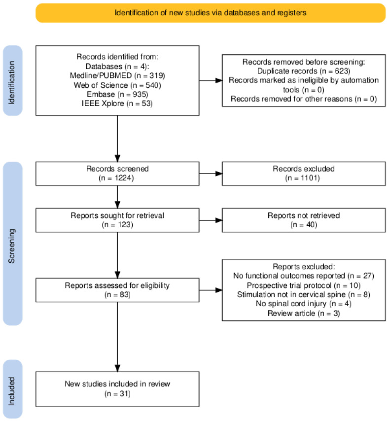 Figure 1
| Authors | Country | Population | Sex | N | Age Range | Injury Level | Injury Duration | Injury Type/Severity | Modality |
|---|---|---|---|---|---|---|---|---|---|
|
| |||||||||
| Kasten et al. (2013) [ | USA | Rats | F | 11 | Adult | C4–C5 | 4 weeks | Contusion injury | ISMS |
| McPherson et al. (2015) [ | USA | Rats | F | 9 | Adult | C4–C5 | >8 weeks | Contusion injury | ISMS |
| Alam et al. (2015) [ | USA | Rats | F | 12 | Adult | C4 | 1–2 weeks | Dorsal funiculi crush | EES |
| Alam et al. (2017) [ | USA | Rats | F | 5 | Adult | C4 | 1 week | Dorsal funiculi crush | EES |
| Bezdudnaya et al. (2018) [ | USA | Rats | M | 20 | Adult | C1 | Acute | Complete transection | EES |
| Yang et al. (2019) [ | USA | Rats | F | 27 | 10–12 weeks | C3 | 11 days | Contusion injury | EES |
| Samejima et al. (2021) [ | USA | Rats | F | 5 | Adult | C4 | 2 weeks | Contusion injury | EES |
| Barra et al. (2022) [ | Switzerland |
| F | 3 | 3–9 years | C5–C6 | 1 week | Unilateral CST transection | EES |
| Pal et al. (2022) [ | USA | Rats | F | 8 | Adult | C4 | 10 days | AAV-based CST inactivation | EES |
| Song et al. (2016) [ | USA | Rats | F | 6 | Adult | C1 | 1 week | Transection at rostral medualla | tSCS |
| Zareen et al. (2017) [ | USA | Rats | F | 13 | 10–12 weeks | C4 | 2 weeks | Contusion injury | tSCS |
|
| |||||||||
| Lu et al. (2016) [ | USA | Human | N.R. | 2 | N.R. | C5–C6 | >18 months | AIS B | EES |
| Murray et al. (2017) [ | USA | Human | M | 1 | 27 years | C6–C7 | 9 years | AIS C | tSCS |
| Inanici et al. (2018) [ | USA | Human | M | 1 | 62 years | C3 | 2 years | AIS D | tSCS |
| Gad et al. (2018) [ | USA | Human | 1F 5M | 6 | 20–62 years | C4–C8 | 1–21 years | AIS A (n = 0), B (n = 2), C (n = 4), D (n = 0) | tSCS |
| Benavides et al. (2020) [ | USA | Human | 4F 6M | 10 | 22–64 years | C4–C6 | >1 year | AIS A (n = 4), B (n = 3), C (n = 2), D (n = 1) | tSCS |
| Zhang et al. (2020) [ | USA | Human | M | 1 | 38 years | C5 | 15 years | AIS A | tSCS |
| Gad et al. (2020) [ | USA | Human | M | 1 | 39 years | C5 | 9 years | AIS A | tSCS |
| Tefertiller et al. (2021) [ | USA | Human | 2F 5M | 7 | 18–55 years | C4–C6 | 15–38 months | AIS A (n = 0), B (n = 4), C (n = 2), D (n = 1) | tSCS |
| Inanici et al. (2021) [ | USA | Human | 2F 4M | 6 | 28–62 years | C3–C5 | 1.5–12 years | AIS A (n = 0), B (n = 2), C (n = 2), D (n = 2) | tSCS |
| Huang et al. (2022) [ | USA | Human | N.R. | 10 | 22–63 years | C3–C7 | 2–14 years | AIS A–B | tSCS |
| McGeady et al. (2022) [ | UK/Hong Kong | Human | M | 1 | 48 years | C4 | 12 years | AIS A | tSCS |
| Zhang et al. (2023) [ | USA | Human | 4M | 4 | 25–78 years | C1–C4 | 4–9 years | AIS A (n = 1), B (n = 1), C (n = 0), D (n = 2) | tSCS |
| Oh et al. (2023) [ | USA | Human | 4M | 4 | 23–29 years | C4–C6 | 2–14 years | AIS A–B | tSCS |
| García-Alén et al. (2023) [ | Spain | Human | 1F 14M | 15 | 18–70 years | C4–C7 | 3–10 months | AIS A–D | tSCS |
|
| |||||||||
| Chandrasekaran et al. (2023) [ | USA | Human | 2M | 2 | 20–39 years | C5 | 4–7 years | AIS A–B | tSCS |
| Moritz et al. (2024) [ | International | Human | 10F 50M | 60 | C2–C7 | AIS B–D | tSCS | ||
| Singh et al. (2024) [ | USA | Human (Children) | 1F 6M | 7 | 6–17 years | C1–C7 | >2 years | AIS A (n = 1), B (n = 4), C (n = 1), D (n = 1) | tSCS |
| Verma et al. (2025) [ | USA | Human | 1F 4M | 5 | 19–67 years | C4–C7 | 3–18 years | AIS A (n = 3), B (n = 1), C (n = 1), D (n = 0) | tSCS |
| Capozio et al. (2025) [ | UK | Human | 5F | 5 | 31–65 years | C3–C8 | 2–30 years | AIS A (n = 0), B (n = 0), C (n = 3), D (n = 2) | tSCS |
| Capozio et al. (2025) [ | UK | Human | 4F 4M | 8 | 22–72 years | C2–C7 | 3–32 years | AIS A (n = 0), B (n = 1), C (n = 4), D (n = 3) | tSCS |
| Authors | Population | Electrode Manufacturer | Electrode Placement | Pulse Width ( | Frequency (Hz) (Carrier, kHz) | Amplitude | Adjunctive Intervention | Stimulation Optimization |
|---|---|---|---|---|---|---|---|---|
| Intraspinal microstimulation | ||||||||
| Kasten et al. (2013) [ | Rats | Custom | C6–T1 ventral horn | 300 |
| N.R. | – | Set at threshold just to evoke forelimb movement when delivered via a single electrode |
| McPherson et al. (2015) [ | Rats | Custom | C6–C7 ventral horn | 200 | 50–100 | 30– | EMG-synchronized | 90% of motor threshold; synchronized ISMS below the injury with volitional motor commands |
| Epidural stimulation | ||||||||
| Alam et al. (2015) [ | Rats | Custom | C6 and C8 | 200 | 40 | 400– | – | 70% of motor threshold for mono- and bipolar electrode pairings |
| Lu et al. (2016) [ | Human | Boston Scientific | C5–T1 | 210 | 2–40 | 0.1–10 mA | – | Individually adjusted for motor evoked response |
| Alam et al. (2017) [ | Rats | Custom | C6 and C8 | 200 | 20, 40, 60 | N.R. | – | 60–70% of sMEP threshold |
| Bezdudnaya et al. (2018) [ | Rats | Custom | C3–C5 | 200–500 | 100–300 | 100– | – | Titrated to elicit phrenic motor responses and entrain breathing |
| Yang et al. (2019) [ | Rats | LGMedSupply | C4–T2 | 200 | 330 | Up to 1.5 mA | Cortical stimulation | 75% of motor threshold for evoked response |
| Samejima et al. (2021) [ | Rats | Custom | C6 | 400 | 50–100 | 300– | BCI | Individually set to invoke elbow extension |
| Barra et al. (2022) [ |
| Custom | C6–T1 | 200–400 | 20–120 | 600– | Cortical stimulation | Stimulation triggered in phase with voluntary movement; individualized optimization |
| Pal et al. (2022) [ | Rats | Custom | C5–C6 | 200 | 5 | N.R. | Cortical stimulation | M1 stimulation at 110% of the threshold for evoking a cortical MEP followed 10 ms later by spinal stimulation at 90% of the threshold for generating a spinal MEP |
| Transcutaneous stimulation | ||||||||
| Song et al. (2016) [ | Rats | StimTentCom | C4–T2; M1 region | 200 | 100; 0.2 | 100–400/ | Cortical stimulation | Paired M1 (0.2 Hz) and spinal stimulation (100 Hz, 5 pulses/burst) with 10 ms interstimulus interval |
| Murray et al. (2017) [ | Human | UniPatch | C5–T2 | 1000 | 0.2 | Up to 150 mA | TMS | Intensities increased from sub-threshold to suprathreshold |
| Zareen et al. (2017) [ | Rats | StimTentCom | C4–T2; M1 region | 200 | 330; 250 | Up to 1.5 mA | Cortical stimulation | 10 biphasic pulses at 250 Hz every 2 s; amplitude set at 75% of motor threshold |
| Inanici et al. (2018) [ | Human | Axelgaard | C3–C4, C6–C7 | 1000 | 30 (10) | 80–120 mA | – | Individually titrated to comfort |
| Gad et al. (2018) [ | Human | Axelgaard | C3–C4, C6–C7 | 1000 | 30 (10) | 10–250 mA | – | Adjusted to enable maximal grip strength without causing discomfort |
| Benavides et al. (2020) [ | Human | Axelgaard | C5–C6 | 200 | 30 (5) | TMS | Intensity set to evoke biceps root-evoked potential | |
| Zhang et al. (2020) [ | Human | STIMEX | C3–C4, C7–T1 | 1000 | 30 (10) | Up to 80 mA | – | Adjusted to enable maximal grip strength without causing discomfort |
| Gad et al. (2020) [ | Human | TESCoN | C5–C6 | 1000 | 30 (10) | 20 mA | – | Adjusted for dose–response curves of inspiratory capacities and forced expiratory volume |
| Tefertiller et al. (2021) [ | Human | Axelgaard | C3–C4, C6–C7, T11–T12 | 1000 | 30 (10) | 45–150 mA | – | Adjusted until optimal functional movement of the targeted area was achieved in a joint below the level of injury |
| Inanici et al. (2021) [ | Human | Axelgaard | C3–C4, C6–C7 | 1000 | 30 (10) | 80–120 mA | – | Individually titrated to comfort |
| Huang et al. (2022) [ | Human | Axelgaard | C4–C5 | 1000 | 30 | 40–180 mA | – | Individually titrated to elicit maximal handgrip; stimulation timed concurrently to voluntary handgrip attempt |
| McGeady et al. (2022) [ | Human | Axelgaard | C4–C6 | 1000 | 30 | 40–55 mA | Motor imagery | Individually titrated to comfort |
| Zhang et al. (2023) [ | Human | STIMEX | C3–C5 | 1000 | 30 | Up to 120 mA | – | Individually adjusted for subthreshold upper extremity muscle activation |
| Oh et al. (2023) [ | Human | Axelgaard | C6–T1 | 500 | 30 | 20–70 mA | – | Adjusted for maximum hand grip |
| García-Alén et al. (2023) [ | Human | Axion | C3–C4, C6–C7 | 1000 | 30 (10) | 45–86 mA | Robotic exoskeleton | 90% of RMT induced by single-pulse tSCS at the abductor pollicis brevis |
| Chandrasekaran et al. (2023) [ | Human | Custom | C4–T1 | 500 | 50 | 140–160 mA | – | Adjusted for maximal tolerable intensity during ABT |
| Moritz et al. (2024) [ | Human | LIFT (ARCex) | C3–C7 | 1000 | 30 (10) | 10–180 mA | – | Individually titrated to comfort; electrode placement individualized based on desired effect |
| Singh et al. (2024) [ | Human (Children) | Syrtenty | C3–C4, C6–C7, T10–T11 | 1000 | 30 (10) | 20–70 mA | – | Adjusted to enable maximal hand-grip force at subthreshold intensity |
| Verma et al. (2025) [ | Human | Anuevo | C3–T1 | 500 | 30 | – | Maximum tolerable intensity just below motor threshold | |
| Capozio et al. (2025) [ | Human | Axelgaard | Above/below lesion | 1000 | 30 | 40–70 mA | TMS | Individually titrated to 80–90% of RMT |
| Capozio et al. (2025) [ | Human | Axelgaard | C5–C6 midline | 1000 | 30 (5) | 30–60 mA | TMS, motor imagery | Individually titrated to 80–90% of RMT |
| Authors | GRASSP | ISNCSCI | Dyn. | Main Finding |
|---|---|---|---|---|
| Epidural stimulation | ||||
| Lu et al. (2016) [ | – | ↑ | ↑ | Immediate and session-to-session motor gains in 2/2 participants; three-fold increase in hand strength with stimulation; upper-extremity motor score increased by 23 and 16 points; improved self-care and mobility. |
| Transcutaneous stimulation | ||||
| Murray et al. (2017) [ | – | – | – | Reduced spasticity and spasms, reversal of anhidrosis. |
| Inanici et al. (2018) [ | ↑ | ↑ | ↑ | Improvements of 52 points on GRASSP, 14 points on UEMS, 2- to 7-fold increase in pinch strength in left and right hand, respectively; overall sensation and neurological level of injury improved from C3 to C4. Functional gains persisted for |
| Gad et al. (2018) [ | – | – | ↑ | Grip strength increased by 325% with stimulation and 225% off stimulation after 8 sessions. |
| Benavides et al. (2020) [ | ↑ | – | – | Immediate GRASSP improvements observed in a single session. |
| Zhang et al. (2020) [ | ↑ | – | ↑ | GRASSP improved by 24 points and handgrip strength increased (left: 283%, right: 30%); gains sustained for 3 months. |
| Gad et al. (2020) [ | – | – | – | Improved breathing and cough effectiveness; benefits persisted beyond the stimulation period. |
| Tefertiller et al. (2021) [ | – | ↑ | – | AIS grade improved in 2/7 participants (B to C; C to D); sensation increased in 5/7; all 4/4 AIS A participants were able to activate lower extremities with stimulation. |
| Inanici et al. (2021) [ | ↑ | ↑ | ↑ | GRASSP scores increased; UEMS improved by up to 8 points. AIS grade improved in 1/6 participants (AIS C to D); |
| Huang et al. (2022) [ | – | – | ↑ | Grip force increased in 5/10 participants with measurable baseline force. |
| McGeady et al. (2022) [ | ↑ | ↑ | ↑ | GRASSP increased by 35 points with associated strength gains and improved neurological level of injury; brain–computer interface priming enhanced gains. |
| Zhang et al. (2023) [ | ↑ | – | ↑ | Five-fold improvement in GRASSP in AIS A; GRASSP increased by 10–33 points; up to three-fold increase in upper-extremity strength; gains maintained for 1 month in all 4/4 participants. |
| Oh et al. (2023) [ | – | – | ↑ | Grip strength increased only with combined stimulation and training. |
| García-Alén et al. (2023) [ | ↑ | ↑ | ↑ | GRASSP strength and pinch improved; also increases in SCIM scores and quality of life. |
| Chandrasekaran et al. (2023) [ | – | ↑ | ↑ | Up to 1136% increase in force; tactile sensation improved. |
| Moritz et al. (2024) [ | ↑ | ↑ | ↑ | Seventy-two percent of participants exceeded MCID; pinch, grip, UEMS, and SCIM scores increased; autonomic function (bladder and blood-pressure regulation) improved in some; significant improvements in patient-reported quality of life. |
| Singh et al. (2024) [ | – | – | ↑ | Handgrip increased in 6/7 participants; more than 20% force increase in 3 participants; safety and tolerability in children confirmed. |
| Verma et al. (2025) [ | – | – | ↑ | Grip strength increased; minimal residual force at baseline predicted response. |
| Capozio et al. (2025) [ | ↑ | – | – | GRASSP improved in 4/5 participants; sensation improved in 3/5. |
| Capozio et al. (2025) [ | – | – | – | Dexterity increased after a single session, with or without stimulation. |
Peer Reviews
No public reviews on file for this paper yet. If you reviewed it on a platform where reviews are public (OpenReview, ICLR, NeurIPS, ICML), you can paste yours below so the community can read it here.
Videos
No videos yet. Explain this paper in a talk, walkthrough, or lecture? Add one.
Taxonomy
TopicsSpinal Cord Injury Research · Pain Management and Treatment · Nerve Injury and Rehabilitation
1. Introduction
Spinal cord injury (SCI) affects primarily young and middle-aged adults, with traumatic cases occurring at a mean age of 35 years and individuals aged 21–40 accounting for over 40% of cases [1,2]. Cervical injuries frequently cause tetraparesis or tetraplegia, resulting in loss of independence in daily activities, caregiver reliance, and, in high cervical lesions, respiratory compromise requiring ventilatory support [3]. Chronic sequelae, including spasticity, neuropathic pain, cardiovascular dysregulation, and pressure ulcers, contribute further to long-term morbidity [4].
The intrinsic potential for recovery after high cervical SCI remains limited. Neurological improvement generally plateaus within the first year, and gains are modest even under intensive rehabilitation [5,6,7,8]. While incremental improvements can meaningfully impact independence, substantial restoration of function is rare in motor-complete or near-complete cervical SCI. Current treatment approaches focus on acute surgical decompression of the spinal cord and stabilization, prevention of secondary injury, pharmacological management, and long-term multidisciplinary rehabilitation. Although advances in rehabilitation techniques have led to incremental improvements, they remain insufficient for restoring meaningful function in patients with severe cervical SCI. Crucially, traditional therapies are limited by the absence of interventions that can directly modulate spinal circuitry and promote recovery beyond the modest natural course.
Spinal cord stimulation (SCS) has emerged as a promising neuromodulation strategy to address this gap. Originally introduced for analgesia in the 1960s based on the gate control theory of pain [9,10], subsequent work showed that activating spinal networks can also influence motor output. In individuals with chronic motor-complete paraplegia, lumbosacral epidural stimulation combined with intensive rehabilitation has enabled weight-bearing standing, volitional control of paralyzed muscles, and even overground walking [11,12,13]. These landmark studies established that residual spinal circuits below the lesion can generate functional motor patterns when appropriately facilitated [14].
Following successful demonstrations of SCS at the lumbosacral cord, attention shifted to the cervical spinal cord, given its critical role in upper limb and respiratory function. Several stimulation modalities have been explored, including intraspinal microstimulation (ISMS) via penetrating microelectrodes, epidural electrical stimulation (EES) using dorsal column arrays, and non-invasive transcutaneous stimulation (tSCS) over the dorsal cervical spine [15,16,17,18,19,20,21,22,23,24,25,26]. ISMS offers high anatomical and functional specificity in preclinical models but relies on penetrating electrodes that currently limit near-term clinical translation; EES enables more targeted segmental recruitment than tSCS but requires invasive surgical implantation; and tSCS provides a non-invasive and readily deployable approach at the expense of reduced spatial specificity. Preclinical studies demonstrate that cervical stimulation can selectively recruit forelimb and respiratory circuits and support recovery of skilled motor behaviors after injury [15,16,19,21,27,28,29,30,31,32,33,34,35,36,37,38]. These findings suggest that cervical neuromodulation may provide a means to restore upper limb and respiratory function beyond the modest natural course of recovery.
Against this background, the present systematic review synthesizes preclinical and clinical evidence on cervical SCS for functional rehabilitation after SCI, with a focus on motor, sensory, respiratory, and autonomic outcomes, and evaluates the methodological quality of the available studies.
2. Materials and Methods
2.1. Search Strategy
This systematic review was conducted and reported in accordance with the Preferred Reporting Items for Systematic Reviews and Meta-Analyses (PRISMA) 2020 guidelines. The review protocol was prospectively registered in PROSPERO (CRD420251088804). Reporting was conducted in accordance with the PRISMA guidelines [39]. The PRISMA 2020 flow diagram was generated using the PRISMA2020 R package and Shiny app version 1.1.1 [40]. A comprehensive search of the published literature was performed in MEDLINE/PubMed, Embase, Web of Science, and IEEE Xplore electronic databases in June–July 2025 by two independent reviewers (M.W. and P.A.), working independently and blinded to each other’s selections to ensure reliability. The systematic search combined both keywords and, where applicable, Medical Subject Headings (MeSH), using the following Boolean string: (“cervical spinal cord stimulation” OR “cervical spinal cord” OR “cervical stimulation”) AND (“spinal cord injury” OR “SCI”) AND (“stimulation” OR “neuromodulation”) AND (“recovery” OR “rehabilitation” OR “function” OR “movement” OR “sensory” OR “motor” OR “autonomic” OR “respiration”). Additionally, the reference lists of all included studies and relevant reviews were manually screened to identify any additional eligible reports not captured by the electronic search.
2.2. Study Selection Criteria
Study selection was guided by predefined PICOS (Population, Intervention, Comparison, Outcome, and Study design) criteria [41]. Studies were eligible for inclusion if they met the following criteria: (1) enrolled humans or animals with spinal cord injury; (2) applied electrical stimulation to the cervical spinal cord; (3) reported outcomes in at least one functional neurological domain (motor, sensory, autonomic, respiratory, or related function); and (4) employed an eligible study design, including case series, case reports, cohort studies, conference abstracts, preprints, or randomized controlled trials (RCTs). Studies were excluded if they (1) focused primarily on chronic pain, spasticity, or other non-functional outcomes; (2) targeted stimulation sites outside the cervical spinal cord and its associated roots; (3) involved participants with peripheral nerve injuries; (4) lacked sufficient extractable data, such as reviews, editorials, or protocol descriptions; or (5) were published in languages other than English.
2.3. Data Extraction
Two independent reviewers (M.W. and P.A.) screened abstracts, titles, and full texts of results yielded by the search strategy. Standardized data were extracted from eligible studies according to the following metrics: first author’s name and year of publication, type of animal in case of non-human studies, age and gender, clinical characteristics (level of injury, time since injury, ASIA Impairment Scale (AIS) grade), stimulator characteristics (type of device/manufacturer, number of leads, location of leads, and method of lead placement), stimulation parameters (frequency, pulse width, amplitude, stimulation time length, and optimization), functional neurological outcomes, and adverse effects.
2.4. Analysis of Neurological Functional Outcomes
Studies reporting functional neurological outcomes were reviewed in detail to identify unique cases and cohorts. Potentially overlapping participants were screened by cross-checking published subject identifiers or explicit references to previously reported cases. When multiple publications described the same cohort, only the most recent or comprehensive report was included. Functional outcomes were synthesized qualitatively across motor, sensory, respiratory, and autonomic domains. Where possible, standardized assessments such as Graded and Redefined Assessment of Strength, Sensibility, and Prehension (GRASSP), ISNCSCI motor scores, and dynamometry were compared descriptively to identify consistent patterns of improvement [42,43]. Due to heterogeneity in study designs, outcome measures, and reporting formats no quantitative pooling or effect size estimation was performed. Instead, findings were narratively summarized to highlight the range and frequency of reported improvements and the contexts in which stimulation was most effective. Functional neurological outcomes in preclinical studies were not systematically analyzed using standardized metrics, as animal experiments employed highly heterogeneous, task-specific outcome measures with incomplete and inconsistent reporting across studies.
To contextualize the magnitude of reported changes, frequently used clinical outcome scales are briefly summarized here. The ISNCSCI Upper Extremity Motor Score (UEMS) ranges from 0 to 50, with higher scores indicating better motor strength across ten key muscles in the upper limbs [43]. GRASSP is reported as separate subscores (not a single total): Strength, 0–50 per limb; Sensibility, 0–12 per limb; Prehension Ability, 0–30 per limb; and Prehension Performance, which is time-based with no maximum score [42]. Neurological severity was categorized using the ASIA Impairment Scale (AIS), where AIS A denotes motor and sensory complete injury and AIS E denotes normal function [44]. Functional independence was commonly measured using the Spinal Cord Independence Measure III (SCIM-III), which ranges from 0 to 100 and captures self-care, respiration, sphincter management, and mobility [45].
2.5. Bias Assessment
Two independent reviewers (M.W. and P.A.) assessed the risk of bias for all included human studies. Version 2 of the Cochrane risk-of-bias tool for randomized trials (Rob-2) was applied to randomized clinical trials and the ROBINS-I tool was used for non-randomized studies [46,47]. Risk of bias for preclinical animal studies was not formally tabulated but common limitations, including small sample sizes, heterogeneous injury models, and absence of blinding, are addressed in the Discussion.
3. Results
3.1. Study Selection
The database search identified 1847 records from PubMed/Medline, Web of Science, Embase, and IEEE Xplore. After removal of 623 duplicate records, 1224 records were screened by title and abstract. Of these, 1101 records were excluded during initial screening. A total of 123 reports were sought for retrieval, of which 40 reports were not retrieved. Eighty-three full-text reports were assessed for eligibility. Fifty-two reports were excluded for the following reasons: no functional outcomes reported (n = 27), prospective trial protocols (n = 10), stimulation not targeting the cervical spine (n = 8), absence of spinal cord injury (n = 4), or review articles (n = 3). In total, 31 studies met the inclusion criteria and were included in the qualitative synthesis. Figure 1 presents the PRISMA 2020 flow diagram summarizing the study selection process [39].
3.2. Study and Patient Characteristics
The included 31 studies were published between 2013 and 2025 and involved a total of 275 participants (156 humans, 119 animals) with cervical SCI. Study designs included five case reports, ten prospective case series, three RCTs, one conference abstract, one preprint, and eleven preclinical animal studies. Each study focused on only one type of interface: ISMS was investigated in two, tSCS in twenty-one, and EES in eight studies. Only one study investigated EES in humans with SCI. Injury chronicity varied from acute (e.g., same day) to 32 years post injury, and neurological severity ranged from AIS A–D, where applicable. Where follow-up assessments were carried out, duration ranged from 2 weeks to 9 months. Table 1 summarizes key study and patient characteristics.
3.3. Stimulation Location and Parameters
Stimulation parameters varied considerably across both animal and human studies (Table 2). Reported frequencies ranged from 20 to 300 Hz, with pulse widths typically between 200 and 1000 µs and amplitudes titrated to elicit visible or EMG-detected motor responses without discomfort or tissue injury. Electrode placement and optimization strategies differed substantially among stimulation modalities. ISMS was applied in preclinical rodent and primate studies using penetrating microelectrodes inserted directly into the ventral horn, most often from C6 to C7 or T1. These sites reliably recruited forelimb and hand muscles, with short pulse widths (200–300 µs) and variable frequencies and currents. Optimization involved systematic mapping of motor pools to identify electrode sites producing selective grasping or reaching movements. EES in animal models employed electrodes placed over the dorsal surface of the cervical enlargement, frequently lateralized to the dorsal root entry zones to maximize segmental recruitment. Frequencies typically ranged from 20 to 100 Hz with pulse widths between 200 and 500 µs. A respiratory-focused study in rats positioned electrodes at C3–C5 to engage phrenic motoneurons and elicit inspiratory activity. In human studies, stimulation optimization was guided by intraoperative neurophysiological monitoring or iterative adjustment to maximize functional responses. Transcutaneous spinal cord stimulation studies placed large surface electrodes over the posterior neck, typically spanning C3–C5, with reference electrodes over the iliac crests or shoulders. Later work often employed two adjacent cervical electrodes or three-patch configurations to broaden current spread. Frequencies ranged from 20 to 50 Hz, sometimes combined with carrier frequencies of 5 kHz to reduce discomfort and improve current penetration. Pulse widths varied from 500 to 1000 µs, and amplitudes reached 40–200 mA depending on electrode configuration and participant tolerance. Optimization strategies included titration to evoke surface motor evoked potentials (sMEPs), motor pool mapping, or real-time adjustments based on voluntary motor output during training tasks. Collectively, these findings indicate that ISMS enables selective recruitment of cervical motor pools but remains limited to preclinical applications. EES allows segmental control of forelimb and respiratory circuits yet requires surgical implantation, whereas tSCS provides a non-invasive alternative that produces broader, less specific activation patterns.
3.4. Outcomes Measured
Motor recovery was the most consistently assessed outcome domain. The synthesis below primarily reflects standardized outcome reporting in human studies, as functional outcomes in animal experiments were heterogeneous and often reported using non-uniform or task-specific measures. Improvements were captured by standardized tools including GRASSP, ISNCSCI upper extremity motor subscores, and dynamometry, supplemented by patient-reported outcomes and task-based assessments (Table 3).
GRASSP outcomes were most frequently reported for tSCS, assessed in nine studies that consistently demonstrated improvements in strength and prehension subscores. Inanici et al. documented a 52-point increase in total GRASSP score, with persistent benefits for more than three months after stimulation ceased; in their later study, similar improvements were maintained up to six months [48,49]. Zhang et al. reported 10–33-point increases in GRASSP scores, with multi-session stimulation plus training producing sustained gains at one to three months [50,51]. Benavides et al. showed measurable GRASSP improvements even after a single tSCS session, while Capozio et al. reported gains in 4 of 5 patients [52,53]. Additional single-patient or small series further reinforced consistent GRASSP improvements when tSCS was paired with adjunctive interventions such as brain–computer interface (BCI) priming or robotic exoskeleton training [54,55]. Similar trends were observed in the EES study by Lu et al., showing improved voluntary control and task performance in two patients after epidural stimulation [18].
ISNCSCI motor scores also demonstrated consistent improvements. Six tSCS studies reported increases of up to 23 points, often exceeding minimally important difference thresholds [48,49,53,54,56,57]. In a multicenter trial of 60 participants, Moritz et al. found that 72% of participants surpassed this threshold [56]. Some studies noted neurological level improvements in one to two segments, though sensory recovery did not consistently follow dermatome maps. Lu et al. documented sustained session-to-session increases in Upper Extremity Motor Scores in two patients treated with cervical EES [18].
PRISMA flow diagram describing the selection process.
Dynamometry outcomes were reported in thirteen tSCS studies, showing consistent gains in voluntary grip and pinch strength. Increases ranged from the first detectable grip force in previously paralyzed muscles to large magnitude gains such as a 325% increase in grip strength and >1000% increases in exerted force [51,58,59,60]. Huang et al. demonstrated that residual baseline grip was a strong predictor of response, with robust gains only in participants who had minimal pre-stimulation grip force [61]. Lu et al. similarly reported improved grip force in implanted participants [18].
Respiratory outcomes were less frequently evaluated. Gad et al. demonstrated in a chronic tetraplegic patient that cervical tSCS improved voluntary breathing and coughing, with effects persisting after stimulation was turned off [62]. In animal models, Bezdudnaya et al. showed that epidural stimulation at C4 maintained breathing with normal end-tidal and improved blood pressure [37].
Autonomic outcomes showed variable but promising findings. Inanici et al. observed improved bladder function, thermoregulation, and heart rate control following tSCS [48]. Singh et al. found cervical tSCS to be hemodynamically safe and feasible in a study of seven children with SCI [63]. Effects were inconsistent across studies, reflecting small sample sizes. Autonomic outcomes were not assessed in the single human EES study [18].
Patient-reported outcomes were included in five tSCS studies, emphasizing improved independence in hand use and quality of life. Moritz et al. reported significant improvements in SCIM-III scores and patient-reported outcome scores in parallel with objective motor gains [64]. EES patients similarly reported improvements in mobility and self-care alongside functional gains [18].
3.5. Combination Interventions
Several studies combined cervical spinal cord stimulation with additional interventions aimed at enhancing neuroplasticity and functional recovery. Four preclinical studies paired motor cortex stimulation with cervical SCS. These studies all demonstrated that concurrent cortical and spinal stimulation produced greater improvements in corticospinal excitability and forelimb motor outcomes than either intervention alone, suggesting synergistic effects on descending motor pathways and spinal circuitry [19,65,66,67].
Multimodal approaches were also explored in human studies. Samejima et al. conducted a human feasibility case study in which motor intention detected by a BCI was used to prime cervical tSCS. The intervention demonstrated safety and short-term improvements in voluntary hand function, supporting the concept that coupling stimulation with brain-driven signals may enhance recovery [68]. Capozio et al. combined cervical tSCS with motor imagery in eight participants. This single-session study showed acute improvements in manual dexterity and cortical excitability, suggesting that cognitive engagement of motor networks may potentiate stimulation effects [69]. García-Alén et al. performed a 15-patient clinical trial combining cervical tSCS with upper limb robotic-assisted rehabilitation. Additive improvements in strength and functional outcomes were seen in robotic exoskeleton training and tSCS compared to exoskeleton-assisted training alone [55].
Several additional studies integrated cervical tSCS with intensive task-specific training. These studies reported more durable functional improvements than stimulation alone, reinforcing the role of activity-dependent plasticity [50,70]. Collectively, these findings suggest that cervical SCS may be most effective not as a stand-alone therapy but when used with neuromodulatory adjuncts.
3.6. Methodological Quality
Risk of bias was high across most human studies (Figure 2). Eleven studies were judged to be at serious risk of bias, primarily due to very small sample sizes, heterogeneous injury cohorts, lack of control groups, and unblinded outcome assessments. An additional seven studies were rated as serious–critical, reflecting the limitations of single-patient case reports, exploratory pilot designs, and sparse reporting (including one conference abstract and one preprint), which further constrain generalizability. These two studies were included to reflect the dynamic and rapidly evolving nature of the field; however, as they have not undergone full peer review, their findings should be interpreted with caution. Only two studies achieved lower overall ratings: Huang et al., a sham-controlled crossover trial in 10 participants, was judged as having moderate risk of bias, and the multicenter trial by Moritz et al. (n = 60) was rated moderate due to its larger sample size, standardized protocol, and comprehensive reporting, though it still lacked a sham-control condition [56,61]. Collectively, the evidence base remains dominated by small, uncontrolled studies. Similarly, while preclinical studies provide compelling mechanistic support for cervical spinal cord stimulation, they are constrained by small sample sizes, heterogeneous injury models, and frequent absence of blinding. Together, these limitations underscore that although findings across both animal and human studies are encouraging, the certainty of evidence remains low and highlights the need for larger, rigorously controlled clinical trials. Full results of the human study assessments are summarized in Appendix A, Table A1.
3.7. Adverse Effects
Adverse events were infrequently reported. Across 31 studies, no serious complications (e.g., permanent neurological worsening, infection requiring device removal) were documented. Mild or transient effects included skin irritation from transcutaneous electrodes, discomfort or muscle spasms during stimulation, and lead migration in epidural implants. Reporting of safety outcomes was inconsistent, and most studies did not include systematic adverse event monitoring.
4. Discussion
The aim of this study was to obtain a validated overview of the current evidence of cervical SCS for functional rehabilitation after SCI, with a focus on motor, sensory, respiratory, and autonomic outcomes, and to evaluate the methodological quality of the available studies. To this end, we conducted a systematic literature review, including human and animal studies, with accompanying risk-of-bias assessment for human studies. Across the included human studies, cervical SCS was generally well tolerated within the limitations of small, largely uncontrolled studies with no major stimulation-related adverse events recorded. Minor side effects, such as transient discomfort, were infrequent and consistent with the established safety profile of SCS in other neurological contexts [10,72].
Preclinical studies offer important mechanistic insights. They demonstrate that cervical stimulation can activate forelimb motor pools, strengthen spared corticospinal connections, and facilitate long-term improvements in skilled motor function [17,27,30,71,73]. Notably, some models demonstrate that stimulation can induce lasting plastic changes rather than only transient facilitation. McPherson et al. showed that activity-dependent stimulation produced durable recovery of reaching ability accompanied by strengthening of spared corticospinal projections, while Zareen et al. demonstrated axonal outgrowth of corticospinal fibers when spinal stimulation was paired with cortical neuromodulation [17,25]. These data suggest that cervical SCS may act not just as an assistive technology but as a therapeutic intervention promoting structural reorganization. Additional work highlights its ability to engage respiratory-related circuits, reinforcing relevance for patients with high cervical injuries [37,62,74,75]. The emerging body of human research reinforces and extends the preclinical evidence base. Across 20 included clinical studies, cervical SCS has been shown to have preliminary assistive and therapeutic effects on upper limb motor function, with occasional reports of enhanced respiratory capacity. However, most investigations were uncontrolled pilot trials or case reports, leaving the certainty of evidence low. Only Huang et al., a sham-controlled crossover trial in 10 participants, and Moritz et al., a multicenter prospective study with 60 participants, were judged to be at a moderate risk of bias [56,61]. These provide early but important steps toward rigorous evaluation. Importantly, aside from the trial by Lu et al., epidural cervical SCS studies in SCI remain limited to animal studies, with no larger clinical series demonstrating sustained functional outcomes [18]. By contrast, epidural approaches at the cervical level have been explored more recently in poststroke hemiparesis, though these still required laminectomy and open surgical implantation, whereas SCI research has largely pivoted toward non-invasive transcutaneous stimulation [20,22]. This shift likely reflects the technical and safety challenges associated with cervical epidural lead implantation in the setting of SCI, including altered anatomy, scarring, and concerns regarding surgical risk in a neurologically vulnerable region. In addition to stimulation alone, several studies integrated adjunctive interventions such as motor cortex stimulation, brain–computer interface priming, motor imagery, robotic exoskeleton training, and task-specific activity-based practice. Although heterogeneous in design, these approaches consistently reported greater or more durable functional improvements than stimulation alone. This supports the hypothesis that cervical SCS works best not as a stand-alone restorative therapy but rather as a facilitator of neuroplasticity, amplifying the effects of motor practice and supraspinal input. These findings corroborate reports on lumbosacral SCS, where task-specific training has been effective in translating the acute effects of stimulation into lasting functional gains [11,12,76]. Future clinical trials should therefore prioritize protocols that systematically pair stimulation with rehabilitation or other neuromodulation methods, as this combined approach is most likely to yield meaningful and durable recovery. A recurring clinical question concerns the degree of residual function required to benefit from cervical SCS. The studies included in this review encompassed both motor-complete (AIS A–B) and motor-incomplete (AIS C–D) injuries. Improvements in hand strength and dexterity were reported even in some participants classified as AIS A; however, these were often single cases and typically involved at least minimal residual motor output detectable on EMG or dynamometry. Larger series and controlled studies suggest that individuals with preserved voluntary activation, even if weak (AIS B–D), are more likely to experience robust gains. For example, Huang et al. and Verma et al. showed that minimal baseline handgrip force was a prerequisite for substantial strength improvements [58,61]. Current evidence therefore indicates that cervical SCS can modulate function across a spectrum of injury severities, but that the predictability and magnitude of response are greatest when some descending drive is preserved. More systematic, stratified trials are needed to define clear thresholds of residual function and to determine whether truly motor-complete injuries without any measurable output can benefit to a similar extent.
Important limitations remain. The current literature is dominated by small sample sizes, heterogeneous injury profiles, variable stimulation protocols, and unblinded outcome assessments. These factors limit generalizability and make it difficult to identify which patients are most likely to benefit. Although motor outcomes in the human studies were assessed using validated measures, substantial variability in the choice of assessments and the frequent use of adjunctive interventions limited comparability across studies and precluded a formal meta-analysis. Few studies incorporated patient-reported outcomes or long-term follow-up, leaving gaps in understanding durability and real-world impact [77].
5. Conclusions
In summary, this systematic review addresses its stated objectives by synthesizing available preclinical and clinical evidence on cervical spinal cord stimulation for functional rehabilitation following SCI, identifying preliminary assistive and therapeutic effects, and evaluating the methodological limitations that currently constrain the strength of this evidence. Based on the reviewed evidence, several priorities for future research emerge. First, larger sham-controlled and blinded clinical trials are essential to establish efficacy and to separate stimulation-specific effects from placebo or training-related gains. To overcome this challenge, future studies may consider alternative electrode placements that preserve cutaneous sensation without engaging spinal circuits or the use of active comparator conditions rather than inert shams. Second, optimization, personalization, and safety of stimulation parameters should be pursued through systematic exploration of electrode placement, optimal stimulation configurations, and activity-dependent paradigms. Third, integration with adjunctive rehabilitation strategies appears critical, as stimulation likely acts as a facilitator of neuroplasticity rather than a stand-alone intervention. Finally, identifying which patient subgroups are most likely to benefit, including potential age- and sex-related differences in functional responsiveness, will be critical for guiding clinical decision-making and resource allocation. Given the profound impact that even modest improvements in hand or respiratory function can have for people with tetraplegia, the continued systematic development and rigorous evaluation of cervical SCS remains a clear research priority.
The reference list from the paper itself. Each links out to its DOI / PubMed record.
- 1Khadour F.A. Khadour Y.A. Meng L. Cui X. Xu T. Epidemiological features of traumatic and non-traumatic spinal cord injury in Wuhan, China Sci. Rep.202414164010.1038/s 41598-024-52210-438238504 PMC 10796334 · doi ↗ · pubmed ↗
- 2Lu Y. Shang Z. Zhang W. Pang M. Hu X. Dai Y. Shen R. Wu Y. Liu C. Luo T. Global incidence and characteristics of spinal cord injury since 2000–2021: A systematic review and meta-analysis BMC Med.20242228510.1186/s 12916-024-03514-938972971 PMC 11229207 · doi ↗ · pubmed ↗
- 3Hagen E.M. Acute complications of spinal cord injuries World J. Orthop.20156172310.5312/wjo.v 6.i 1.1725621207 PMC 4303786 · doi ↗ · pubmed ↗
- 4Sezer N. Akkus S. Ugurlu F.G. Chronic complications of spinal cord injury World J. Orthop.20156243310.5312/wjo.v 6.i 1.2425621208 PMC 4303787 · doi ↗ · pubmed ↗
- 5Kirshblum S. Snider B. Eren F. Guest J. Characterizing natural recovery after traumatic spinal cord injury J. Neurotrauma 2021381267128410.1089/neu.2020.747333339474 PMC 8080912 · doi ↗ · pubmed ↗
- 6Steeves J.D. Kramer J.K. Fawcett J.W. Cragg J. Lammertse D.P. Blight A.R. Marino R.J. Ditunno J.F. Coleman W.P. Geisler F.H. Extent of spontaneous motor recovery after traumatic cervical sensorimotor complete spinal cord injury Spinal Cord 20114925726510.1038/sc.2010.9920714334 · doi ↗ · pubmed ↗
- 7Ter Wengel P.V. De Haan Y. Feller R.E. Oner F.C. Vandertop W.P. Complete Traumatic Spinal Cord Injury: Current Insights Regarding Timing of Surgery and Level of Injury Glob. Spine J.20201032433110.1177/2192568219844990 PMC 716080932313798 · doi ↗ · pubmed ↗
- 8Dengler J. Steeves J.D. Curt A. Mehra M. Novak C.B. Fox I.K. Spontaneous motor recovery after cervical spinal cord injury: Issues for nerve transfer surgery decision making Spinal Cord 20226092292710.1038/s 41393-022-00834-635896613 · doi ↗ · pubmed ↗