Histopathological and Immunohistochemical Findings in Postmortem Lungs from Mexican Patients with Severe COVID-19
Laura Guadalupe Chávez Gómez, Diana Gabriela Ríos Valencia, Tania Lucía Madrigal-Valencia, Lilian Hernández Mendoza, Armando Pérez-Torres, Rocio Tirado Mendoza

TL;DR
This study examines lung tissue from Mexican patients who died of severe COVID-19 to identify histopathological and immunohistochemical changes caused by the virus.
Contribution
The study provides new insights into the lung pathology of severe COVID-19 in Mexican patients using postmortem samples and immunohistochemistry.
Findings
All nine samples showed diffuse alveolar damage, with some showing no alveolar space and pleural fibrosis.
Immunohistochemistry revealed Spike-positive inclusion bodies in type I pneumocytes in several samples.
H&E staining showed eosinophilic inclusion bodies and vascular congestion, suggesting tissue damage linked to severe symptoms.
Abstract
During the COVID-19 pandemic, SARS-CoV-2 quickly spread all over the world in a pattern of waves. In Mexico, the first wave was from March 2020 to September 2020, and during this time autopsies were forbidden. After that, the postmortem lung samples allowed us to identify histological alterations because of COVID-19. Moreover, SARS-CoV-2 infections are characterized by the manifestation of cytopathic effects like inclusion bodies, and multinucleated cells in alveolar spaces and alveolar walls. Additionally, atypical, enlarged cells, presence of macrophages in alveolar spaces, and congestion of vascular vessels were the other histopathologic alterations of the lung. Our study covered the analysis of nine postmortem lung samples from patients with severe COVID-19 diagnosed by qRT-PCR. The samples were stained with Hematoxylin-Eosin to identify the histological alterations related to lung…
Genes, proteins, chemicals, diseases, species, mutations and cell lines named across the full text — each resolved to its canonical identifier and authoritative record.
Click any figure to enlarge with its caption.
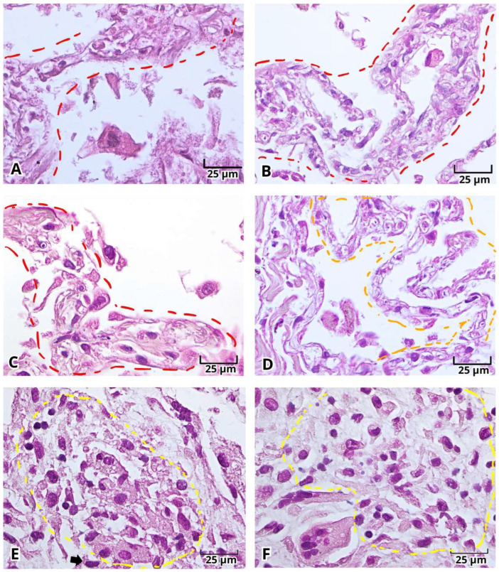 Figure 1
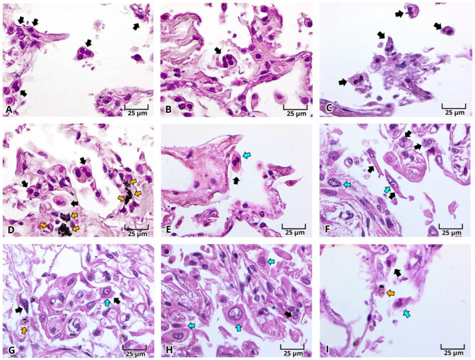 Figure 2
 Figure 3
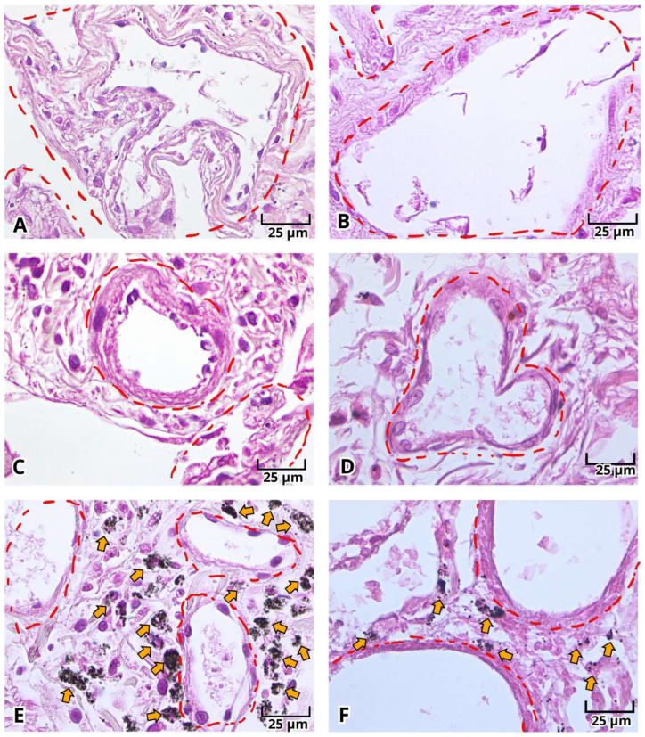 Figure 4
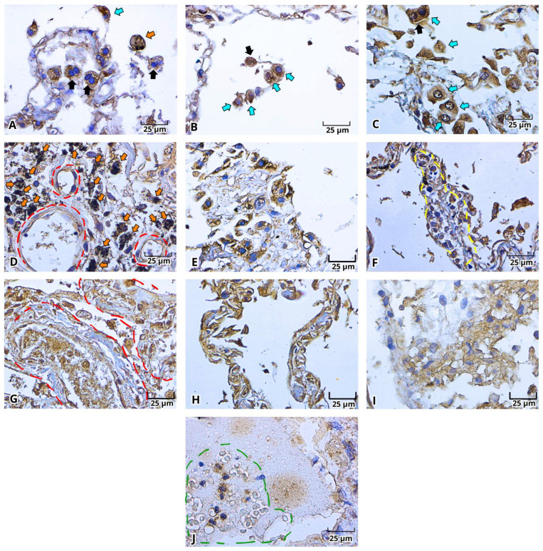 Figure 5
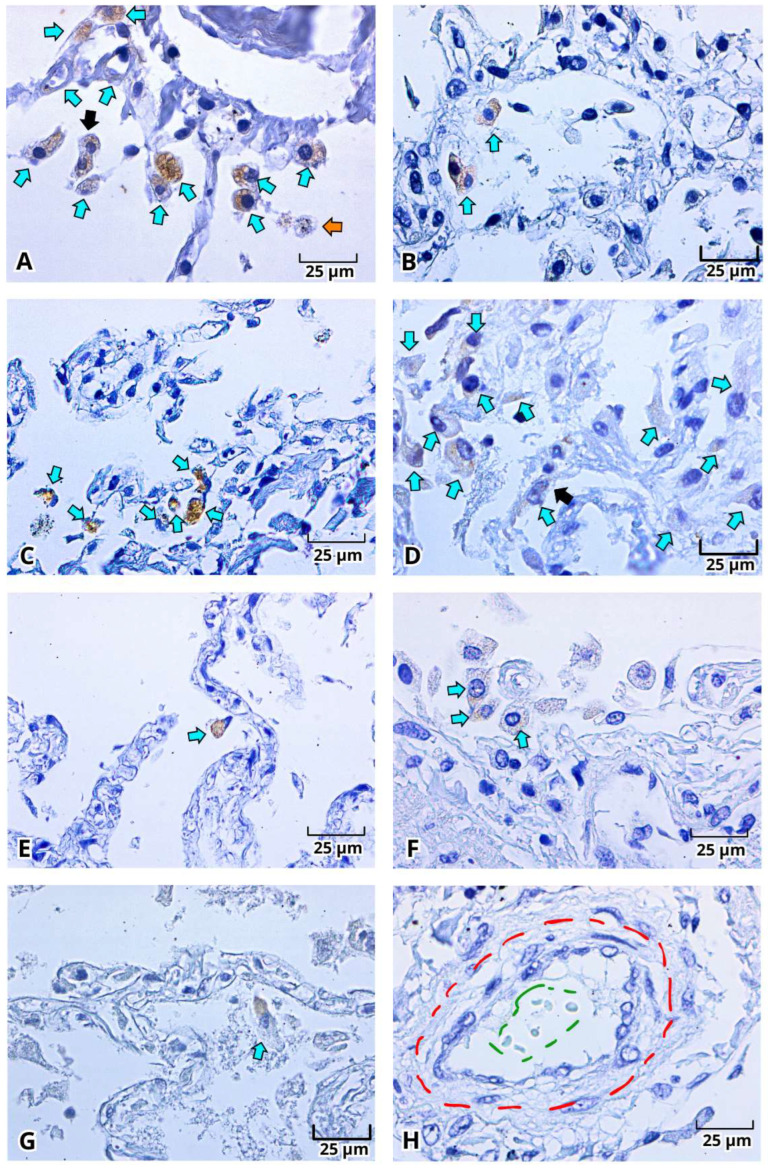 Figure 6
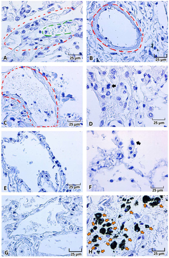 Figure 7
Peer Reviews
No public reviews on file for this paper yet. If you reviewed it on a platform where reviews are public (OpenReview, ICLR, NeurIPS, ICML), you can paste yours below so the community can read it here.
Videos
No videos yet. Explain this paper in a talk, walkthrough, or lecture? Add one.
Taxonomy
TopicsCOVID-19 Clinical Research Studies · Dermatological and COVID-19 studies · Long-Term Effects of COVID-19