Extracellular Vesicular Proteins in Plasma from Patients with Cutaneous Lupus Correlate with Disease Activity
Mariko Ogawa-Momohara, Avital Baniel, Nilesh Kodali, Fazelinia Hossein, Hua Ding, Spruce Lynn, Julianne Kleitsch, DeAnna Diaz, Thomas Vazquez, Victoria P. Werth

TL;DR
This study found that proteins in extracellular vesicles from the blood of cutaneous lupus patients correlate with disease activity, offering potential biomarkers for monitoring the condition.
Contribution
The study identifies specific extracellular vesicle proteins in cutaneous lupus that correlate with disease activity, providing new insights into biomarker development.
Findings
Four proteins (mimecan, IFI27, fibulin-2, and snRNP B/B′) were uniquely present in CLE extracellular vesicles and increased with systemic lupus activity.
Lysozyme C and hyaluronan-binding protein 2 levels in EVs correlated with cutaneous disease activity but not systemic activity.
CLE EVs showed enrichment in antigen-presenting cell markers and proinflammatory proteins, suggesting a role in local inflammation.
Abstract
Cutaneous lupus erythematosus (CLE) can occur independently of lupus erythematosus. SLE, and its responsiveness to treatment, does not necessarily align with that of coexisting SLE. Extracellular vesicles (EVs) allow communication between cells and rapid delivery throughout the body. We hypothesized that EVs may support disease-specific inflammation in CLE and SLE patients. Plasma EVs from healthy controls (n = 5), CLE (n = 6), and dermatomyositis (n = 17) were purified by ultracentrifugation and size-exclusion chromatography, phenotyped by flow cytometry, and profiled by LC-MS/MS. Circulating EVs were mainly platelet-, endothelial-, and antigen-presenting cell-derived examples. CLE EVs harbored four proteins absent in the controls—mimecan, IFI27, fibulin-2, and snRNP B/B′ (anti-Sm an-tigens)—and their cumulative number increased with SLEDAI. Relative to the controls, 18 proteins were…
Genes, proteins, chemicals, diseases, species, mutations and cell lines named across the full text — each resolved to its canonical identifier and authoritative record.
Click any figure to enlarge with its caption.
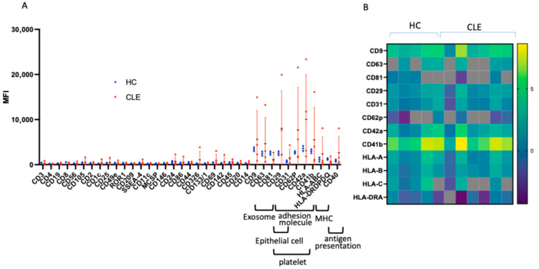 Figure 1
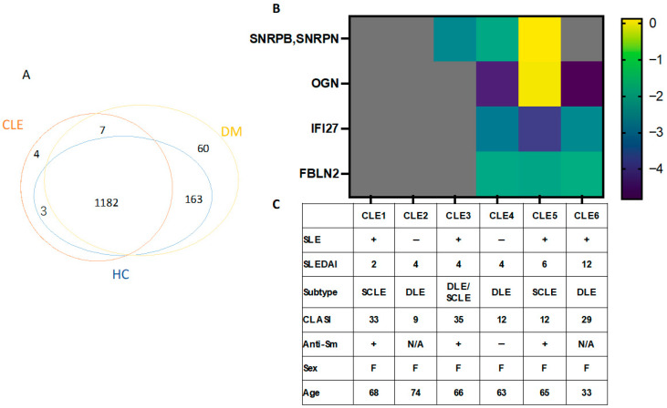 Figure 2
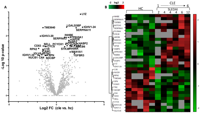 Figure 3
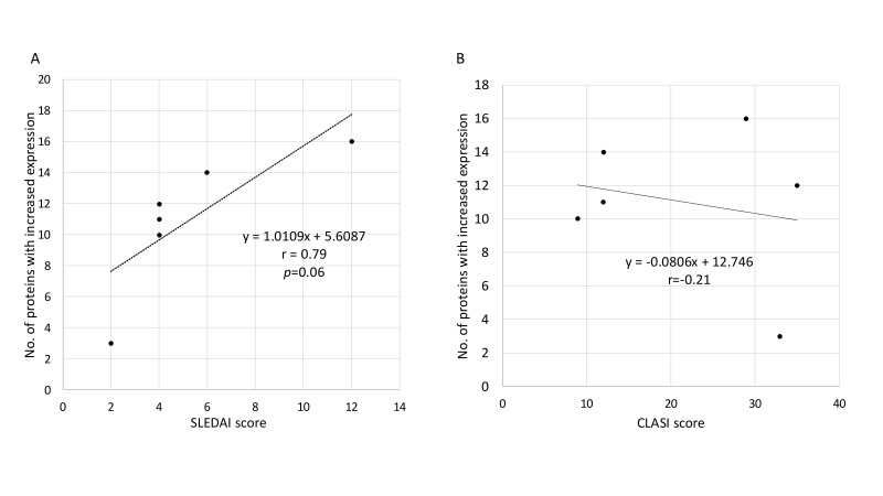 Figure 4
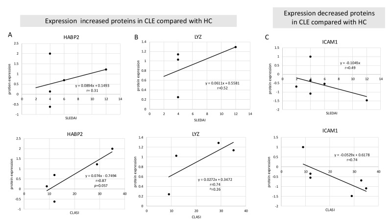 Figure 5
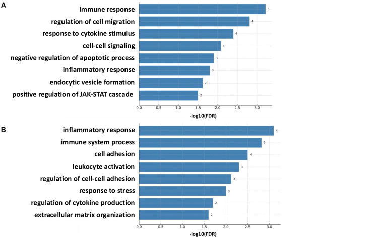 Figure 6
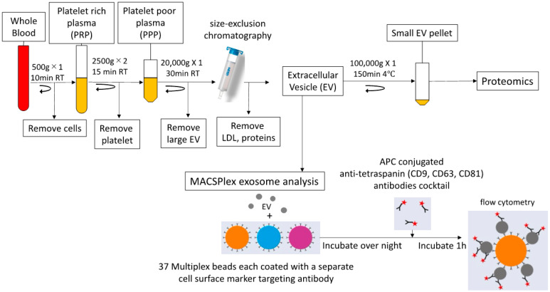 Figure 7
Peer Reviews
No public reviews on file for this paper yet. If you reviewed it on a platform where reviews are public (OpenReview, ICLR, NeurIPS, ICML), you can paste yours below so the community can read it here.
Videos
No videos yet. Explain this paper in a talk, walkthrough, or lecture? Add one.
Taxonomy
TopicsExtracellular vesicles in disease · Systemic Lupus Erythematosus Research · interferon and immune responses