Renal-AI: A Deep Learning Platform for Multi-Scale Detection of Renal Ultrastructural Features in Electron Microscopy Images
Leena Nezamuldeen, Walaa Mal, Reem A. Al Zahrani, Sahar Jambi, M. Saleet Jafri

TL;DR
This paper introduces Renal-AI, a deep learning platform that automates the detection of kidney ultrastructural features in electron microscopy images, improving diagnostic accuracy and efficiency.
Contribution
The study introduces architectural refinements to YOLOv8-OBB for detecting small, low-contrast renal ultrastructural features in TEM images.
Findings
A modified YOLOv8-OBB model achieved an F1-score of 0.93 and [email protected] of 0.953 for detecting renal ultrastructural features.
The new model (Pretrained + FPRbl) achieved an F1-score of 0.92 and [email protected] of 0.941, showing strong performance.
Expert evaluation showed complementary strengths between the two models in detecting subtle and frequent features.
Abstract
Background/Objectives: Transmission electron microscopy (TEM) is an essential tool for diagnosing renal diseases. It produces high-resolution visualization of glomerular and mesangial ultrastructural features. However, manual interpretation of TEM images is labor-intensive and prone to interobserver variability. In this study, we introduced and evaluated deep learning architectures based on YOLOv8-OBB for automated detection of six ultrastructural features in kidney biopsy TEM images: glomerular basement membrane, mesangial folds, mesangial deposits, normal podocytes, podocytopathy, and subepithelial deposits. Methods: Building on our previous work, we propose a modified YOLOv8-OBB architecture that incorporates three major refinements: a grayscale input channel, a high-resolution P2 feature pyramid with refinement blocks (FPRbl), and a four-branch oriented detection head designed to…
Genes, proteins, chemicals, diseases, species, mutations and cell lines named across the full text — each resolved to its canonical identifier and authoritative record.
Click any figure to enlarge with its caption.
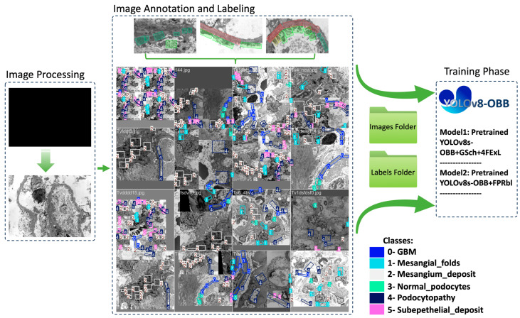 Figure 1
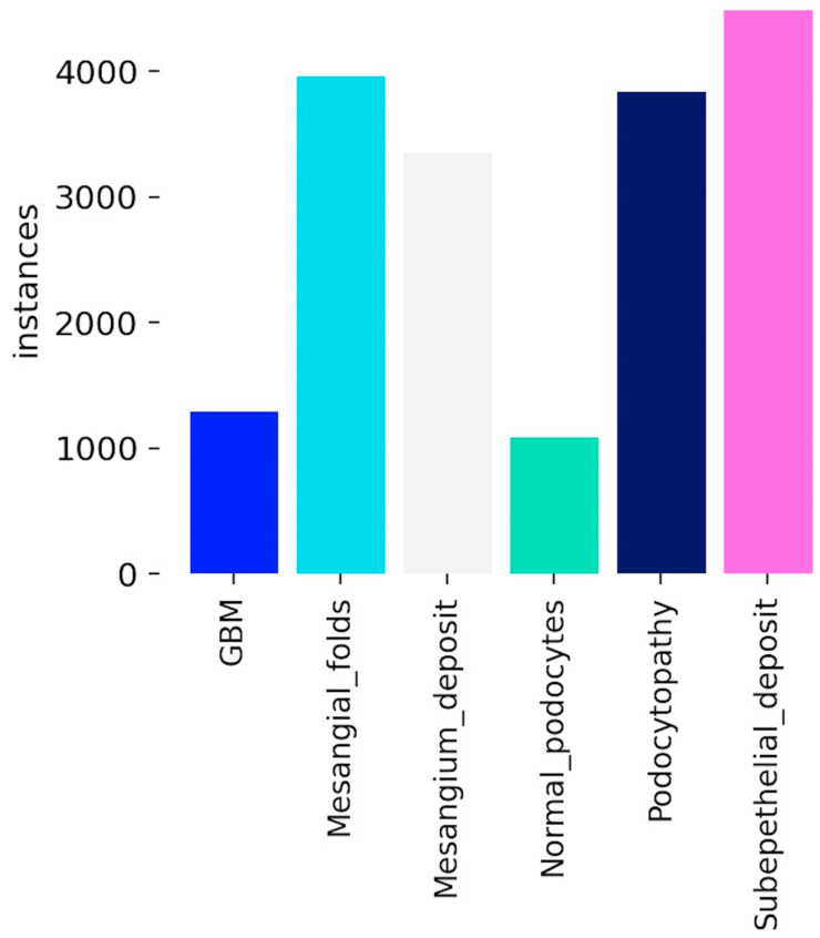 Figure 2
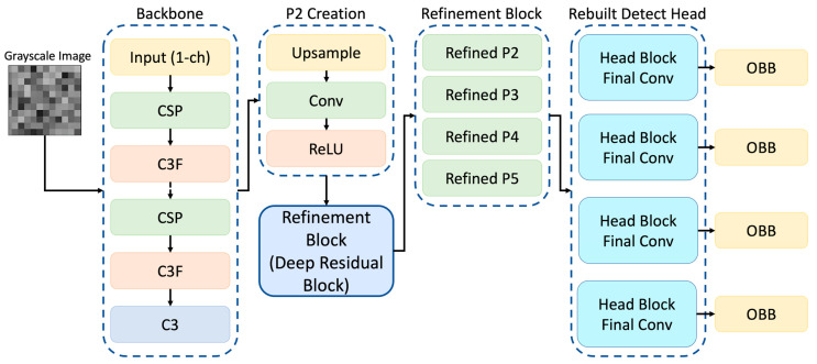 Figure 3
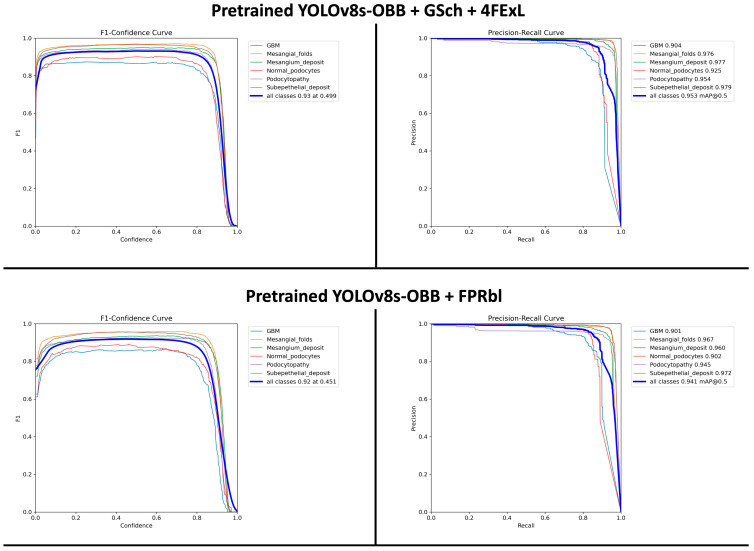 Figure 4
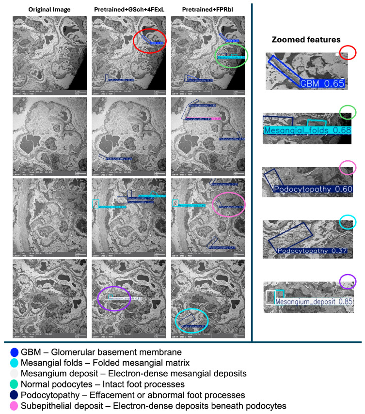 Figure 5
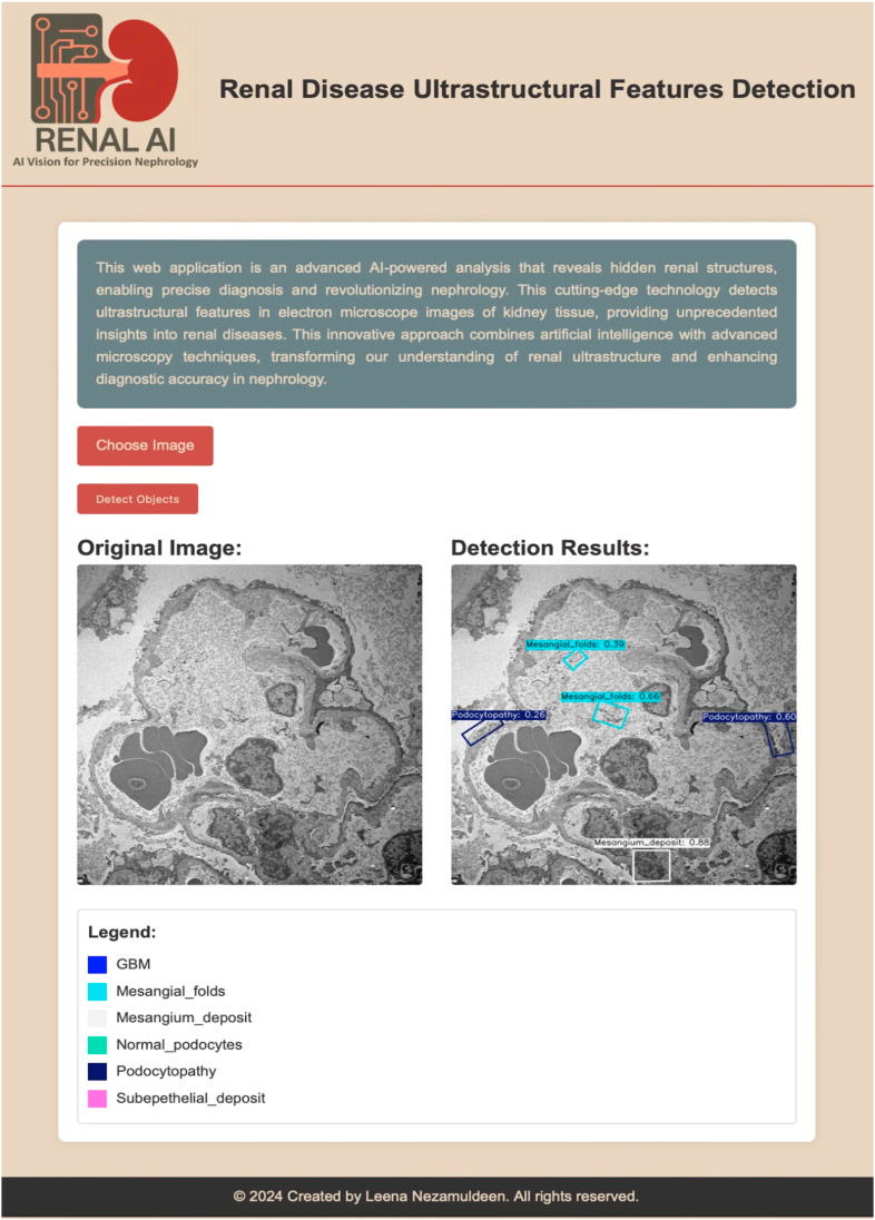 Figure 6
Peer Reviews
No public reviews on file for this paper yet. If you reviewed it on a platform where reviews are public (OpenReview, ICLR, NeurIPS, ICML), you can paste yours below so the community can read it here.
Videos
No videos yet. Explain this paper in a talk, walkthrough, or lecture? Add one.
Taxonomy
TopicsAI in cancer detection · Retinal Imaging and Analysis · Renal cell carcinoma treatment