Optimizing Layer Thickness in Multi-Planar Volume Reconstruction for Distinguishing Invasive Adenocarcinoma from Non-Invasive and Minimally Invasive Lesions in Pulmonary Nodules (≤15 mm): A Comparative Study with Conventional Lung Window Settings
Ke Zhang, Wen-Tao Zhang, Ji-Wen Huo, Wei-Wei Jing, Si-Fan Chen, Mao-Lu Tan, Fa-Jin Lv

TL;DR
This study finds that a 10 mm layer thickness in multi-planar volume reconstruction improves accuracy in distinguishing invasive lung cancer from less severe lesions in small nodules.
Contribution
The study identifies 10 mm as the optimal layer thickness for MPVR in diagnosing invasive adenocarcinoma in small pulmonary nodules.
Findings
The 10 mm MPVR model achieved the highest AUC of 0.910 for differentiating invasive from non-invasive lesions.
MPVR with 10 mm thickness outperformed conventional lung window settings in diagnostic accuracy.
Performance metrics declined when layer thickness exceeded 10 mm.
Abstract
Objective: To determine the optimal layer thickness for multi-planar volume reconstruction (MPVR) in differentiating invasive adenocarcinoma from non-invasive and minimally invasive lesions in pulmonary nodules (≤ 15 mm). Materials and Methods: This retrospective study enrolled a total of 601 solitary pulmonary nodules (≤15 mm) between June 2020 and February 2024, including 404 invasive adenocarcinomas (IAC), 80 micro-invasive adenocarcinomas (MIAs), 96 adenocarcinomas in situ (AISs), and 21 atypical adenomatous hyperplasias (AAHs). Thin-section computed tomography (TSCT) images with lung window settings and MPVR images with varying layer thicknesses (ranging from 2 to 14 mm with intervals of 2 mm) were analyzed for their morphological characteristics. Multivariate logistic regression analysis was employed to develop models for differentiating invasive adenocarcinoma from non-invasive…
Genes, proteins, chemicals, diseases, species, mutations and cell lines named across the full text — each resolved to its canonical identifier and authoritative record.
Click any figure to enlarge with its caption.
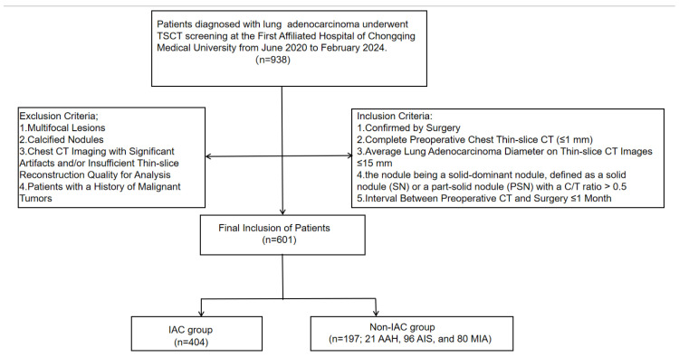 Figure 1
 Figure 2
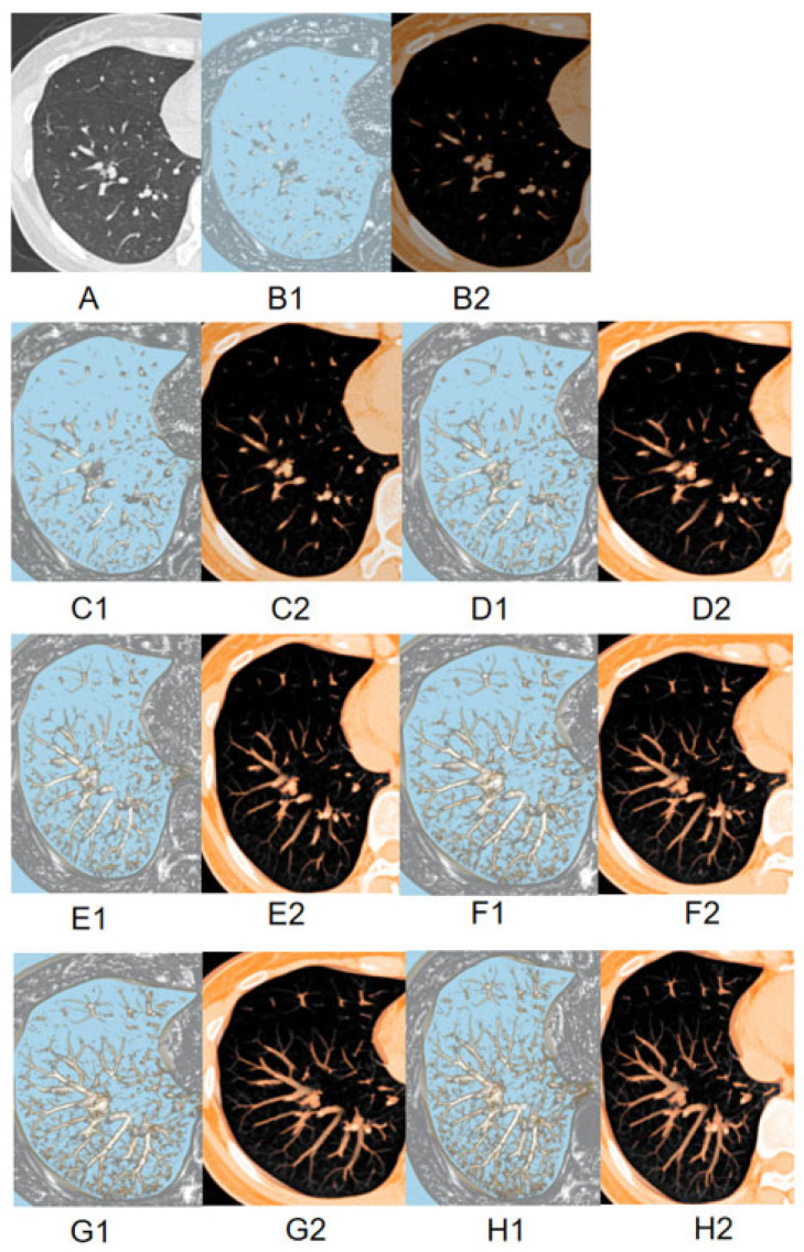 Figure 3
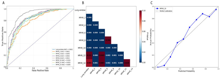 Figure 4
Peer Reviews
No public reviews on file for this paper yet. If you reviewed it on a platform where reviews are public (OpenReview, ICLR, NeurIPS, ICML), you can paste yours below so the community can read it here.
Videos
No videos yet. Explain this paper in a talk, walkthrough, or lecture? Add one.
Taxonomy
TopicsLung Cancer Diagnosis and Treatment · Radiomics and Machine Learning in Medical Imaging · Advanced X-ray and CT Imaging