Occipital Pial AVM Rupture in a Young Adult: Dual Intranidal Aneurysms, Solitary Parasagittal SSS Drainage, and Hematoma-Corridor Microsurgical Cure
Alexandru Breazu, Stefan Oprea, Nicolaie Dobrin, Ionut Bogdan Diaconescu, Octavian Munteanu, Matei Șerban, Răzvan-Adrian Covache-Busuioc, Corneliu Toader, Mugurel Petrinel Rădoi, Cosmin Pantu

TL;DR
A young adult with a ruptured brain AVM underwent successful microsurgical removal through a precise approach based on the AVM's location and structure.
Contribution
A novel microsurgical approach using hematoma-corridor access and feeder-first removal for posterior convexity AVMs is described.
Findings
A single-stage microsurgical removal of a ruptured AVM with dual intranidal aneurysms was successfully performed.
The patient recovered nearly to premorbid function after the procedure.
The approach preserved en-passage arteries and parasagittal veins while targeting weak points in the AVM.
Abstract
Background and Clinical Significance: Focal hemorrhagic severity associated with posterior convexity pial brain arteriovenous malformation (AVM) cases can be exacerbated by hemodynamic stress focusing on focal areas of architectural weakness and by superficial venous outflow being restricted by non-redundant superficial venous drainage. This clinical case report exemplifies how bedside neurologic localization and angioarchitectural characteristics can inform the selection of microsurgical approaches for the treatment of ruptured AVMs that are directed at reducing hemorrhage recurrence risk through corridors based on rupture location. Case Presentation: An otherwise healthy young adult male (modified Rankin scale [mRS] pre-morbid = 0) initially presented with a thunderclap headache, emesis, photophobia, decreased level of consciousness (admitted Glasgow Coma Score [GCS] = 11; E3V3M5),…
Genes, proteins, chemicals, diseases, species, mutations and cell lines named across the full text — each resolved to its canonical identifier and authoritative record.
Click any figure to enlarge with its caption.
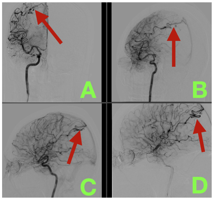 Figure 1
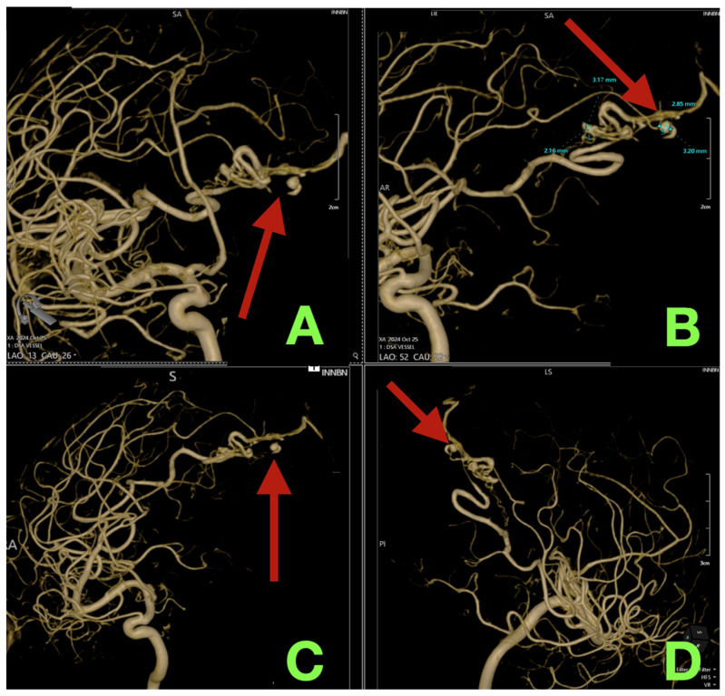 Figure 2
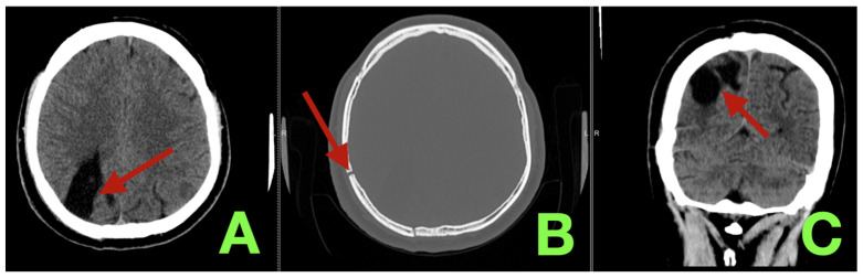 Figure 3
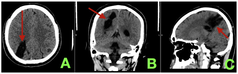 Figure 4
Peer Reviews
No public reviews on file for this paper yet. If you reviewed it on a platform where reviews are public (OpenReview, ICLR, NeurIPS, ICML), you can paste yours below so the community can read it here.
Videos
No videos yet. Explain this paper in a talk, walkthrough, or lecture? Add one.
Taxonomy
TopicsVascular Malformations Diagnosis and Treatment · Intracranial Aneurysms: Treatment and Complications · Vascular Malformations and Hemangiomas