Improved Quantification of ICG Perfusion Through Motion Compensation in Fluorescence-Guided Surgery
Sermed Ellebæk Nicolae, Thomas Baastrup Piper, Nikolaj Albeck Nerup, Michael Patrick Achiam, Morten Bo Søndergaard Svendsen

TL;DR
This paper introduces a method to reduce motion artifacts in fluorescence-guided surgery, improving the accuracy of perfusion measurements during operations.
Contribution
The study presents an automated motion compensation technique that effectively reduces motion artifacts in ICG perfusion imaging during surgery.
Findings
Automated motion compensation successfully corrected motion artifacts in 67.5% of frame sequences.
PCA analysis showed a clear separation between successful and unsuccessful motion compensation (AUC = 0.80).
Individual-level perfusion metrics showed significant changes after motion compensation, with large percentage differences observed.
Abstract
Background/Objectives: Motion artifacts significantly distort fluorescence measurements during surgical perfusion assessment, potentially leading to incorrect clinical decisions. This study evaluates the efficacy of automated motion compensation (MC) in quantitative indocyanine green (q-ICG) imaging to improve the accuracy of perfusion assessment. Methods: Frames from ICG perfusion assessment during 17 pancreaticoduodenectomies were analyzed. Regions of interest (ROIs) were systematically placed on each frame series, and automated MC was applied to track tissue movement. Performance was evaluated by comparing MC with surgeon-adjusted placement using multiple image quality metrics and analyzing perfusion metrics on time–intensity curves. Principal Component Analysis (PCA) was applied to explore whether image patterns could distinguish between successful and unsuccessful motion…
Genes, proteins, chemicals, diseases, species, mutations and cell lines named across the full text — each resolved to its canonical identifier and authoritative record.
Click any figure to enlarge with its caption.
 Figure 1
 Figure 2
 Figure 3
 Figure 4
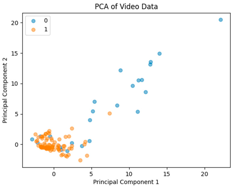 Figure 5
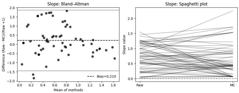 Figure 6
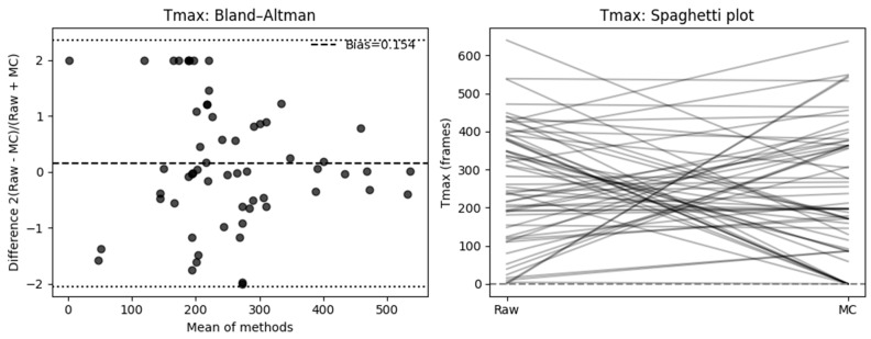 Figure 7
Peer Reviews
No public reviews on file for this paper yet. If you reviewed it on a platform where reviews are public (OpenReview, ICLR, NeurIPS, ICML), you can paste yours below so the community can read it here.
Videos
No videos yet. Explain this paper in a talk, walkthrough, or lecture? Add one.
Taxonomy
TopicsOptical Imaging and Spectroscopy Techniques · MRI in cancer diagnosis · Pancreatic and Hepatic Oncology Research