Breathprints for Breast Cancer: Evaluating a Non-Invasive Approach to BI-RADS 4 Risk Stratification in a Preliminary Study
Ashok Prabhu Masilamani, Jayden K. Hooper, Md Hafizur Rahman, Romy Philip, Palash Kaushik, Geoffrey Graham, Helene Yockell-Lelievre, Mojtaba Khomami Abadi, Sarkis H. Meterissian

TL;DR
A breath test could help avoid unnecessary breast cancer biopsies by identifying high-risk cases non-invasively.
Contribution
This study introduces breath analysis as a potential non-invasive tool for BI-RADS 4 risk stratification in breast cancer screening.
Findings
The breath test model achieved 88% sensitivity and 75% specificity in distinguishing cancerous from benign cases.
The high negative predictive value (97%) suggests breath analysis could reliably rule out cancer in BI-RADS 4 cases.
Results were consistent across BI-RADS 4 subcategories, especially effective in higher-risk groups.
Abstract
Breast cancer screening often identifies findings that are suspicious but uncertain, especially those labeled as BI-RADS 4. While doctors usually recommend a biopsy for these cases, most turn out to be benign, meaning many women go through an invasive procedure unnecessarily. This study explored whether a simple breath test could help better identify high-risk patients. By analyzing patterns of natural chemicals in exhaled breath, we trained a computer model to distinguish between cancerous and non-cancerous findings. The model was able to correctly identify most cancers while also giving strong reassurance when no cancer was present. These results suggest that a breath test could be used alongside mammography to provide patients and doctors with clearer information. If confirmed in larger studies, this approach could spare many women from unnecessary biopsies, lower healthcare costs,…
Genes, proteins, chemicals, diseases, species, mutations and cell lines named across the full text — each resolved to its canonical identifier and authoritative record.
Click any figure to enlarge with its caption.
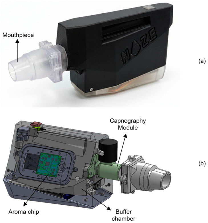 Figure 1
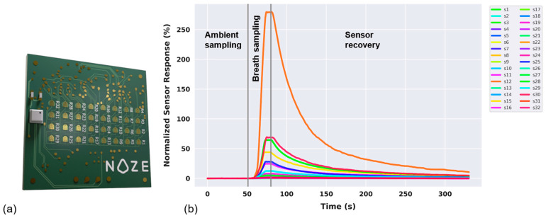 Figure 2
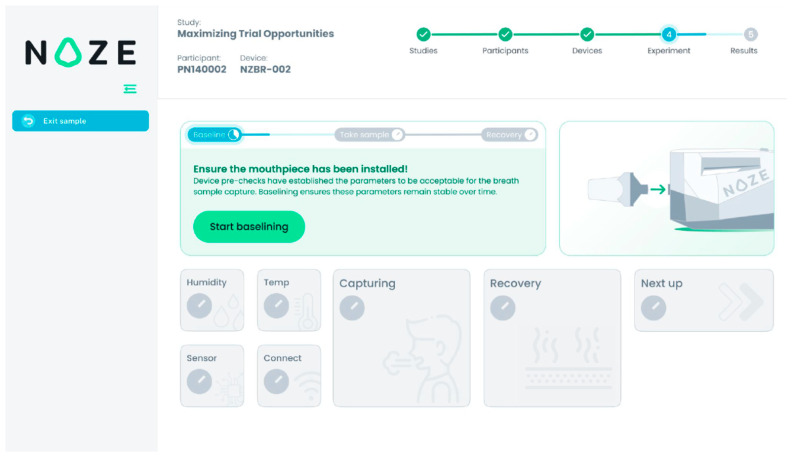 Figure 3
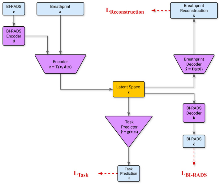 Figure 4
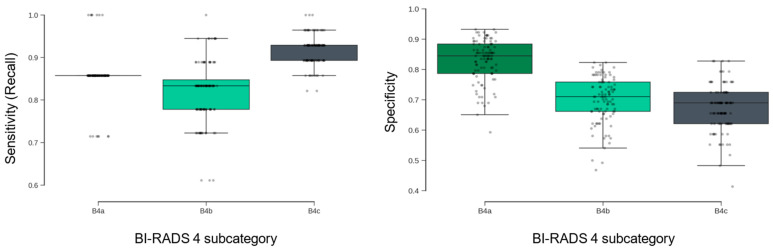 Figure 5
- —Noze
Peer Reviews
No public reviews on file for this paper yet. If you reviewed it on a platform where reviews are public (OpenReview, ICLR, NeurIPS, ICML), you can paste yours below so the community can read it here.
Videos
No videos yet. Explain this paper in a talk, walkthrough, or lecture? Add one.
Taxonomy
TopicsAdvanced Chemical Sensor Technologies · Traditional Chinese Medicine Studies · Biochemical Analysis and Sensing Techniques
1. Introduction
Breast cancer remains a major global health challenge, and early and accurate diagnosis is crucial for improving survival [1,2,3]. The current standard for detection relies on screening mammography and a standardized classification system developed by the American College of Radiology called Breast Imaging Reporting and Data System (BI-RADS) to interpret and report breast imaging findings [4,5]. The purpose of BI-RADS is to ensure consistent reporting and to provide clear management recommendations for patients, with a classification ranging from BI-RADS 1 up to BI-RADS 6. The rankings BI-RADS 1 (negative), 2 (benign) and 3 (probably benign) are generally associated with very low risk of cancer and do not require a follow-up tissue biopsy. Higher rankings, such as BI-RADS 4 (suspicious abnormality) and 5 (highly suggestive of malignancy), strongly recommend a biopsy, while with BI-RADS 6, malignancy is already confirmed via biopsy.
While the BI-RADS system is effective at identifying lesions requiring further investigation, it presents a significant diagnostic dilemma, particularly for lesions classified as BI-RADS 4. This “suspicious” category recommends an invasive tissue biopsy as the standard practice across all its subcategories. However, the BI-RADS 4 category is so broad (with a malignancy risk ranging from just above 2% to as high as 95% [6] across its subcategories 4A, 4B and 4C) that published data show that 70–80% of biopsies performed for these lesions ultimately yield a benign result [7,8,9]. This ambiguous category is also where the BI-RADS system, despite its goal of standardization, is subject to the highest variability in how different radiologists interpret the same mammogram.
Although the BI-RADS 4 subcategories are intended to reflect a gradient of malignancy risk, this category remains a significant diagnostic challenge in clinical practice. Inter-reader variability in the interpretation and classification of imaging findings can lead to inconsistencies in subcategory assignment and subsequent management decisions. As a result, patients are subjected to the anxiety, pain, and potential complications of an invasive biopsy procedure that, in retrospect, could have been avoided. Furthermore, a study demonstrated that women who had a BI-RADS 4 assessment that turned out to be benign (given a subsequent negative biopsy result) are about 10% less likely to return for a subsequent screening [10], demonstrating the potential for a false diagnosis to compromise long-term engagement in recommended cancer monitoring. These downstream effects of false-positive assessments not only places a heavy emotional and physical burden on patients [11] but also impose a substantial economic cost on the healthcare system [12]. There is a clear and urgent need for a non-invasive tool to better stratify risk and reduce the number of unnecessary biopsies.
Our research addresses this challenge by exploring the diagnostic potential of Volatile Organic Compounds (VOCs) in exhaled breath. The distinctive metabolic processes of malignant tumors are known to produce a distinct profile of VOCs, which are released into the bloodstream and subsequently expelled in the breath, creating a unique “breathprint” that can serve as a non-invasive biomarker [13,14,15,16,17,18]. This paper presents a proof-of-concept study using the DiagNoze breathalyzer (Noze Inc., Montreal, QC, Canada) designed to differentiate between malignant and benign pathologies in the challenging BI-RADS 4 cohort. We hypothesize that a predictive model, developed using machine learning on a combination of breath data and BI-RADS classification, could more accurately stratify risk than using BI-RADS assessment alone.
2. Materials and Methods
2.1. Study Design and Population
2.1.1. Study Design
This study was conducted at the McGill University Health Centre (MUHC) Breast Center, a tertiary referral clinic specializing in breast cancer diagnosis and treatment. The study received full approval from the MUHC Research Ethics Board (REB), and written informed consent was obtained from all participants prior to enrollment. The study was guided by three core design principles: scientific rigor, respect for patient experience, and clinical workflow integration. First, the study was designed to be subtype-agnostic, which included enrolling participants with any form of breast imaging abnormality requiring biopsy, to ensure that the resulting VOC breathprint would be broadly representative and clinically applicable across the disease spectrum. A dedicated breath sample collection room was set up in the Breast Center of MUHC for collecting breath samples from the subtype-agnostic participants. To limit the burden on participants, they were enrolled from the waiting room on the day of their scheduled procedures, such as diagnostic mammogram, ultrasound or biopsy. Further, the breath sample collection was restricted to a single-day procedure, during which the entire sample collection was completed within 1 h–1.5 h, allowing up to 5 samples collected per participant. The ambient humidity and temperature were observed to be <50% RH and <27 °C. Second, stringent inclusion and exclusion criteria were implemented to control for known metabolic confounders, thereby enhancing the specificity of the breath VOC signature. Third, the protocol was designed to align seamlessly with standard clinical pathways, minimizing disruption to care and imposing no additional burden on participants.
2.1.2. Study Population
To ensure the integrity of the breath-based biomarker data, stringent eligibility criteria were applied to control for both internal metabolic and external environmental confounders. Inclusion criteria required participants to be biologically female, between 18 and 80 years of age, recently referred to the clinic for a suspicious breast imaging finding, and capable of providing informed consent. Exclusion criteria were implemented to eliminate factors known to influence breath VOC composition. Individuals were excluded if they had a medical history of asthma, chronic obstructive pulmonary disease (COPD), or diabetes—conditions known to significantly alter endogenous VOC profiles [19,20,21]. Additional exclusions included current smoking [22], consumption of alcohol, tobacco (in a consumable format), vaping, marijuana, or other recreational narcotics eight (8) h prior to the breath test. An additional 1 h fasting limitation that excluded consumption of food, coffee, chewing gum, or consuming any substance other than water was implemented. These measures were essential to ensure that the collected data reflected physiological signals specific to breast pathology, free from confounding metabolic noise.
Eligible participants were recruited from among patients referred to the MUHC Breast Center following abnormal findings on screening mammography. Recruitment occurred during natural waiting periods within the standard diagnostic pathway, such as while awaiting imaging or consultation, allowing for a seamless integration into clinical workflows without disrupting care. Breath sampling was completed in a single visit and required no special preparation or follow-up. The target sample size was 176 participants, reflecting the typical diagnostic distribution observed at the clinic. Group 1 (Controls) included individuals with comprehensive diagnostic evaluation, who were confirmed via biopsy to have benign findings. Group 2 comprised individuals with biopsy-confirmed breast cancer. Importantly, the control group (patients presenting with comparable clinical suspicion but ultimately non-malignant outcomes) provided a highly relevant benchmark for evaluating model performance.
Each participant was asked to provide four to five replicate breath specimens using the DiagNoze breathalyzer (manufactured by Noze Inc., Montreal, QC, Canada), enabling generation of high-resolution VOC breathprint profiles. In parallel, clinical data were extracted from electronic medical records, including diagnostic outcomes from imaging and pathology, tumor characteristics, and relevant genetic information (e.g., BRCA1/2 mutation status).
2.2. Device Description
Breathprints were recorded from participants using the DiagNoze (manufactured by Noze Inc., Montreal, QC, Canada), an eNose-powered breathalyzer device, as shown in Figure 1a, which digitizes alveolar biomarkers including volatile organic compounds (VOCs) in an exhaled breath specimen. The breathalyzer is composed of two primary components: the mouthpiece and the main unit. The single-patient, disposable mouthpiece streamlines the exhaled breath into the device, filters humidity and is restricted to five exhalation cycles. Each mouthpiece is vacuum packed individually in a clean and sterile environment. The main unit contains three sequential modules: a capnography [23] module, a buffer chamber, and the aroma chip module, as shown in Figure 1b.
To isolate the physiologically relevant portion of the breath, the device has integrated an in-line capnography logic to detect and discard the initial phase of exhalation (commonly referred to as dead space air), which originates from the upper airways and contains minimal metabolic information. Only the alveolar fraction, drawn from the deeper lungs and enriched in endogenous VOCs reflective of systemic metabolism [24], is retained in the subsequent buffer chamber for digitization. This selective sampling process enhances the reliability and biological relevance of the resulting VOC fingerprint.
The alveolar breath specimen is then transferred from the buffer chamber to the aroma chip [25] (Figure 2a). The aroma chip comprises a cross-reactive chemiresistive sensor array of 32 thin films, each made from a different proprietary polymer-carbon black nanocomposite material [26]. In these thin films, the conductive carbon black network is embedded within chemically diverse polymer matrices, enabling reversible changes in electrical resistance upon exposure to VOCs [27,28]. Differences in polymer chemistry confer varying affinities toward broad classes of VOCs relevant to human metabolism, including aliphatic and aromatic hydrocarbons, alcohols, aldehydes and ketones. Rather than targeting individual molecules, the array is designed to generate overlapping yet distinct response patterns across the thin films, allowing discrimination of complex biological mixtures. This cross-reactive sensing strategy is well suited to capturing disease-associated metabolic signatures, including those linked to oxidative stress, lipid peroxidation, and altered cellular metabolism in cancer [14,15].
As VOCs in the breath interact with the sensor surfaces, they induce specific changes in electrical impedance, which are captured in real time. The result is a high-dimensional, time-resolved dataset (“digital breathprint”) captured at a frequency of 1 Hz across all sensing elements simultaneously (Figure 2b). This dynamic output reflects the full course of the breath-sensor interaction, including ambient-sampling, breath-sampling, and sensor-recovery phases. The multidimensional nature of this data provides a rich foundation for machine learning-based pattern recognition and classification. The DiagNoze breathalyzer and the embedded aroma chip module have been applied and tested in other studies. The performance of the DiagNoze breathalyzer in a peppermint breath study, based on benchmarking protocol for breath analysis developed by Henderson et al., has been reported [25,29] where the benchmark was done against a GC-MS system. Also, a ketone breath analysis study performance with DiagNoze breathalyzer has been reported where the results were benchmarked against a Biosense ketone breath analyzer (Readout Inc., St. Louis, MO, USA) [25].
2.3. Breath Sampling Protocol
To ensure reproducibility and data integrity, breath collection was conducted using a standardized, operator-guided protocol via the DiagNoze web-based user interface (UI) (Figure 3). Each participant was asked to provide four to five replicate breath specimens to account for natural physiological variability in the breath composition [25].
For each replicate, the UI guided the clinical coordinator through a structured, three-phase measurement cycle:
- Ambient Sampling Phase (30 s): The device initially sampled ambient air, through the mouthpiece, to establish a stable response with respect to the ambient. This step calibrates the sensor array to the room’s background VOC composition, ensuring accurate differential detection during breath sampling. In Figure 3, this phase is referred to as “Baselining”.
- Breath Sampling Phase (5–15 s): With the participant’s nose gently occluded to prevent nasal breathing, a single full exhalation was performed into the mouthpiece. The integrated capnography [23] module automatically identifies the end-tidal (alveolar) portion of the breath and triggers its capture in the buffer chamber. In Figure 3, this phase is referred to as “Capturing”.
- Sensor Recovery Phase (250 s): Following sample capture, ambient air was drawn through the system to facilitate desorption of VOCs from the sensor surfaces, allowing the array to return to the ambient state in preparation for the next measurement. In Figure 3, this phase is referred to as “Recovery”.
Between adjacent specimens, a high-flow purge fan actively evacuated residual VOCs and moisture from the internal components, preventing signal carryover and ensuring full sensor recovery to its equilibrium with the ambient air as the reference state prior to the next measurement. This automated cleaning process preserved the independence and integrity of each breathprint. The entire sampling workflow—including participant instructions, real-time monitoring, and device readiness—was orchestrated through the DiagNoze UI, streamlining operations and optimizing data quality across all replicates. All breathprint data were anonymized at the point of collection and transmitted securely to a managed-access cloud platform for analysis, in compliance with institutional data governance policies. Detailed security protocols are described in the Appendix A.
To assess the reproducibility of measurements across different analytical sessions, we conducted a statistical analysis of the aroma chip data over time. The collected breathprints were chronologically divided into four subsets. For each breathprint, the extremum of normalized responses was calculated, and the distribution of these measurements was analyzed for shifts using t-tests. The results indicated no significant shifts (p-value > 0.05) between any of the consecutive subset pairs.
2.4. Data Preprocessing and Model Building
Developing a malignancy classifier model capable of interpreting complex breathprint data requires a structured, multi-phase approach. The methodology comprised three main stages: (1) preprocessing the raw sensor data into a standardized analytical format; (2) training a machine learning model optimized for clinical relevance; and (3) evaluating model performance using a rigorous, multilayered validation framework.
Breathprints were included in the model development, blind to patient information and biopsy outcome, if they passed the following criteria: (i) ensuring stable sensor responses during ambient sampling, with changes less than 0.01%, (ii) successful breath sample collection confirmed by statistical analysis of sensor response during the breath sampling interval, (iii) validating no data loss during digitization, and (iv) verifying that the device’s mechanism worked as expected for sensor recovery by validating that the most-responsive sensing elements recovering to less than 20% of their peak response.
2.4.1. Data Preprocessing
To prepare the data for building an AI model, we applied a standardized preprocessing protocol involving time-series normalization with respect to the ambient air to account for environmental variability following [30]. The dataset is then partitioned using stratification into multiple folds in order to enable a nested cross-validation [31] to evaluate the model development process. To preserve clinical representativeness and avoid sampling bias, the folds are stratified by diagnostic category, including intermediate BI-RADS 4 subgroups, ensuring consistent outcome distribution across both sets.
2.4.2. Model Architecture and Clinically Optimized Training
To learn the key attributes (features) of the digital breathprint that jointly represent its time-series dynamics, we engineered a semi-supervised model based on the Autoencoder (AE) architecture [32]. The Autoencoder architecture facilitates the extraction of key features by compressing the information content of the data into a condensed vector representation within its latent space, Z. The modified architecture shown in Figure 4 incorporates encoders designed to jointly map the breathprint time-series (d) and the associated BI-RADS score (c) into a shared latent vector (z). The encoders/decoders are learned to ensure the latent representation encapsulates the necessary information by reconstructing both the breathprint and the BI-RADS score from (z). Concurrently, a multi-layer perceptron block optimizes the latent space Z, suited most for the malignancy classification task in a supervised fashion.
The model is optimized using a composite loss function comprising L_Task_, L_Breathprint_, and L_BI-RADS_, respectively, representing:
- L_Task_: the error for performing the malignancy classification task
- L_Breathprint_: the error for decoding the breathprint from the latent vector
- L_BI-RAD_: the error for decoding the BI-RADS score from the latent vector
To ensure that the model is aligned with clinical priorities, the training process incorporated several safety-focused optimizations. First, a class-weighted loss function addressed the natural imbalance in the dataset, where benign findings were more prevalent than malignant ones. Second, we explicitly prioritized sensitivity by assigning higher penalties to false negatives (missed cancers), thereby reducing the likelihood of underdiagnosis. Third, model selection is based on a custom clinical utility metric that emphasized generalizability and diagnostic robustness over raw accuracy. A full description of the model architecture, loss functions, and training procedures is available in the Appendix.
2.4.3. Model Cross-Validation
To ensure a fair and clinically meaningful assessment of model performance, we implemented a robust evaluation framework emphasizing reproducibility and diagnostic safety. Rather than relying on a single train–test split, we used the nested cross-validation [31] framework to evaluate the model across multiple partitions of the dataset, reducing the risk of overfitting or optimistic bias. Model predictions were generated using an ensemble approach, where multiple independently trained models contributed to the final output via majority vote. In alignment with our safety-first principle, any vote ties were resolved conservatively in favor of a positive (malignant) classification.
Final performance metrics are reported as the mean and standard deviation across 100 independent runs, providing a stable and reliable estimate of the performance of the generated model following the model development process. During each run, the folds were generated using the original dataset independently. See the Appendix A for more details on the cross-validation structure and the employed ensemble procedure.
3. Results
3.1. Study Population and Data Distribution
The study enrolled 176 participants. Four to five breath specimen digitization attempts were performed per patient, of which three breathprints were successfully recorded on average across the patients. Fifty-one participants were excluded due to an inconclusive (n = 15) or unreported (n = 13) BI-RADS score, or having a BI-RADS score not among 3, 4A, 4B, 4C, or 5 (n = 6). For 17 participants, sampling the digitized breath aroma failed across all attempts.
The analysis included 125 participants who provided a total of 437 successful breathprints. Of these, 72 participants had confirmed benign findings, contributing 270 successfully recorded breathprints. The remaining 53 participants had biopsy-confirmed breast cancer, from whom 167 breathprints were successfully recorded.
The BI-RADS 4 group (A, B, and C) included 85 participants. Among these, 17 had biopsy-confirmed breast cancer, and 68 had biopsy-confirmed benign findings. A total of 53 and 256 successful breathprints were recorded from these participants, respectively.
Table 1 summarizes the distribution of these 309 successful breathprints.
3.2. Predictive Performance in the BI-RADS 4 Cohort
The malignancy rate for all BI-RADS 4 cases combined is 17%, meaning most of these cases are benign. To create a model that can meaningfully differentiate between malignant and benign tumors, we included breathprints from patients with BI-RADS scores of 3 and 5 as well. This broader dataset of 437 breathprints was used to train the model, enabling it to learn the data dynamics required for accurate classification. However, the core of this analysis was concentrated on participants whose mammography results were classified as BI-RADS 4 (A, B, and C). These categories represent patients with findings that are considered suspicious. Across the full BI-RADS 4 group, the model achieved a mean sensitivity of 88% ± 3% (Table 2), indicating a high capacity to correctly identify malignant cases. The metric is consistent across BI-RADS 4 subcategories, with particularly high sensitivity maintained in the 4C group. The results are stable across the 100 randomized cross-validation runs. As shown in Figure 5, sensitivity distributions were tightly clustered across subgroups, suggesting minimal variance in model behavior across different partitions of the dataset.
3.2.1. Specificity and Sensitivity Trade-Offs Across Subcategories
Specificity is the ability of a test to correctly identify those without the disease. The model’s specificity demonstrated a decreasing trend from BI-RADS 4A to 4C subgroups (Figure 5, right), with the highest specificity observed in the 4A group. This pattern reflects the model’s sensitivity-focused training protocol, which prioritized the reduction of false negatives in higher-risk categories. While specificity was lower in BI-RADS 4B and 4C, this trade-off was made to preserve high sensitivity in patients with a higher pre-test probability of malignancy.
3.2.2. Summary Metrics and Negative Predictive Value
Table 2 presents comprehensive model performance across all BI-RADS 4 subgroups. The model achieved an overall NPV of 97% ± 1%, indicating a high level of confidence in negative test results. Specificity was maintained at 75% ± 7%, indicating that the model correctly identified approximately three out of four benign cases. This balance between high sensitivity and moderate specificity supports the model’s potential utility as a rule-out tool in diagnostic workflows.
3.2.3. Additional Observations
A strong correlation was observed between mammography lesion size and biopsy outcome, where the median maximum size for malignant cases (21.0 mm) is more than double that of non-malignant cases (10.0 mm), though overlapping minimum sizes suggest size is not the sole diagnostic factor. The patient cohort is skewed towards early-stage disease, with Stage I (45.8%), Stage II (33.3%), and Stage 0, Ductal Carcinoma In Situ (DCIS) (14.6%), while advanced stages are minimally represented. Furthermore, the molecular profile of positive cases is predominantly Luminal A (74.2%), a key finding that guides clinical standard of care, including the application of endocrine or targeted anti-HER2 therapy.
4. Discussion
This proof-of-concept study demonstrates that exhaled breath contains a detectable and clinically informative signal capable of distinguishing between malignant and benign findings in women with BI-RADS 4 mammographic assessments. Using a digital olfaction platform and a machine learning model trained with clinically informed constraints, we achieved a mean sensitivity of 88% and an overall NPV of 97%. These results suggest that breath-based diagnostics may offer a promising non-invasive approach for stratifying risk in patients with indeterminate imaging findings.
The clinical implications are particularly relevant for managing BI-RADS 4A and 4B lesions, which account for a high volume of benign biopsies. A high NPV in this setting may enable more conservative management strategies, such as short-interval imaging follow-up, potentially reducing unnecessary procedures and associated patient anxiety. Additionally, according to a previously published study [33], performance remains strong in participants with dense breast tissue, a population in which mammographic sensitivity is known to be reduced. This suggests that a breath-based approach could serve as a complementary modality in cases where traditional imaging has limitations.
The model’s diagnostic behavior was shaped by a deliberate training strategy that prioritized sensitivity. Through the use of class weighting and penalty adjustments, we explicitly reduced the likelihood of false-negative results. This design choice aligns with clinical priorities, particularly in early cancer detection where the cost of a missed diagnosis is high. While this approach resulted in a moderate reduction in specificity, the observed trade-off is clinically appropriate in a rule-out context.
Importantly, the model’s performance was validated using a rigorous cross-validation framework, including 100 independent runs and ensemble-based predictions, ensuring robustness across multiple data partitions. However, the study is limited by its single-center design and the use of a single device platform. Although the results are internally consistent and statistically stable, external validation in multi-center cohorts will be essential to confirm generalizability. These findings support further investigation of breath-based diagnostics in breast cancer. Future studies should aim to evaluate the reproducibility of these results across different clinical settings, devices, and populations, and explore integration with existing diagnostic pathways.
The low positive predictive value (PPV) for categories 4A (28%) and 4B (29%) is largely attributed to the low prevalence of malignancy within these subgroups. For instance, the malignancy rate for BI-RADS 4A is only 6%, indicating that the majority of these cases are benign. This low prevalence inflates the false positive rate, thereby decreasing the PPV, as defined by the formula PPV = TP/(TP + FP). Conversely, the PPV for category 4C (73%) is considerably higher due to a significantly greater malignancy prevalence in that group, which increases the likelihood that a positive prediction is correct.
These results also open the door to a complementary diagnostic paradigm, in which breath analysis is used not as a standalone tool but as a tandem modality alongside mammography and ultrasound. In this scenario, the breath-based test could be administered immediately after a suspicious BI-RADS 4 mammogram, offering an additional layer of risk stratification prior to biopsy. For example, a negative breath test result in a BI-RADS 4A or 4B case, especially given the model’s high negative predictive value, could support a more conservative management approach such as short-term imaging follow-up rather than immediate biopsy. This combinatorial use of imaging and breath diagnostics may help reduce inter-reader variability, alleviate patient anxiety, and optimize clinical decision-making by tailoring biopsy recommendations to a more individualized risk profile.
5. Conclusions
This study establishes proof-of-concept that a non-invasive breath test, analyzed using a digital olfaction platform and a clinically optimized machine learning model, can differentiate benign from malignant breast lesions in women with BI-RADS 4 findings. The model achieved high sensitivity (88%) and a negative predictive value of 97%, supporting its potential use as a rule-out tool in the diagnostic workup of suspicious mammograms. Our findings demonstrate that this model achieves high sensitivity and an exceptionally high NPV. These results establish a strong foundation for a tool that could confidently rule out malignancy, potentially sparing a majority of women from an unnecessary biopsy.
By reducing reliance on invasive biopsy in low-risk cases, this technology could alleviate patient burden and streamline clinical decision-making and provide cost savings to an already overburdened healthcare system. The results warrant further validation in larger, multi-center studies to confirm generalizability and evaluate integration into real-world diagnostic workflows.
Despite the good outcomes observed, this study has limitations. The study was designed as a single-center, proof-of-concept with a relatively small sample size, and the performance was evaluated using nested cross-validation within the existing dataset. In the study, we did not evaluate generalizability across different clinical settings or different demographics. Although exclusion criteria were applied to reduce known metabolic confounders, co-morbidities and concomitant conditions may influence breath volatile organic compound profiles and should be more comprehensively evaluated in future studies. Future studies should focus on prospective, multi-center validation in larger and more diverse populations, assessment of inter-site reproducibility, and refinement of system calibration to optimize sensitivity-specificity trade-offs for different clinical use cases. Integration with additional clinical and imaging features may further enhance performance. Successful clinical translation will depend on robust validation, seamless workflow integration, and clear definition of how breath-based testing complements existing diagnostic pathways.
The reference list from the paper itself. Each links out to its DOI / PubMed record.
- 1Caswell-Jin J.L. Sun L.P. Munoz D. Lu Y. Li Y. Huang H. Hampton J.M. Song J. Jayasekera J. Schechter C. Analysis of breast cancer mortality in the US-1975 to 2019 JAMA 202433123324110.1001/jama.2023.2588138227031 PMC 10792466 · doi ↗ · pubmed ↗
- 2Ellison L.F. Saint-Jacques N. Five-year cancer survival by stage at diagnosis in Canada Health Rep.20233431510.25318/82-003-x 202300100001-eng 36716075 · doi ↗ · pubmed ↗
- 3Sung H. Ferlay J. Siegel R.L. Laversanne M. Soerjomataram I. Jemal A. Bray F. Global cancer statistics 2020: GLOBOCAN estimates of incidence and mortality worldwide for 36 cancers in 185 countries CA Cancer J. Clin.20217120924910.3322/caac.2166033538338 · doi ↗ · pubmed ↗
- 4Liberman L. Menell J.H. Breast imaging reporting and data system (BI-RADS)Radiol. Clin. N. Am.20024040943010.1016/S 0033-8389(01)00017-312117184 · doi ↗ · pubmed ↗
- 5Spak D. Plaxco J. Santiago L. Dryden M. Dogan B. BI-RADS ® fifth edition: A summary of changes Diagn. Interv. Imaging 20179817919010.1016/j.diii.2017.01.00128131457 · doi ↗ · pubmed ↗
- 6Elezaby M. Li G. Bhargavan-Chatfield M. Burnside E.S. De Martini W.B. ACR BI-RADS assessment category 4 subdivisions in diagnostic mammography: Utilization and outcomes in the National Mammography Database Radiology 201828741642210.1148/radiol.201717077029315061 PMC 6413875 · doi ↗ · pubmed ↗
- 7Liu C. Sun M. Arefan D. Zuley M. Sumkin J. Wu S. Deep learning of mammogram images to reduce unnecessary breast biopsies: A preliminary study Breast Cancer Res.2024268210.1186/s 13058-024-01830-938790005 PMC 11127450 · doi ↗ · pubmed ↗
- 8Meng M. Li H. Zhang M. He G. Wang L. Shen D. Reducing the number of unnecessary biopsies for mammographic BI-RADS 4 lesions through a deep transfer learning method BMC Med. Imaging 2023238210.1186/s 12880-023-01023-437312026 PMC 10265786 · doi ↗ · pubmed ↗