Structural Insights into the Staphylococcus aureus DltC-Mediated D-Alanine Transfer
Hanul Jeon, Hyebin Lee, Chiman Song, In-Gyun Lee

TL;DR
This study reveals the structure of a key protein in Staphylococcus aureus that helps it resist antibiotics, offering new ways to combat infections.
Contribution
The paper presents the first crystal structure of SaDltC and identifies a critical protein interface for D-alanylation in S. aureus.
Findings
The crystal structure of SaDltC was determined, providing structural insights into D-alanine transfer.
Mutations at the SaDltA-SaDltC interface disrupt catalytic activity, highlighting its importance.
Structural data suggest targeting the DltA-DltC interface as a potential anti-Staphylococcus strategy.
Abstract
Staphylococcus aureus (S. aureus) is a major Gram-positive pathogen, and treatment of S. aureus infections is often challenging due to widespread antibiotic resistance. In Gram-positive bacteria such as S. aureus, D-alanylation of teichoic acids (TA) reduces the net negative charge of the cell envelope and contributes to resistance to diverse antibiotics, particularly cationic antimicrobial peptides. D-alanylation is mediated by the dltABCD operon, which encodes four proteins (DltA, DltB, DltC, and DltD), all of which is essential for the multistep transfer of D-alanine to teichoic acids. Here, we present the first crystal structure of the S. aureus D-alanyl carrier protein DltC and analyze its interaction with DltA using AlphaFold3 and all-atom molecular dynamics simulations. We further show that single substitutions of SaDltA-SaDltC interface residues abolish SaDltC mediated…
Genes, proteins, chemicals, diseases, species, mutations and cell lines named across the full text — each resolved to its canonical identifier and authoritative record.
Click any figure to enlarge with its caption.
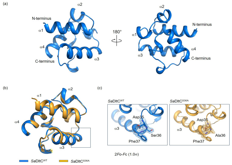 Figure 1
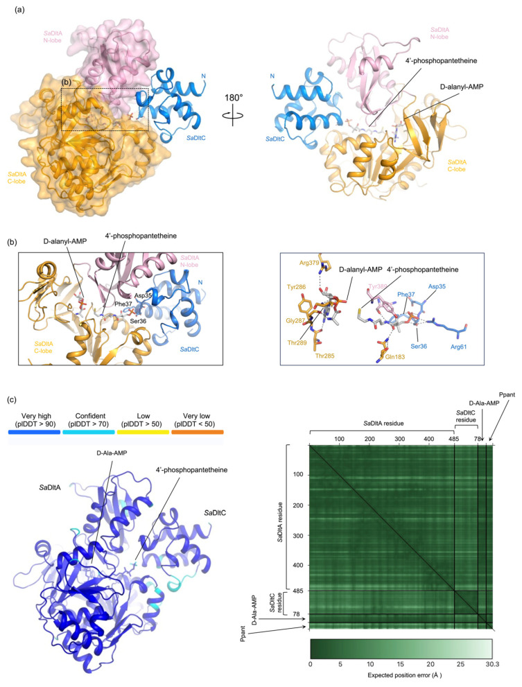 Figure 2
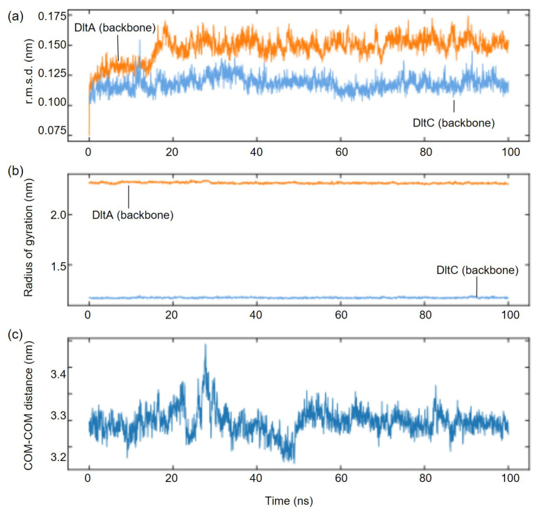 Figure 3
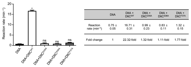 Figure 4
- —Korean government (MSIT)
- —National Research Foundation of Korea (NRF)
- —Korean government (MSIT)
- —Korea Basic Science Institute (National research Facilities and Equipment Center)
- —Korean government (MSIT)
- —Korea Health Technology R&D Project
- —Korea Health Industry Development Institute (KHIDI)
- —Ministry of Health & Welfare, Republic of Korea
- —National Research Council of Science and Technology (NST)
- —Korean government (MSIT)
Peer Reviews
No public reviews on file for this paper yet. If you reviewed it on a platform where reviews are public (OpenReview, ICLR, NeurIPS, ICML), you can paste yours below so the community can read it here.
Videos
No videos yet. Explain this paper in a talk, walkthrough, or lecture? Add one.
Taxonomy
TopicsAmino Acid Enzymes and Metabolism · Enzyme Structure and Function · Cancer Research and Treatments
1. Introduction
Staphylococcus aureus (S. aureus) is a Gram-positive human pathogen that causes a wide spectrum of diseases, from skin and soft tissue infections to life-threatening nosocomial infections [1,2,3]. The global spread of antibiotic-resistant strains—most notably methicillin-resistant S. aureus (MRSA) and vancomycin-resistant S. aureus (VRSA)—has made the development of new therapeutic strategies urgent [4,5]. One of the key cell envelope mechanisms that contributes to virulence and resistance in Gram-positive bacteria, including S. aureus, is D-alanylation of teichoic acids (Tas) [4,5]. This modification attenuates the negative charge of the cell wall, reducing binding of cationic antimicrobial peptides and decreasing susceptibility to several antibiotic classes [6].
Tas are abundant anionic polymers in the Gram-positive cell wall. In S. aureus, wall teichoic acid (WTA) is covalently linked to peptidoglycan, whereas lipoteichoic acid (LTA) is anchored at the cytoplasmic membrane interface [6]. D-alanine is incorporated into these Tas by the sequential action of four proteins, DltA, B, C, and D, which are encoded by the dltABCD operon [6,7]. Because D-alanylation neutralizes the cell surface charge and modulates antibiotic sensitivity, the Dlt pathway has been considered as an attractive drug target [8,9,10,11,12]. Here, we focus on S. aureus DltC, the D-alanyl carrier protein, and its interaction with SaDltA, the D-alanine-D-analyl carrier protein ligase. Among the four Dlt proteins involved in the D-alanylation process, DltA and DltC operate in the cytosol and initiate the process; DltA activates D-alanine using ATP and subsequently transfers the D-alanine adenylate to the conserved serine residue of DltC (Ser36 in S. aureus DltC), which is post-translationally modified with 4′-phosphopantetheine (Ppant) cofactor, forming DltC-Ppant-D-alanine. DltC is crucial for the transfer of D-alanine to the cell surface with the aid of DltB and DltD, through a not yet fully understood mechanism [3,13,14,15]. Thus, understanding the interaction between DltA and DltC is critical to comprehending the D-alanylation pathway.
Here, we present the crystal structure of SaDltC and analyze its interface with SaDltA with AlphaFold3 modeling and all-atom molecular dynamics simulations. We further show that single-point substitutions of Ser36 in SaDltC—the Ppant attachment site that accepts D-alanine from DltA—as well as of Asp35 and Phe37, residues that lie at the predicted SaDltA-SaDltC interface, abolish the SaDltC-mediated catalytic enhancement of SaDltA activity. Together, these results define a catalytically essential S. aureus DltA-DltC interface and provide structural insight for targeting TA D-alanylation as a potential anti-Staphylococcus strategy.
2. Materials and Methods
2.1. Gene Cloning and Protein Expression, and Purification
The cDNAs encodes SaDltC (UniProt ID: P0A018) was amplified and expressed as His_6_-tagged protein using the pET28a expression vector (Takara Bio, Kusatsu, Japan). The cDNA that encodes Escherichia coli (E. coli) acyl carrier protein synthase (AcpS, UniProt P24224) was amplified and cloned into the pBAD expression vector (New England Biolabs [NEB], Ipswich, MA, USA), resulting in non-tagged AcpS (for co-expression with wild-type SaDltC). The point mutations were introduced using a QuikChange site-directed mutagenesis kit (Agilent Technologies, Santa Clara, CA, USA). SaDltC^WT^ was co-expressed with AcpS in E. coli Rosetta (DE3) cells (Thermo Fisher Scientific, Waltham, MA, USA) to produce Ppant-attached SaDltC. The E. coli cells were grown in Luria–Bertani (LB) medium at 37 °C until the OD_600_ reached 0.5, and the protein expression was induced with 0.25 mM IPTG. The bacterial cell culture was further grown at 37 °C for 4 h. The cells were centrifuged at 8000× g and resuspended in purification buffer A (50 mM Tris–HCl pH 8.0, 500 mM NaCl). After cell lysis by ultrasonication, cell lysate was centrifuged at 20,000× g and bound to an Ni-NTA affinity column. After washing with Buffer A, the bound fractions were eluted with Buffer B (Buffer A supplemented with 300 mM imidazole). The His_6_-tag was removed by the addition of TEV protease overnight. The non-tagged protein was further purified via size-exclusion chromatography using a HiLoad 16/60 Superdex 75 pg column (GE Healthcare, Chicago, IL, USA) equilibrated with 20 mM Tris pH 7.5, 200 mM NaCl. The purified protein was concentrated to 20 mg ml^−1^. SaDltC mutant proteins were also expressed in E. coli Rosetta (DE3) and purified essentially identically to wild-type SaDltC. The purity of the proteins was analyzed by SDS-PAGE.
2.2. Crystallization and Determination of Crystal Structure
Recombinant SaDltC^WT^ protein at a concentration of 20 mg mL^−1^ in 20 mM Tris, pH 7.5, and 200 mM NaCl was used for initial screening of crystallization conditions. A total of 1 µL of protein solution was mixed with an equal volume of reservoir solution consisting of 30% PEG 400, 0.2 M sodium thiocyanate, 0.2 M magnesium chloride, and 0.1 M HEPES, pH 7.5, at 16 °C. X-ray diffraction data was collected at 100 K using an ADSC Quantum 315r CCD detector system at the BL-7A beamline of Pohang Light Source (Republic of Korea). The SaDltC^WT^ crystal belonged to the monoclinic space group P2_1_, with unit-cell parameters a = 33.14 Å, b = 63.97 Å, c = 77.14 Å, α = γ = 90.00°, β = 93.95°. The raw data were processed and scaled using HKL-2000 [16]. SaDltC^S36A^ protein crystal was obtained from 14% PEG 8 K, 18% PEG 400, 0.1 M magnesium chloride, and 0.1 M Tris-HCl pH 7.5 at 16 °C. A set of X-ray diffraction data was collected at 100 K, also at the BL-7A beamline of Pohang Light Source. The SaDltC^S36A^ crystal belonged to the orthorhombic space group P2_1_2_1_2, with unit cell parameters a = 32.73 Å, b = 157.96 Å, c = 27.8 Å, and α = β = γ = 90.00°. The raw data were processed and scaled as described above (Table 1).
2.3. Molecular Dynamics
MD simulations of the SaDltA-SaDltC complex were performed with GROMACS 2024.3 [17]. The Alphafold3-predicted SaDltA-SaDltC complex was used as an initial structure [18]. The AMBER99SB-ILDN force field and TIP3P water were employed for protein parameters [19]. Nonbonded interactions were calculated using the Particle Mesh Ewald (PME) method with a 1.0 nm cutoff [20]. Newton’s equations of motion were integrated using the leap-frog algorithm with a 2 fs time step, and all hydrogen bonds were constrained using Linear Constraints Solver (LINCS) algorithm [21]. After energy minimization, the system was equilibrated for 1 ns in the NVT ensemble at 300 K using the V-rescale thermostat with backbone position restraints [22], followed by 2 ns NPT equilibration at 300 K and 1 bar using the Parrinello–Rahman barostat [23]. Production MD simulations of 100 ns was then carried out in the NPT ensemble without restraints. Structural analysis including r.m.s.d., Rg, and inter-chain distances were calculated using GROMACS tools. The inter-chain distance was evaluated as the Euclidean distance between the mass-weighted centers of mass of SaDltA and SaDltC using gmx distance after PBD correction.
2.4. Pyrophosphatedetection Assay
The adenylation activity of recombinant SaDltA was assessed using a pyrophosphate detection assay. Reactions were carried out in the presence of 5 μM SaDltA, 5 units ml^−1^ inorganic pyrophosphatase, 5 mM ATP, 100 mM KCl, 10 mM MgCl_2_, 50 mM Tris–HCl (pH 7.4), and 5 mM D-alanine at 37 °C. The reaction was initiated by the adding D-alanine, and 20 μL aliquots were collected at 0, 1, 2, 4, 6, and 10 min. Each aliquot was immediately mixed with 380 μL dye solution containing 0.033% (w/v) malachite green and 1.3% (w/v) ammonium molybdate in 1.0 M HCl and incubated for 90 s. Absorbance at 620 nm was measured using a BioSpectrometer (Eppendorf, Hamburg, Germany), and reactions lacking 5 μM SaDltA served as blanks. Determination of the adenylation activity of SaDltA in the presence of 300 μM WT or D35A, S36A, F37A mutant SaDltC was determined under essentially identical conditions. Initial rates of the enzyme reaction were derived from phosphate accumulation over time.
3. Results
3.1. Crystal Structures of Wild-Type and Ser36Ala Mutant S. aureus DltC
For SaDltC to accept the D-alanine adenylate, Ser36 of SaDltC must be post-translationally modified with a Ppant group by the phosphopantetheinyl transferase AcpS [3]. DltA then transfers the D-alanyl moiety from D-Ala-AMP to the thiol of this Ppant prosthetic group covalently attached to Ser36 of SaDltC, forming a thioester [3]. To understand the structural basis of SaDltC recognition by SaDltA, we determined the crystal structures of the wild-type (SaDltC^WT^) and non-modifiable Ser36Ala mutant (SaDltC^S36A^). Both structures were solved by the molecular replacement using B. subtilis DltC [24] as the search model (Table 1). The SaDltC^WT^ contained four DltC molecules in the asymmetric unit (chains A–D), whereas contains SaDltC^S36A^ contains two molecules (chains A and B). Chains within each asymmetric group are essentially identical, with Cα root mean square deviation (r.m.s.d.) values of ~0.14–0.28 Å for SaDltC^WT^ and ~0.24 Å for SaDltC^S36A^, so unless otherwise noted, we describe chain A for both structures.
The overall structure of SaDltC^WT^ adopts a compact, globular fold consisting of four α-helices: α1 (residues 2–15), α2 (residues 19–22), α3 (residues 38–50), and α4 (residues 67–75) (Figure 1a). Several DltC structures have been elucidated, including the Ppant-loaded form (PDB ID: 4BPH), the apo (non-Ppant modified) form (PDB ID: 4BPG), and the DltB-complexed structures (PDB ID: 8JF2 and 6BUG) [13,24,25]. In all contexts, DltC adopts a highly similar globular fold, with small overall Cα r.m.s.ds between structures, indicating that its compact, globular architecture is strongly conserved during acceptance and handover of D-alanine (Supplementary Table S1). Also, the SaDltC^Ser36Ala^ structure was highly similar to SaDltC^WT^ with Cα r.m.s.d. of 0.19 Å, indicating that the mutation does not perturb the global fold of the protein (Figure 1b,c).
3.2. AF3 Prediction- and MD Simulation-Based Architecture of the SaDltA-SaDltC Interface
Although recent studies have elucidated complex structures of DltC with other Dlt-family proteins, such as DltB and DltD [13,25]—clarifying the architecture of Dlt-mediated D-alanylation and the transfer of D-alanine from the cytosol to TAs—the high-resolution DltA-DltC complex has not been characterized, presumably due to the transient and weak nature of its interaction. SaDltA comprises two structurally distinct lobes, an N-terminal lobe (residues 1–377) and a C-terminal lobe (residues 382–485), linked by a flexible interdomain hinge region (residues 378–381) [26]. Following ATP-dependent adenylation of D-alanine, the C-lobe rotates from the adenylation state to the thiolation state, positioning the D-alanyl-AMP for transfer to DltC [14,15]. We therefore hypothesized that DltC binds DltA in the thiolation state, with the Ppant on Ser36 positioned near the D-alanyl group of D-Ala-AMP.
To test this, we used AlphaFold3 (AF3) to predict the complex structure, using SaDltA, SaDltC, D-alanyl-AMP, and 4′-phosphopantetheine as input. The resulting model revealed SaDltA in a thiolation conformation, similar to thiolation conformation of B. subtilis DltA (PDB ID: 3E7W) [14], with D-alanyl-AMP bound in the active site (Figure 2a). According to PLIP analysis [27], D-Ala-AMP was located in the active site of SaDltA, making extensive hydrogen bonds and hydrophobic interactions (Figure 2b). Notably, the D-alanyl moiety was positioned adjacent to the thiol group of Ppant, while the phosphate end of the Ppant localizes near Ser36 of SaDltC (Figure 2b). Confidence metrics were high for both proteins (average pLDDT 0.94 for SaDltA; average pLDDT 0.89 for SaDltC) with a low interfacial PAE (~6.0 Å), supporting the reliability of the predicted SaDltA-SaDltC interface (Figure 2c). We also performed an AlphaFold3 prediction of the DltC-DltA complex from B. subtilis, and the predicted overall architecture is similar; the Ppant-accepting serine in B. subtilis DltC (Ser36) adopts a comparable orientation, positioning its side chain toward the phosphopantetheine phosphate head group (Supplementary Figure S1).
To further assess the interaction between SaDltA and SaDltC, we carried out 100 ns all-atom molecular dynamics (MD) simulations using AF3-predicted complex structure as an initial model (Figure 3). Throughout the trajectory, the complex assembly remained stable and compact, without signs of dissociation. Backbone r.m.s.d. values for each SaDltA and SaDltC proteins were stable (SaDltA, ~1.2–1.8 Å; SaDltC, ~0.7–1.1 Å), radii of gyration were also constant (SaDltA, ~23 Å; SaDltC, 11 Å), and the inter-chain center-of-mass distance fluctuated narrowly around 3233 Å (Figure 3). Also, the Ser36 of DltA remained stable during the course of MD simulations (Supplementary Figure S2). Collectively, these AF3-predicted structures, as well as the MD simulation data, support the conclusion that (i) SaDltA in the thiolation state engages DltC and (ii) the SaDltC Ser36-centered interface is geometrically compatible with D-alanyl transfer.
3.3. Mutations of SaDltA-SaDltC Interface Residues Abolish SaDltC-Mediated Enhancement of SaDltA Catalysis
To test the functional relevance of residues highlighted by the AF3 model and MD simulations, we generated alanine substitution mutants at the interface hotspot (Asp35, Ser36, and Phe37) of SaDltC. Adenylation activity of SaDltA was assessed using pyrophosphate detection assays in the presence of WT or mutant SaDltC. SaDltC^WT^ significantly stimulated SaDltA adenylation activity (22.3-fold increase), whereas the Ser36Ala mutant, lacking the serine required for Ppant modification, showed no activation, consistent with a previous report [26] (Figure 4). In addition to the Ser36Ala mutant, mutations of the neighboring residues (Asp35Ala and Phe37Ala) also abolished the significant stimulatory effect, indicating that a SaDltA-SaDltC interaction interface centered around Ser36 of SaDltC is critical for enzymatic function (Figure 4).
4. Discussion
S. aureus is a major opportunistic human pathogen that employs diverse immune evasion strategies to cause infections ranging from skin infections to severe, invasive diseases. Further, the emergence of antimicrobial resistance, including methicillin-resistant S. aureus (MRSA), makes S. aureus notorious and complicates its treatment. Current therapeutic options remain limited, underscoring the urgent need for development of new antibacterial strategies [28]. In this context, we studied the structural and functional interplay between two proteins, S. aureus DltA and DltC, components of the Dlt protein family that confer resistance to multiple antibacterials in S. aureus. Dlt proteins catalyze the D-alanylation of teichoic acids in Gram-positive bacteria, which reduces net charge and thus promotes resistance to cationic antimicrobial peptides. In this pathway, DltA adenylates the D-alanine to form D-ala-AMP and transfers the D-alanyl moiety to the 4′-phosphopantethein (Ppant) arm attached to Ser36 of SaDltC, the acyl carrier protein. Thus, the DltA-DltC interaction is central to D-alanyl transfer to the cell wall and, consequently, to antimicrobial resistance.
Here, we report two crystal structures of S. aureus DltC (wild-type and Ser36Ala mutant). To our knowledge, these represent the first DltC structure from S. aureus. Both SaDltC crystal structures—wild-type and Ser36Ala mutant—reveal the similar compact globular fold composed of mainly four alpha-helices. DltA is known to transfer D-alanyl-AMP to DltC, initializing subsequent D-alanine transfer from cytosolic space to TAs in the bacterial cell wall. However, this DltA-DltC interaction is thought to be weak, and no high-resolution complex structure has been available. To characterize the weak, transient SaDltA-SaDltC interaction, we combined AF3 structure prediction with all-atom MD simulations. The AF3-predicted model places the Ser36-linked Ppant arm of SaDltC within reach of the D-alanyl group of D-Ala-AMP bound to the catalytic site of SaDltA, consistent with a thiolation-competent configuration. High confidence metrics of the AF3 prediction supported the model’s reliability. To further characterize the interaction, we performed MD simulations and revealed a stable assembly over 100 ns, with SaDltC Ser36 maintaining interactions at the DltA-DltC interface. Mutation of interface residues impaired SaDltA catalytic activity, further validating this binding mode. In the DltB-DltC complex structure, DltC interacts with DltB with an interface that involves not only the Ppant-attached serine residue but also additional structural elements, including the long α3-α4 loop. Similarly, Ser36 together with adjacent structural elements is likely to contribute to DltA-binding surface, providing additional stability and specificity for productive handover during the D-alanylation cycle. Overall, our study specifically shows the overall plausible architecture of the SaDltA-SaDltC interaction, pinpointing Ser36 for the thiolation and D-alanine transfer reaction. Recently, D-Ala-AMP analogs that inhibit DltA enzymatic activity have been shown to re-sensitize methicillin-resistant S. aureus to antibiotics, validating the Dlt pathway as a promising target for novel anti-staphylococcus strategy [29]. In this regard, our structural insight may help to design new classes of antibiotics targeting S. aureus.
5. Conclusions
Our X-ray crystal structures of SaDltC^WT^ and SaDltC^S36A^ revealed that the DltC protein adopts a compact globular fold that is not perturbed by the Ser36Ala mutation, confirming the stability of the core structure. Using an AlphaFold3 prediction and molecular dynamics (MD) simulations, we provided a model architecture for the transient SaDltA–SaDltC complex. Further, the functional relevance of this predicted interface was validated by in vitro biochemical assays. In conclusion, using combined structural and biochemical approaches, we provide insights for the rational design of new antibacterial agents specifically targeting the dlt operon-mediated cell envelope modification pathway in antibiotic-resistant S. aureus.
The reference list from the paper itself. Each links out to its DOI / PubMed record.
- 1Lowy F.D. Staphylococcus aureus Infections N. Engl. J. Med.199833952053210.1056/NEJM 1998082033908069709046 · doi ↗ · pubmed ↗
- 2Tong S.Y. Davis J.S. Eichenberger E. Holland T.L. Fowler V.G.Jr. Staphylococcus aureus Infections: Epidemiology, Pathophysiology, Clinical Manifestations, and Management Clin. Microbiol. Rev.20152860366110.1128/cmr.00134-1426016486 PMC 4451395 · doi ↗ · pubmed ↗
- 3Nikolopoulos N. Matos R.C. Courtin P. Ayala I. Akherraz H. Simorre J.-P. Chapot-Chartier M.-P. Leulier F. Ravaud S. Grangeasse C. Dltc Acts as an Interaction Hub for Acps, Dlta and Dltb in the Teichoic Acid D-Alanylation Pathway of Lactiplantibacillus plantarum Sci. Rep.202212131333590794910.1038/s 41598-022-17434-2PMC 9338922 · doi ↗ · pubmed ↗
- 4Coupri D. Verneuil N. Hartke A. Liebaut A. Lequeux T. Pfund E. Budin-Verneuil A. Inhibition of D-Alanylation of Teichoic Acids Overcomes Resistance of Methicillin-Resistant Staphylococcus aureus J. Antimicrob. Chemother.2021762778278610.1093/jac/dkab 28734450626 PMC 8521394 · doi ↗ · pubmed ↗
- 5Peschel A. Otto M. Jack R.W. Kalbacher H. Jung G. Gotz F. Inactivation of the Dlt Operon in Staphylococcus aureus Confers Sensitivity to Defensins, Protegrins, and Other Antimicrobial Peptides J. Biol. Chem.19992748405841010.1074/jbc.274.13.840510085071 · doi ↗ · pubmed ↗
- 6Neuhaus F.C. Baddiley J. A Continuum of Anionic Charge: Structures and Functions of D-Alanyl-Teichoic Acids in Gram-Positive Bacteria Microbiol. Mol. Biol. Rev.20036768672310.1128/MMBR.67.4.686-723.200314665680 PMC 309049 · doi ↗ · pubmed ↗
- 7Perego M. Glaser P. Minutello A. Strauch M.A. Leopold K. Fischer W. Incorporation of D-Alanine into Lipoteichoic Acid and Wall Teichoic Acid in Bacillus subtilis: Identification of Genes and Regulation (∗)J. Biol. Chem.1995270155981560610.1074/jbc.270.26.155987797557 · doi ↗ · pubmed ↗
- 8Kovács M. Halfmann A. Fedtke I. Heintz M. Peschel A. Vollmer W. Hakenbeck R. Brückner R. A Functional Dlt Operon, Encoding Proteins Required for Incorporation of D-Alanine in Teichoic Acids in Gram-Positive Bacteria, Confers Resistance to Cationic Antimicrobial Peptides in Streptococcus pneumoniae J. Bacteriol.20061885797580510.1128/JB.00336-0616885447 PMC 1540085 · doi ↗ · pubmed ↗