The Antimicrobial Peptide CRAMP-34 Eradicates Escherichia coli Biofilms by Interfering with the kduD-Dependent Network
Hongzao Yang, Jing Xiong, Sisi Su, Zhuo Yang, Wu Yang, Lianci Peng, Suhui Zhang, Jinjie Qiu, Yuzhang He, Hongwei Chen

TL;DR
CRAMP-34, an antimicrobial peptide, effectively destroys E. coli biofilms by targeting the kduD gene, which is crucial for biofilm formation.
Contribution
The study identifies kduD as a novel regulator of E. coli biofilms and reveals CRAMP-34's mechanism of action through this gene.
Findings
CRAMP-34 eradicates E. coli biofilms and promotes wound healing in a mouse model.
kduD is essential for biofilm formation and its deletion mimics CRAMP-34's effects.
CRAMP-34 inhibits motility and represses biofilm-related genes similar to kduD deletion.
Abstract
Background/Objectives: Bacterial biofilms formed by Escherichia coli pose a significant challenge in veterinary medicine due to their intrinsic resistance to antibiotics. Antimicrobial peptides (AMPs) represent a promising alternative. AMPs exert their bactericidal activity by binding to negatively charged phospholipids in bacterial membranes via electrostatic interactions, leading to membrane disruption and rapid cell lysis. Methods: In vitro assays including MIC determination, biofilm eradication testing (crystal violet, colony counts, and CLSM), swimming motility, and EPS quantification were performed. CRISPR/Cas9 was used to construct and complement a kduD mutant. A transposon mutagenesis library was screened for biofilm-defective mutants. In an in vivo murine excisional wound infection model treated with the mouse cathelicidin-related antimicrobial peptide (CRAMP-34), wound closure…
Genes, proteins, chemicals, diseases, species, mutations and cell lines named across the full text — each resolved to its canonical identifier and authoritative record.
Click any figure to enlarge with its caption.
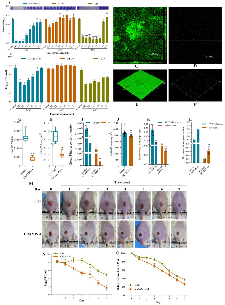 Figure 1
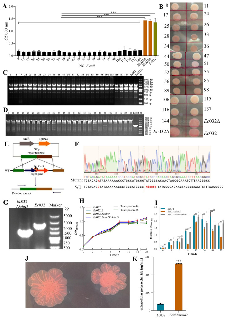 Figure 2
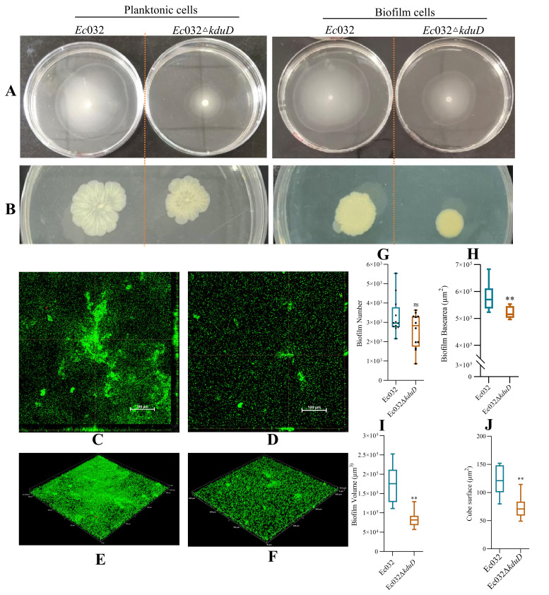 Figure 3
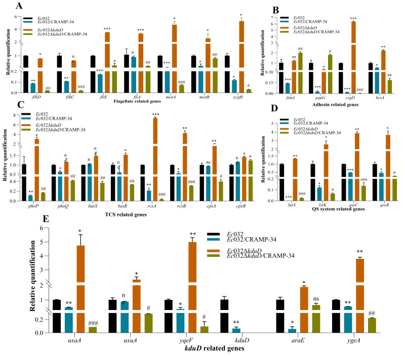 Figure 4
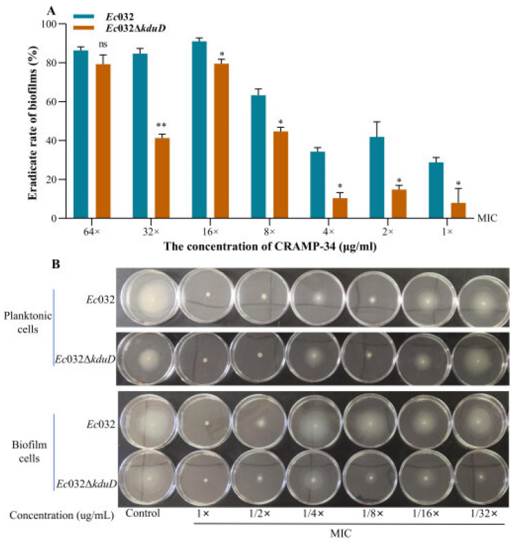 Figure 5
- —National Center of Technology Innovation for Pigs
- —Yunnan Province Science and Technology Department Key Research Development Plan
- —Chongqing Natural Science Foundation
- —Science and Technology Research Program Project of the Chongqing Municipal Education Commission
Peer Reviews
No public reviews on file for this paper yet. If you reviewed it on a platform where reviews are public (OpenReview, ICLR, NeurIPS, ICML), you can paste yours below so the community can read it here.
Videos
No videos yet. Explain this paper in a talk, walkthrough, or lecture? Add one.
Taxonomy
TopicsAntimicrobial Peptides and Activities · Bacterial biofilms and quorum sensing · Pediatric health and respiratory diseases
1. Introduction
Escherichia coli (E. coli) causes persistent diarrhea and antimicrobial resistance due to its propensity to form aggressive and dense bacterial biofilms [1]. The bacteria in biofilms secrete various components, such as extracellular polymeric substances (EPSs) that cause bacterial accumulation, forming multiple layers, which protect the embedded cells from antimicrobial agents and host immune defenses; these infections are called biofilm-associated infections (BAIs) [2,3]. Previous studies have reported that the tolerance of biofilms to various antibiotics is 10–1000 times greater than that of planktonic cells [4]. The life cycle of biofilms includes the following stages: reversible/irreversible adhesion, formation, maturation, and dispersion [5]. In the initial attachment stage, flagella and fimbriae facilitate mechanical attachment to the surface. As the cell density increases, the autoinducer concentration reaches a threshold, and the autoinducer/regulatory protein complex induces or represses the expression of target genes [6]. Pili-mediated twitching motility and a high concentration of cyclic diguanylic acid (c-di-GMP) enable the attached cells to aggregate to form microcolonies that then produce EPSs, thereby ensuring the adherence of the bacteria to the surface of a highly complex bio-molecular layer [7]. Fully mature biofilms typically have a three-dimensional mushroom-like structure [8]. These highly structured biofilms are difficult to eradicate.
Genes that are directly involved in E. coli biofilm formation include the synthetic operon pgaABCD for Poly-β-1,6-N-acetyl-d-glucosamine (poly-β-1,6-GlcNAc; PGA), glycogen synthesis operon glgCAP, autoaggregation adhesin Artigen 43, wza gene for synthesizing the multimeric outer membrane protein complex, operons bcsABZC and bcsEFG that encode the cellulose synthase complex, and operons csgBAC and csgDEFG that are involved in curli fimbria protein synthesis and assembly [9,10,11]. Another study showed that more than 60 genes are directly involved in the regulation of E. coli biofilm formation via signal transduction pathways, such as the quorum sensing (QS) system, second messenger c-di-GMP system, two-component regulatory system (TCS), transcription regulatory protein pathways, and non-coding small RNA (sRNA) pathways [12,13,14]. Nonetheless, it is not known if there are other genes that regulate E. coli biofilm formation.
Our studies have shown that the mouse cathelicidin-related antimicrobial peptide (CRAMP-34) inhibits biofilm formation and can eradicate mature biofilms formed by the biofilm model strain Pseudomonas aeruginosa [15]. However, whether CRAMP-34 has the same inhibitory effect on E. coli biofilms has yet to be clarified. In addition, the key regulatory genes for biofilm formation and maturation in E. coli also need to be explored further. Therefore, the current study focused on a strain of E. coli isolated from a porcine source that has a strong biofilm formation ability and carries the mobilized colistin resistance (mcr) gene. Despite the well-characterized roles of operons such as bcsABZC and csgDEFG in biofilm formation, the complete genetic network regulating this process in E. coli remains incompletely understood [16,17]. To systematically identify novel regulators, we employed a functional screen using transposon mutagenesis. This approach, combined with subsequent CRISPR/Cas9-mediated validation, led to the identification of an previously unknown key gene, kduD, which encodes 2-dehydro-3-deoxy-D- gluconate 5-dehydrogenase (KduD), as a critical regulator of biofilm formation in this strain. Furthermore, given the central role of KduD, we investigated its potential as a target for anti-biofilm agents and specifically evaluated whether it mediates the anti-biofilm effect of the antimicrobial peptide CRAMP-34.
2. Results
2.1. CRAMP-34 Demonstrates Potent Biofilm-Eradicating Activity and Promotes Wound Healing In Vivo
Different concentrations of CRAMP-34 were used on 1-day-old preformed biofilms; ciprofloxacin (CIP) and human antibacterial peptide LL-37 were used as controls. The MIC values of CRAMP-34, LL-37, and CIP were 7.8125 μg/mL, 15.625 μg/mL, and 0.015625 μg/mL, respectively. The results of the crystal violet staining showed that CRAMP-34 and CIP significantly reduced the biomass of Ec032 biofilms at a concentration of 4–64× MIC. However, CRAMP-34 exerted a significant effect on reducing the biomass of Ec032 biofilms at 16× MIC, with a rate of 92.95% (Figure 1A). The results of the colony-counting assay showed that CRAMP-34 killed more biofilms than LL-37 and CIP; the biofilms decreased by 2.76 log values with a killing rate of 99.83% at 16× MIC (Figure 1B).
To visualize the anti-biofilm activity of CRAMP-34, SYTO 9 (living) and propidium iodide (PI) (dead) staining was performed and analyzed using CLSM. As shown in Figure 1 C–F, the CRAMP-34 intervention group biofilms were significantly reduced compared to Ec032. The results showed that CRAMP-34 remarkedly reduced Ec032 biofilms at a concentration of 125 µg/mL, decreasing the cell number (75.90%), volume (97.69%), and base area (8.69%) (Figure 1H–I). Furthermore, the fluorescence intensity of SYTO 9- and PI-positive bacteria decreased by 89.11% and 92.28%, respectively (Figure 1J). In addition, the ratio of fluorescence intensity of SYTO 9 and PI per unit base area of the biofilms decreased by 88.09% (p < 0.01) and 91.63% (p < 0.01), respectively (Figure 1K), and the ratio of fluorescence intensity of SYTO 9 and PI per unit volume of the biofilms increased by 77.68% (p < 0.01) and 70.91% (p < 0.01), respectively (Figure 1L).
The wounds of each group of mice were photographed regularly to observe the healing process. As shown in Figure 1M, compared with the PBS group, the wound healing rate in the CRAMP-34 treatment group was significantly accelerated, and the wound area was significantly reduced. Residual bacteria at the wound site were sampled daily and counted using the plate method. Compared with the PBS group, the number of bacterial colonies in the CRAMP-34 treatment group was significantly reduced (Figure 1N) and the relative wound closure rate became increasingly faster (both differences were statistically significant), which further verified the antibacterial effect of CRAMP-34 (Figure 1O). In summary, CRAMP-34 significantly promoted the healing of wounds caused by E. coli.
2.2. A Genome-Wide Screen Identifies kduD as a Novel Essential Gene for Robust Biofilm Formation
An Ec032 random Mariner transposon mutagenesis library comprising 141,667 mutants was constructed to identify genes involved in biofilm formation. From this library, 25 independent mutants exhibiting a pronounced reduction in biofilm formation were isolated (Figure 2A). Subsequent phenotypic characterization via the Congo red assay revealed that these mutants collectively displayed a loss of colony wrinkling, a transition to a smoother morphology, and diminished pigmentation compared to the wild-type Ec032 and an empty vector control strain (Ec032Δ) (Figure 2B). ERIC-PCR analysis confirmed that all 25 strains were genuine transposon mutants, ruling out contamination (Figure 2C). Whole-genome sequencing and comparative analysis pinpointed the transposon insertion site in all the mutants to a single specific locus, which we designated as kduD (Figure 2D).
To definitively establish the role of kduD in biofilm formation, an in-frame deletion mutant (Ec032ΔkduD) was generated using the CRISPR/Cas9 system (Figure 2E), with the deletion verified by Sanger sequencing and electrophoretic analysis (Figure 2F,G). The deletion of kduD had no discernible impact on bacterial growth kinetics (Figure 2H). However, quantitative assessment of biofilm formation using crystal violet staining demonstrated a significant impairment in the Ec032ΔkduD mutant across multiple time points. The most severe defect, a reduction of 60.13%, was observed at 9 h. This phenotype was successfully complemented as biofilm formation was restored to near wild-type levels when the mutant was transformed with a plasmid carrying the kduD gene (pBAD24-kduD) (Figure 2I).
Further analysis using the Congo red-binding assay indicated that the deletion of kduD did not abolish the production of curli fimbriae and cellulose, which are key biofilm matrix components, and the colony morphology of the defined mutant was distinct from that of the initial transposon mutants (Figure 2J). Intriguingly, quantification of EPSs revealed a significant increase in EPS production in the Ec032ΔkduD mutant at equivalent bacterial densities (Figure 2K). These results demonstrated that kduD plays a critical role in mediating biofilm formation.
Collectively, these findings demonstrate that kduD is a key regulator of biofilm development in Ec032, likely through a mechanism involving the modulation of EPS production.
2.3. kduD Regulates Biofilm Formation by Modulating Flagellar Motility and EPS Production
Swimming and twitching motility assays were used to investigate the flagella and pili movement of the biofilm cells. The results showed that the swimming motility of the planktonic and biofilm cells of the Ec032ΔkduD strain decreased by 46.43% and 77.46% (Figure 3A), and the twitching motility decreased by 32.50% and 36.17%, respectively (Figure 3B). These results suggested that kduD mediates the formation of E. coli biofilms by regulating motility.
CLSM was utilized to observe the effect of kduD deletion on biofilm formation. As shown in Figure 2C–F, Ec032ΔkduD biofilms were significantly reduced compared to Ec032 biofilms. In addition, Ec032ΔkduD did not affect the cell number and base area (Figure 3G,H), but significantly reduced the volume and surface area of the biofilms (Figure 3I,J).
We found that the mRNA expression levels of the biofilm-related genes in Ec032ΔkduD were upregulated compared to Ec032, as assessed by RT-qPCR. These biofilm-related genes included flagella-related genes (fliE, fliA, motA, motB, and ycgR) (Figure 4A), adhesion-related genes (csgD and bcsA) (Figure 4B), QS-related genes (luxS, lsrK, qseC, and qseB) (Figure 4C), TCS-related genes (phoP, rcsA, rcsB, and cpxR) (Figure 4C), and kduD-related genes (uxaA, uxuA, yqeF, araE, and ygeA) (Figure 4D). These results demonstrated that kduD plays a critical role in mediating biofilm formation.
Moreover, the mRNA expression levels of biofilm-related genes in CRAMP-34-treated Ec032 and Ec032ΔkduD were measured using RT-qPCR. The results showed that the mRNA levels of the flagella-related genes (flhD, flhC, fliE, fliA, motA, motB, and ycgR) (Figure 4A), adhesion-related genes (csgD and bcsA, but not fimA and papG) (Figure 4B), QS-related genes (luxS, lsrK, qsEc, and qseB) (Figure 4C), TCS-related genes (phoP, phoQ, basR, rcsA and rcsB, but not basS and cpxR) (Figure 4D), and kduD-related genes (uxaA, uxuA, yqeF, kduD, araE, and ygeA) (Figure 4E) were downregulated in Ec032 and Ec032ΔkduD after treatment with CRAMP-34. These results indicated that the scavenging effect of CRAMP-34 on mature biofilms of E. coli is related to EPSs and the kduD metabolic pathway.
The results of the crystal violet staining revealed that the biofilm inhibition rate of CRAMP-34 on Ec032ΔkduD was lower at all concentrations compared to Ec032 (Figure 5A). The effect of CRAMP-34 on the flagella motility of Ec032 and Ec032ΔkduD was observed by spotting planktonic and biofilms cells in liquid onto agar plates containing different concentrations of CRAMP-34. The results showed that CRAMP-34 inhibited the flagellar movement, which was dependent on the concentration of CRAMP-34 (Figure 5B).
3. Discussion
E. coli biofilms represent a serious public and animal health concern, yet the regulatory mechanisms governing their development remain incompletely elucidated. Our data demonstrate that the antimicrobial peptide CRAMP-34 effectively eliminates mature biofilms formed by pathogens such as Pseudomonas aeruginosa, Acinetobacter baumannii, and E. coli, while the present study elucidates the specific molecular mechanism of CRAMP-34 against a model E. coli biofilm; however, the full scope and clinical relevance of CRAMP-34 require further investigation in diverse infection models [15,18]. The observed requirement for relatively high concentrations (≥16× MIC) of CRAMP-34 to eradicate mature biofilms reflects both the intrinsic tolerance mechanisms of biofilms and the peptide’s unique mode of action. Unlike CIP, which primarily inhibits DNA replication, CRAMP-34 operates through membrane disruption combined with metabolic interference, specifically targeting the kduD-dependent network that is essential for biofilm maturation. This dual mechanism likely underlies its superior efficacy against established biofilms compared to conventional antibiotics. Although effective concentrations are higher under in vitro conditions, studies on CRAMP family peptides have demonstrated favorable safety profiles with low cytotoxicity and immunomodulatory potential at relevant doses, supporting their therapeutic feasibility [18]. Importantly, such concentration requirements can be achieved with localized sustained-release delivery systems (e.g., hydrogels and advanced dressings), which can maintain effective drug levels at infection sites while minimizing systemic exposure [19]. Structural advantages over human LL-37, including optimized hydrophobicity and helical stability, may enhance CRAMP-34′s penetration through biofilm matrices and interaction with bacterial membranes. In addition, CRAMP-34 significantly accelerated the healing of Escherichia coli-infected wounds in mice. This therapeutic effect can be primarily attributed to its direct antimicrobial activity, as evidenced by the marked reduction in bacterial load at the wound site. Given its potent anti-biofilm efficacy against mature biofilms in vitro, it is reasonable to infer that CRAMP-34 may also contribute to wound healing by disrupting biofilm structures in vivo, thereby enhancing bacterial clearance. The accelerated wound closure may partly result from the potential immunomodulatory functions of CRAMP-34 as a host defense peptide, which could indirectly promote healing by modulating local inflammatory responses or facilitating tissue repair. It should be noted, however, that the present study primarily focused on validating its antimicrobial and anti-biofilm effects. The specific mechanisms and relative contribution of its immunomodulatory role need to be further elucidated through follow-up experiments. Collectively, these properties position CRAMP-34 as a promising next-generation anti-biofilm agent, with its multi-mechanistic action, compatibility with targeted delivery approaches, and demonstrated efficacy supporting its potential for clinical translation to treat persistent biofilm-mediated infections.
To identify key the genetic determinants of biofilm formation and assess whether CRAMP-34 targets these pathways, we performed a transposon mutagenesis screen. All 25 isolated biofilm-deficient mutants mapped to kduD, showing that deletion of kduD severely compromised biofilm formation. As E. coli forms air–liquid interface biofilms—a process dependent on oxygen, flagellar motility, and cellulose—and given the established role of flagellar function in approximately half of E. coli biofilms mutants [20,21], we investigated the motility of Ec032ΔkduD. The mutant showed markedly impaired swimming motility despite the upregulation of flagellar assembly (fliE and fliA), motor genes (motA and motB), and the c-di-GMP-responsive brake protein gene ycgR. Since YcgR binds to c-di-GMP to mechanically inhibit flagellar rotation, its overexpression likely explains the motility defect [22]. Furthermore, the upregulation of the flagellar regulatory system qseBC suggests that KduD functions within or downstream of the QseBC circuit, potentially influencing motility via c-di-GMP and YcgR [23]. In summary, KduD emerged as a novel essential regulator of biofilm formation in Ec032, likely through the QseBC system and a c-di-GMP–YcgR-mediated braking mechanism. Its exact molecular role and therapeutic relevance as a target of CRAMP-34 warrant further study.
The established model of biofilm development proceeds through attachment, microcolony formation, and maturation into complex structures such as mushroom-like macrocolonies. We observed that the Ec032ΔkduD mutant retained the ability to adhere and form microcolonies but failed to develop architecturally mature biofilms. This defect occurred despite the upregulation of the key matrix genes csgD and bcsA and a measurable increase in EPSs under a standardized bacterial density [24,25]. Notably, while cellulose and curli fibers—encoded by bcsA and csgD, respectively—are essential for structural integrity, particularly under nutrient limitations; their overproduction can disrupt surface adhesion and aggregation [26,27]. In E. coli MG1655, overexpression of csgD was shown to inhibit biofilm formation on hydrophobic surfaces due to excessive cellulose production [28]. Similarly, our findings demonstrate that deletion of kduD leads to a significant increase in total EPS production and alters colony morphology, indicating its essential role in biofilm matrix regulation. However, we recognize that the proposed link between KduD-mediated metabolism and biofilm matrix organization remains largely inferential as the composition of EPSs was not directly characterized in this study. We explicitly state that while kduD clearly influences EPS accumulation, the specific metabolic pathways and compositional shifts underlying this phenotype warrant further investigation. Future studies including EPS profiling, metabolomic analysis, and high-resolution imaging of the biofilm matrix will be essential to elucidate the precise mechanism through which kduD coordinates EPS synthesis and biofilm architecture. In support of this, we observed concomitant upregulation of the RcsAB, a known repressor of csgD, likely reflecting a compensatory cellular response to restore regulatory balance [29,30]. Thus, KduD appears to act as a metabolic modulator whose activity fine-tunes matrix biosynthesis, ensuring the appropriate spatial and temporal deposition of the polysaccharide and protein components required for a stable biofilm architecture [31].
KduD, a 2-dehydro-3-deoxy-D-gluconate 5-dehydrogenase, participates in pentose and glucuronate metabolism [32]. Our analysis revealed that deletion of kduD triggered upregulation of the adjacent genes ygeA and yqeF, which are involved in glucose metabolism, indicating a compensatory metabolic adaptation. Given that KduD is involved in the metabolism of galacturonic and glucuronic acids [33]—key precursors for EPS biosynthesis—we hypothesize that it regulates biofilm architecture by controlling the supply of essential carbon substrates. This is supported by the observation that the Ec032ΔkduD mutant, similar to colanic acid (CA)-deficient strains, failed to form mature biofilms with the typical mushroom-like structure. CA, a branched EPS composed of glucose, galactose, and glucuronic acid, is critical for building the three-dimensional matrix of E. coli biofilms [34]. The inability to form structured biofilms may thus stem from a shortage of these vital EPS building blocks, impairing bacterial colonization and survival in the mammalian intestine. Furthermore, the upregulation of ygeA, which encodes an amino acid racemase that produces biofilm-dispersing D-amino acids, may represent a concurrent disassembly signal that further destabilizes the biofilm matrix [35,36]. In conclusion, we propose that KduD influences biofilm formation not through direct regulatory means, but by being a critical metabolic node that fuels the biosynthesis of structural EPSs, thereby linking central carbon metabolism to biofilm structural integrity.
Our data demonstrate that CRAMP-34 exerts potent anti-Ec032 biofilm activity by inhibiting flagellar motility and eradicating mature biofilms. The observation that kduD expression is drastically suppressed following CRAMP-34 treatment prompted us to investigate a potential functional link. The finding that CRAMP-34′s Ec032ΔkduD biofilm clearance efficacy was significantly attenuated, though not entirely abolished, provides direct genetic evidence that KduD is a critical target for its action. This partial persistence of activity suggests that CRAMP-34, like many effective antimicrobial peptides, employs a polypharmacological strategy, engaging multiple cellular targets to disrupt the complex biofilm network. The RT-qPCR analysis results supported this model by revealing a marked downregulation of the kduD-associated metabolic operon (which includes uxaA and uxuA) and the glucose transporter araE upon CRAMP-34 treatment. This indicates a targeted disruption of a specific metabolic pathway centered on KduD. Furthermore, the significant suppression of the key biofilm regulators csgD and bcsA aligns with the proposed role of KduD in maintaining EPS stability and suggests a cascade of transcriptional repression. The temporal disconnect between this rapid transcriptional reprogramming and the subsequent phenotypic collapse is a hallmark of targeted anti-virulence strategies, where disabling master regulators leads to a progressive dismantling of the biofilm structure.
4. Materials and Methods
4.1. Bacterial Strains, Plasmids, Primers, and Growth Conditions
E. coli (No. Ec032) was isolated from porcine sources and identified as a colistin-resistant strain carrying the mcr-1 gene of the plasmid IncX4 with a strong biofilm formation ability [1]. The bacteria were grown overnight at 37 °C in Brain Heart Infusion (BHI) broth (Haibo Co., Ltd., Qingdao, China), harvested by centrifugation at 6000 rpm for 10 min, and resuspended in BHI broth. The optical density of the bacterial suspension was measured at 600 nm (OD600 nm). This suspension was used in the subsequent experiments. The strains, plasmids, and primers are listed in Table 1, Table 2 and Table 3, respectively.
4.2. Determination of Minimum Inhibitory Concentration (MIC)
The MIC values of the antibiotics (ciprofloxacin, CIP; Yuanye Co., Ltd., Shanghai, China) and antibacterial peptides (AMPs; CRAMP-34 and LL-37; synthesized by ChinaPeptides Co., Ltd., Shanghai, China) were determined using the microbroth dilution technique described by the Clinical and Laboratory Standards Institute (CLSI).
4.3. Biofilm Formation and Anti-Biofilm Assays
For the microtiter plate test (in 96-well plates), mature biofilms were formed by adding 100 μL the test bacterial solution into each well of the 96-well plates and incubating them at 37 °C for 24 h. Then, the plates were washed with phosphate-buffered saline (PBS). Two-fold dilutions of CRAMP-34, LL-37, and CIP ranging from 64 to 0.5× MIC were prepared with sterile water. Next, each antimicrobial compound was added to the corresponding plate and incubated at 37 °C for 3 h. The appropriate concentration of CRAMP-34 was selected and tested in a 6-well plate (Corning^®^3516, Corning Incorporated, Corning, NY, USA) to confirm the results; PBS was used as a control. For selective growth of the bacteria, the following antibiotics and substances were added: 2 μg/mL colistin, 30 μg/mL apramycin, 100 μg/mL rifampin, 57 μg/mL diaminopimelic acid, 5% sucrose, and 2 mg/mL L-arabinose.
4.4. Skin Wound Infection Model Was Established
The animal experiments utilized male Balb/c mice (6–8 weeks old, weighing 20–23 g; Dashuo Co., Ltd., Chengdu, China). All housing, handling, and experimental procedures strictly followed the national standard GB/T 35823-2018; Laboratory Animal-Guidelines for Ethical Review of Animal Welfare (National Standards of the People’s Republic of China; Beijing, China, 2018). The protocol was approved by the Institutional Animal Care and Use Committee of Southwest University (Permit No.: LAC2025-1-0259). Initially, the mice were subjected to general anesthesia. Subsequently, a 1 cm diameter artificial wound was created on the dorsal surface of each mouse using surgical scissors. A volume of 200 µL of bacterial suspension (108 CFU/mL) was applied topically to the wound immediately post-injury and allowed to colonize for 24 h prior to the initiation of treatment. After successful establishment of the skin infection model, the mice were randomly assigned to two groups (n = 10 per group): the PBS treatment control group that was infected with the Ec032 strain and the CRAMP-34 treatment group that was infected with the Ec032 strain. The treatment was administered as a single topical application on day 1 post-infection. A dose of 50 µg/wound of CRAMP-34 (dissolved in PBS) or an equal volume of PBS was applied directly to the wound surface. No further treatment was administered on the subsequent days. The total observation period was 7 days post-infection. Throughout the treatment period, wound changes were documented daily via photography, and samples were collected for bacterial counting.
4.5. Crystal Violet Staining and Colony Count Assay
The biofilm formation assay was performed using crystal violet, and the viable bacteria were counted on trypticase soy agar plates. Briefly, the supernatant was discarded, and the cells were washed twice with sterile PBS, fixed in methanol for 10 min, and stained with crystal violet for 20 min. After washing, acetic acid was used to dissolve the bound crystal violet, and the absorbance was measured at OD600 nm. To count the number of bacterial biofilm cells, 100 µL of Triton-100X was added to disrupt the biofilms, and the bacteria were spread on TSA plates at a 10-fold dilution at 37 °C for 12 h.
4.6. Swimming Motility Assay
LB medium plates with 0.25% (wt/vol) agar were used for the swimming motility assay. One-microliter aliquots of mid-log-phase bacteria in BHI broth were spotted onto a control agar plate and an agar plate containing decreasing concentrations of CRAMP-34. The diameters of the swimming zones were measured after incubation at 37 °C for 15 h.
4.7. Confocal Laser Scanning Microscopy (CLSM)
The morphological features of biofilms were observed using CLSM as described previously with some modifications [1]. In this experiment, 500 µL of the test bacterial solution (OD600 nm = 0.1) was added to an 8-well chambered coverglass (Lab-Tek II, Rochester, NY, USA), and the medium was replaced every 24 h. After incubation at 37 °C for 2 days, the biofilms were treated with CRAMP-34 (125 μg/mL) at 37 °C for 3 h, washed with 0.9% (wt/vol) NaCl, and stained at room temperature for 20 min in the dark using a Filmtracer™ LIVE/DEAD™ Biofilm Viability Kit. After rinsing with sterile water, the biofilm samples were imaged with a point-scanning confocal microscope (Eclipse Ti2; Nikon, Tokyo, Japan). The signals were recorded using the green (SYTO9, excitation wavelength of 488 nm) and red (PI, excitation wavelength of 561 nm) channels. The field of view under the ×20 objective was randomly selected, and a three-dimensional image was constructed by stacking multiple images with different Z values (z-stack). The images were acquired using NIS Viewer v5.21.00 software. Four representative images were selected from each biofilm, and each experiment was repeated at least three times. The biofilm-related test parameters, such as the cell number, volume, base area, and fluorescence intensity, were analyzed using BiofilmQ software version 2.1.0 (The authors, Vienna, Austria).
4.8. Screening of Gene Mutants with Decreased Biofilm Formation
For the primary screening of the transposon library, the transconjugants were cultured to the mid-log phase, and then 5 μL was added to Corning 3595 microtiter plates containing 100 μL of BHI broth and incubated at 37 °C for 24 h. The mature biofilms were assessed by crystal violet staining. Isolates exhibiting reduced biofilm accumulation (<75% of the wild-type level) in the primary screen were retested individually, as described previously [1]. For all mutants with a confirmed defect in biofilm accumulation, the growth kinetics were evaluated based on the changes in the bacteria in BHI broth over time at OD600 nm. The formation of biofilms is generally divided into four grades based on the average OD value of the negative control (ODc): OD ≤ ODc is non-adherent, ODc < OD ≤ 2× ODc is weakly adherent, 2× ODc < OD ≤ 4× ODc is moderately adherent, and 4× ODc < OD is strongly adherent.
4.9. Generation of a Ec032 Mutagenesis Library
E. coli WM3064 (pCure-oriT-GFP-MCR) and Ec032 were used as the donor (carrying Mariner transposons with a GFP gene) and the recipient, respectively. After the donor and recipient bacteria were cultured to the logarithmic growth stage, they were conjugated at 37 °C at a ratio of 1:1 for 8–12 h. The transconjugants with GFP were selected on LB agar containing 30 μg/mL apramycin and induced in sugar-free LB broth (Yuanye Co., Ltd., Shanghai, China) with 2 mg/mL L-arabinose (Shenggong Co., Ltd., Shanghai, China) for 6 h to obtain a plasmid-free Ec032 strain containing the pCure-oriT-GFP-MCR plasmid. These strains were cultured on LB agar plates containing 5% sucrose (Maclin Co., Ltd., Shanghai, China) at 37 °C for 24 h to obtain a plasmid-free Ec032 strain (Ec032Δ). E. coli WM3064 (pCat-arr) (donor carrying Mariner transposons) and Ec032Δ (recipient) were conjugated at 37 °C at a ratio of 1:1 for 8–12h. The transconjugants were obtained in a total volume of 1 mL. The transconjugants were selected on LB agar containing 100 μg/mL rifampin to positively select for arr transconjugants. A 100 μL aliquot was diluted to a suitable gradient and used to coat a plate; the number of mutants was calculated and a transposon mutation library was obtained.
4.10. Identification of Transposon Insertion Sites
ERIC-PCR was used to detect any contamination of the screened transposon mutants with decreased biofilm formation. The genomic DNA of Ec032 and the transposon insertion mutants with reduced biofilm formation was extracted using the TaKaRa MiniBEST Bacteria Genomic DNA Extraction Kit Ver. 3.0 (TaKaRa Co., Ltd., Beijing, China) and subjected to whole-genome sequencing at Novogene Co., Ltd., Beijing China. The insertion site of the Mariner transposon was identified using SnapGene software and verified using PCR validation with the primers Ec032-F-XJ and Ec032-R-XJ. The sequence was compared to the NCBI database to identify the gene name and base sequence for the subsequent gene editing.
4.11. kduD Mutant Construction Using CRISPR/Cas9 System
Plasmids and genomic DNA were extracted using the TaKaRa MiniBEST Bacteria Genomic DNA Extraction Kit Ver. 3.0 (Takara Biomedical Technology (Beijing) Co., Ltd., Beijing, China). In this study, we used two plasmid systems (which express Cas9 and the sgRNA in pCasKp-OriT and pSGKp, respectively) for genome editing using the CRISPR-Cas9 system in E. coli DH5α. First, pSGKp-Ec032-kduD, which encodes the sgRNA targeting the kduD gene (target sequence selected by SnapGene software), was constructed. Briefly, the guide RNA target sequence in pSGKp was mutagenized using PCR to target kduD. Primers kduD-F1, kduD-R1, kduD-F2, and kduD-R2 were used to amplify left homologous arm H1 and right homologous arm H2. The pSGKp-Ec032-kduD plasmid was cut into linear DNA using BamHI and XbaI enzymes. The left and right homologous arms H1 and H2, and linear pSGKp-Ec032-kduD plasmids were connected using the NEBuilder HiFi DNA Assembly Master Mix (NEB Biological Technology (Beijing) Co., Ltd., Beijing, China) and then transformed into E. coli DH5α to obtain pSGKp-Ec032-kduD-500 plasmids. We performed cloning for a pSGKp-Ec032-kduD-500 plasmid harboring a spacer (for guiding Cas9 cleavage of the wild-type Ec032) in E. coli DH5α, which was selected on LB plates with 100 μg/mL rifampin (Shanghai Maclin Biochemical Technology Co., Ltd., Shanghai, China). The transformants were confirmed by sequencing.
E. coli WM3064 (pCasKp-OriT) and Ec032 (pSGKp-Ec032-kduD-500) were used as the donor and recipient, respectively. After conjugation, the plasmid pCasKp-OriT from E. coli WM3064 was introduced into Ec032. The transconjugants were selected on LB agar containing 30 μg/mL apramycin (Shanghai Maclin Biochemical Technology Co., Ltd., Shanghai, China) at 30 °C for 8 h–12 h. Then the transconjugants were confirmed by colony PCR and DNA sequencing. The gene deletion strains were plated onto 5% sucrose LB agar plates and cultured at 37 °C to eliminate plasmids pSGKp-Ec032-kduD-500 and pCasKp-OriT.
4.12. Obtaining Ec032ΔkduD Plasmid Deletion Strains
The construction of kduD complementation strains followed the design for plasmid pBAD24. The kduD gene fragment, amplified from the genomic DNA of Ec032, was inserted into the multiple cloning site (MCS) of plasmid pBAD24 using the ClonExpress^®^ Ultra One Step Cloning Kit (Vazyme Biotech Co., LTD, China). Then, the recombinant product pBAD24-kduD was transformed into E. coli DH5α chemically competent cells and inoculated on transformed LB plates supplemented with 100 μg/mL ampicillin. Positive clones were identified through colony PCR and sequencing. Finally, the recombinant plasmid was transferred into a kduD mutant strain via electroporation. The constructed complementation strain, named Ec032-ΔkduD/pkduD, was induced with arabinose during culturing and was used to ascertain the target of CRAMP-34 in Ec032.
4.13. Growth Curves
The bacteria were cultured in BHI broth to the mid-log phase and diluted to equal optical densities (OD600 nm = 1). A 200 µL volume of these cultures was added to 96-well microtiter plates (Corning^®^3599; Corning Inc., Corning, NY, USA) and incubated at 37 °C. The growth of these cultures was monitored by determining the absorbance at 600 nm every 2 h for 24 h. The OD600 nm was measured using an Infinite^®^ M Plex microplate reader (Tecan, Switzerland) after shaking. Three independent experiments were performed.
4.14. Congo Red-Binding Assay and EPS Assay
One microliter of the test bacterial suspensions of Ec032 and Ec032ΔkduD was spotted onto 40 μg/mL Congo red salt-free LB agar plates, which were incubated at 37 °C for 2–4 days. Subsequently, their colony morphologies were compared.
EPSs were determined using the phenol–sulfuric acid method. The working bacterial suspension was inoculated into 6-well plates and cultivated at 37 °C for 24 h to form biofilms. Then, the planktonic bacteria were discarded, the wells were rinsed with PBS, normal saline was added to scrape up the biofilms, which were homogenized by repeated and even blowing. The resulting suspension was clarified by centrifugation at 4000 rpm at 4 °C for 20 min. The supernatant was filtered through a 0.22 μm filter membrane; 100 μL was added into a new centrifuge tube and mixed with 200 μL of concentrated sulfuric acid and then 25 μL of a 6% phenol solution in a water bath at 90 °C for 5 min. After cooling to room temperature, 200 μL was dispensed into a 96-well plate to measure the absorbance at the wavelength of 490 nm. Each experiment was set up with three parallel groups and was repeated three times.
4.15. Real-Time Fluorescence Quantitative PCR (RT-qPCR)
The CRAMP-34 treatment of the biofilms was carried out as described above. Total RNA was extracted using the Total RNA Isolation Reagent (Biosharp Co., Ltd., Beijing, China) according to manufacturer’s instructions. RNA was reverse transcribed using the PrimeScript™ RT reagent Kit (Takara Bio Inc., Shiga, Japan)according to the manufacturer’s instructions. Primers and probes were designed using Primer 5.0 and synthesized by Tsingke Biotechnology (Beijing, China). RT-qPCR was conducted on a CFX Connect qPCR instrument. The reaction conditions were as follows: 95 °C for 30 s and 40 cycles of 95 °C for 5 s and 60 °C for 30 s. The relative expression levels of each target gene were computed using the 2−ΔΔCt method.
4.16. Statistical Analysis
All experiments were conducted in triplicate, and the data represent a minimum of three biological replicates. The statistical analyses were carried out using Microsoft Excel and GraphPad Prism 8.0 software. An unpaired t-test (two-tailed) was used to calculate the statistical significance. Significant differences are indicated as * p < 0.05, ** p < 0.01, and *** p < 0.001.
5. Conclusions
This study demonstrated that CRAMP-34 can effectively eradicate mature E. coli biofilms through mechanisms involving suppression of flagellar motility and disruption of biofilm structural integrity. We further identified kduD as a previously unrecognized key regulatory gene controlling bacterial motility and biofilm development. While the experiments primarily focused on 1-day-old biofilms—a standard model for assessing early biofilm susceptibility—the persistent role of kduD-dependent metabolic regulation throughout biofilm maturation suggests broader relevance to more established biofilms. Collectively, these findings not only highlight the therapeutic potential of CRAMP-34 as a dual-function antimicrobial and anti-biofilm agent, but also establish kduD as a central metabolic regulator in biofilm formation, offering a promising new target for innovative anti-biofilm strategies.
The reference list from the paper itself. Each links out to its DOI / PubMed record.
- 1Yang H. Liang Y. Yang Z. Liu L. Ran L. Liu J. Ma C. Wei W. Zhang S. Zhu M. Paeonol eradicates biofilm in porcine-source Escherichia coli by targeting the quorum sensing system BMC Vet. Res.20252164410.1186/s 12917-025-05095-y 41188864 PMC 12584432 · doi ↗ · pubmed ↗
- 2Thakur D. Kumar L. Biofilm-associated Escherichia coli infections: Pathogenesis, clinical implications, and treatment strategies Crit. Rev. Microbiol.202514510.1080/1040841 X.2025.254849040832759 · doi ↗ · pubmed ↗
- 3Maric T. Tanaka Y. Li Z. Wu J. Li J. Guan J. Boisen A. Micro/nanomotors for biofilm remediation: Nanoarchitectonic breakthroughs with a focus on regulatory and clinical translation challenges Biomaterials 202632612371510.1016/j.biomaterials.2025.12371540976139 · doi ↗ · pubmed ↗
- 4Prosswimmer T. Nick S.E. Bryers J.D. Daggett V. Designed De Novo α-Sheet Peptides Destabilize Bacterial Biofilms and Increase the Susceptibility of E. coli and S. aureus to Antibiotics Int. J. Mol. Sci.202425702410.3390/ijms 2513702439000131 PMC 11241457 · doi ↗ · pubmed ↗
- 5Sauer K. Stoodley P. Goeres D.M. Hall-Stoodley L. Burmølle M. Stewart P.S. Bjarnsholt T. The biofilm life cycle: Expanding the conceptual model of biofilm formation Nat. Rev. Microbiol.20222060862010.1038/s 41579-022-00767-035922483 PMC 9841534 · doi ↗ · pubmed ↗
- 6Gao S. Yuan S. Quan Y. Jin W. Shen Y. Li R. Liu B. Wang Y. Yi L. Wang S. Targeting AI-2 quorum sensing: Harnessing natural products against Streptococcus suis biofilm infection Vet. Res.2025562610.1186/s 13567-025-01450-x 39905565 PMC 11796197 · doi ↗ · pubmed ↗
- 7Tang A. Li C. Feng D. Li A. Deciphering the code of temperature rise on aerobic granular sludge stability: A DSF-c-di-GMP mediated regulatory mechanism Environ. Res.202526712070510.1016/j.envres.2024.12070539732421 · doi ↗ · pubmed ↗
- 8Djermoun S. Rode D.K.H. Jiménez-Siebert E. Netter N. Lesterlin C. Drescher K. Bigot S. Biofilm architecture determines the dissemination of conjugative plasmids Proc. Natl. Acad. Sci. USA 2025122 e 187751517410.1073/pnas.2417452122 PMC 1205480240279390 · doi ↗ · pubmed ↗