Image reconstruction and elongation artifact reduction for a dual‐panel dedicated prostate PET scanner
Abdollah Saberi Manesh, Mehdi Amini, Yazdan Salimi, Katayoun Doroud, Crispin Williams, Themistoklis Williams, Hossein Arabi, Habib Zaidi

TL;DR
This paper introduces new image reconstruction techniques for a prostate-specific PET scanner, showing that deep learning improves image quality and lesion detection.
Contribution
The study introduces a deep learning-enhanced reconstruction method for a prostate-dedicated PET scanner, demonstrating improved image quality and lesion detection.
Findings
Swin-UNETR-based deep learning model achieved highest CNR and CNC for small lesions in simulations.
Experimental results showed Swin method outperformed others in CNR for both large and small prostate lesions.
Model-based and learned methods showed complementary strengths depending on lesion contrast and size.
Abstract
The development of PET scanners dedicated to high temporal and spatial resolution organ‐specific imaging is an active research area, motivated by the need for cost reduction, improved lesion detectability and quantification in specific clinical scenarios, as well as by ongoing hardware and software innovations. This study investigates and compares various image reconstruction strategies for a dual‐panel prostate‐dedicated PET scanner (ProVision), which features four‐layered dual‐readout time‐of‐flight depth‐of‐interaction detectors and a 22‐position acquisition protocol to improve angular coverage. A list‐mode MLEM algorithm with multi‐ray modeling was developed and optimized using a scaled NEMA image quality phantom to determine optimal number of rays and iterations. These parameters were then used to reconstruct data from both simulation and experimental acquisitions, including an…
Genes, proteins, chemicals, diseases, species, mutations and cell lines named across the full text — each resolved to its canonical identifier and authoritative record.
Click any figure to enlarge with its caption.
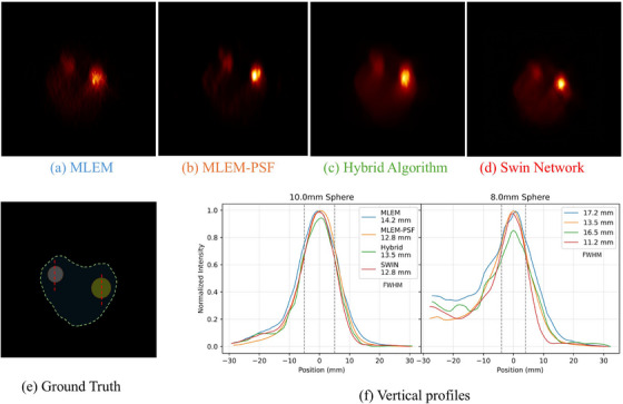 Figure 1
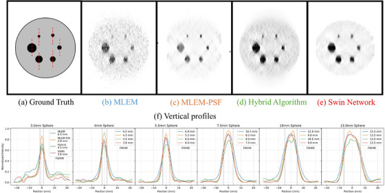 Figure 2
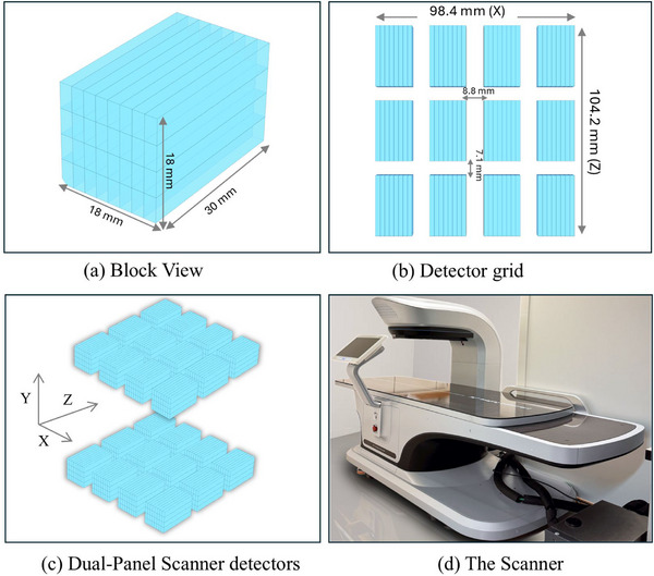 Figure 3
 Figure 4
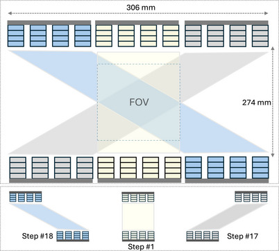 Figure 5
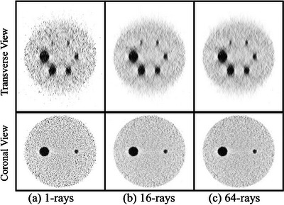 Figure 6
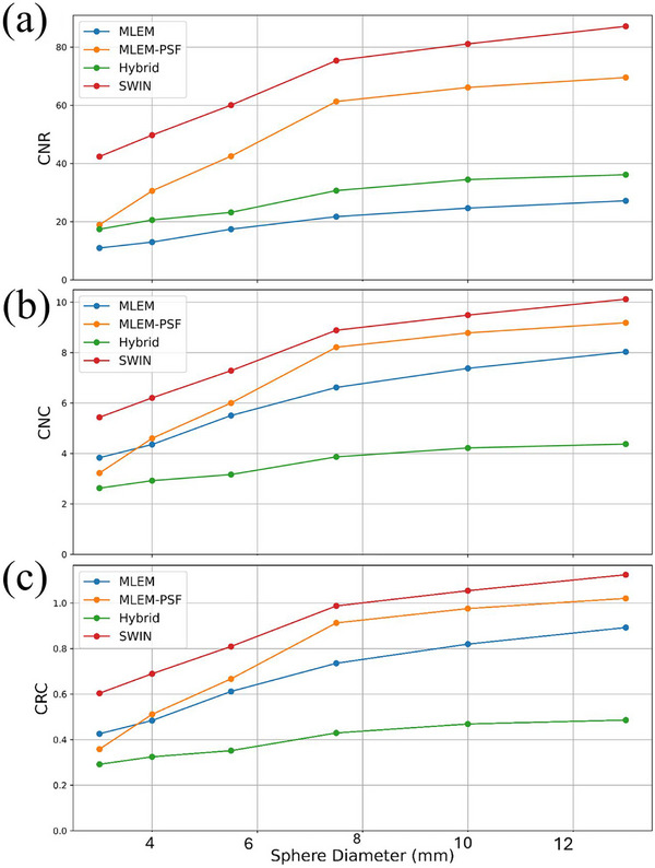 Figure 7
 Figure 8
 Figure 9
Peer Reviews
No public reviews on file for this paper yet. If you reviewed it on a platform where reviews are public (OpenReview, ICLR, NeurIPS, ICML), you can paste yours below so the community can read it here.
Videos
No videos yet. Explain this paper in a talk, walkthrough, or lecture? Add one.
Taxonomy
TopicsMedical Imaging Techniques and Applications · Radiation Detection and Scintillator Technologies · Advanced Radiotherapy Techniques