Micro‐Computed Tomography Based Whole Block Imaging of Asthma‐Associated Airway Remodeling With Mycobacterium avium‐Induced Cavity Formation: 3‐Dimensional Nondestructive Analysis
Tetsuya Tsukamoto, Yasushi Hoshikawa, Alexei Teplov, Eiko Sakurai, Yasushi Matsuda, Hisato Ishizawa, Emmy Yanagita, Kaori Ushida, Naoya Asai, Kazuyoshi Imaizumi, Yukako Yagi

TL;DR
This study uses 3D imaging to show how asthma-related airway changes may help spread Mycobacterium avium infections in the lungs.
Contribution
First 3D visualization of a continuous airway route from the hilum to a subpleural cavity in M. avium infection.
Findings
Merged whole block imaging revealed a remodeled airway connecting the hilum to a subpleural cavity with granulomas.
The airway intertwined with granulomatous tissue, suggesting a structural pathway for mycobacterial spread.
WBI also showed minor granulomas and calcifications, providing detailed anatomical insights.
Abstract
Asthma is a recognized risk factor for nontuberculous mycobacterial (NTM) pulmonary infections, yet the precise routes of infection and dissemination remain visually unclear. We analyzed a rare surgical case of asthma complicated by Mycobacterium avium–associated lung cavity formation in a young adult. Following antibiotic treatment, the patient underwent left upper segmentectomy, with successful subsequent treatment of residual lesions using amikacin liposome inhalation suspension. To precisely visualize the infection route, we utilized a three‐dimensional merged whole block imaging (WBI) technique. Fifteen formalin‐fixed paraffin‐embedded tissue blocks from the resected lung were individually scanned using a custom‐built micro‐computed tomography system, reconstructed, and combined to generate merged‐WBI. Three core merged‐WBIs revealed that the remodeled asthma‐associated airway…
Genes, proteins, chemicals, diseases, species, mutations and cell lines named across the full text — each resolved to its canonical identifier and authoritative record.
Click any figure to enlarge with its caption.
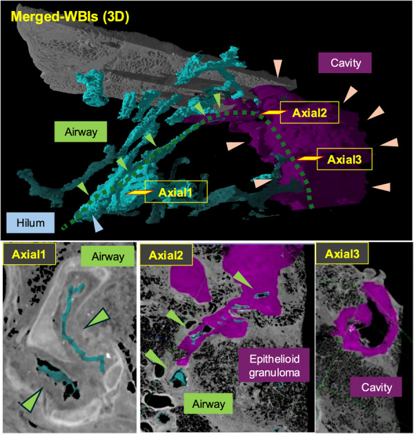 Figure 1
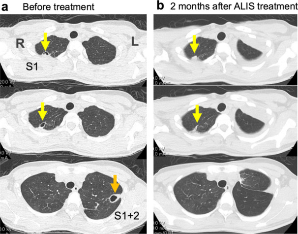 Figure 2
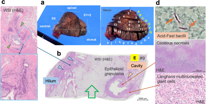 Figure 3
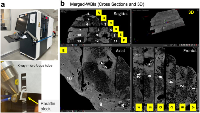 Figure 4
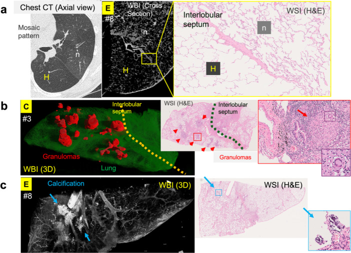 Figure 5
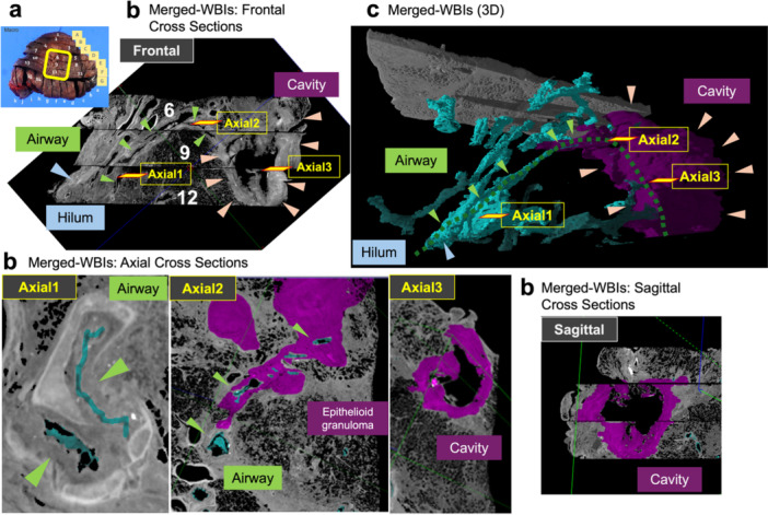 Figure 6
Peer Reviews
No public reviews on file for this paper yet. If you reviewed it on a platform where reviews are public (OpenReview, ICLR, NeurIPS, ICML), you can paste yours below so the community can read it here.
Videos
No videos yet. Explain this paper in a talk, walkthrough, or lecture? Add one.
Taxonomy
TopicsMycobacterium research and diagnosis · Tuberculosis Research and Epidemiology · Inhalation and Respiratory Drug Delivery