Optical coherence tomography angiography reveals insights into complementary vascular and neurodegenerative mechanisms in multiple sclerosis
Charmaine Hiu-Ying Yam, Danuta M Sampson, Andreia Marques Elias, Jed Wingrove, Baris Kanber, Ronja Christensen, Pryanka Sood, Riccardo Nistri, Anna He, Alyssa A Toorop, Elena Panella, Dimitrios Champsas, Suraya Mohamud, Weaam Hamed, Ferran Prados Carrasco, Frederik Barkhof

TL;DR
The study shows that retinal blood vessel changes in multiple sclerosis patients are linked to brain atrophy and vision loss, suggesting new ways to track disease progression.
Contribution
The study introduces optical coherence tomography angiography as a complementary biomarker for assessing neurovascular mechanisms in multiple sclerosis.
Findings
Reduced retinal vessel density in MS patients correlates with atrophy in higher-order visual regions and worse vision.
Peripapillary retinal nerve fibre layer thinning is associated with white matter and optic chiasm volume loss.
Retinal microvasculature abnormalities are linked to grey matter atrophy in visual processing regions.
Abstract
Optical coherence tomography angiography quantifies retinal microvasculature biomarkers, offering insights into neurovascular mechanisms underlying brain damage in multiple sclerosis. This study evaluated these potential mechanisms of neurodegeneration by examining associations between optical coherence tomography and optical coherence tomography angiography metrics, brain volumes and clinical outcomes in people with multiple sclerosis. This cross-sectional study included multiple sclerosis patients from a prospective cohort. Participants underwent optical coherence tomography/optical coherence tomography angiography, vision and clinical assessments and brain MRI. Age- and sex-matched controls underwent optical coherence tomography/optical coherence tomography angiography. The OCTA Vascular Analyser toolbox was used to derive metrics that reflect superficial plexus retinal vessel…
Genes, proteins, chemicals, diseases, species, mutations and cell lines named across the full text — each resolved to its canonical identifier and authoritative record.
Click any figure to enlarge with its caption.
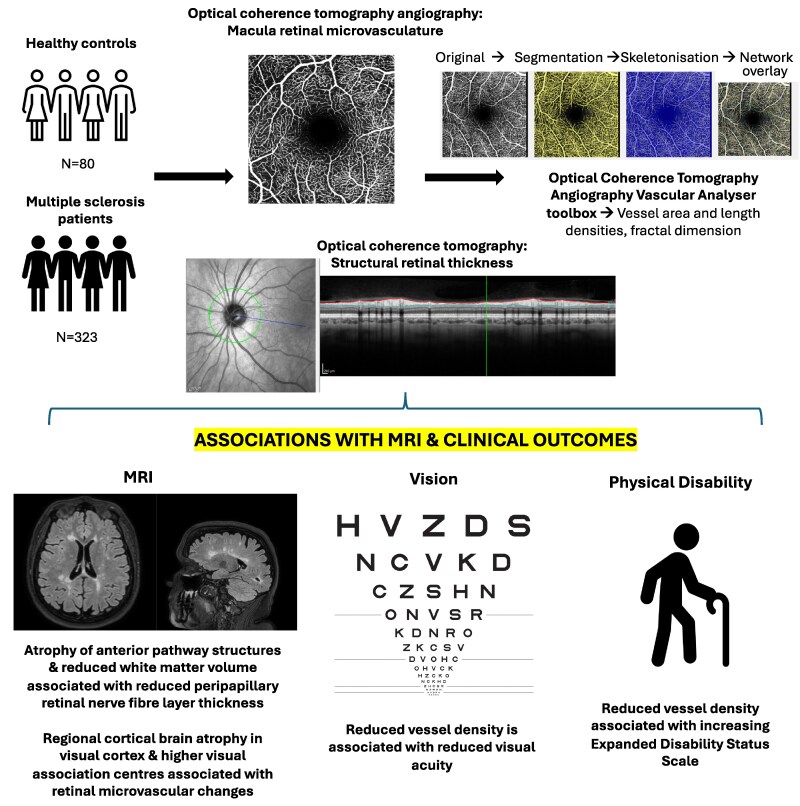 Figure 1
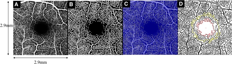 Figure 2
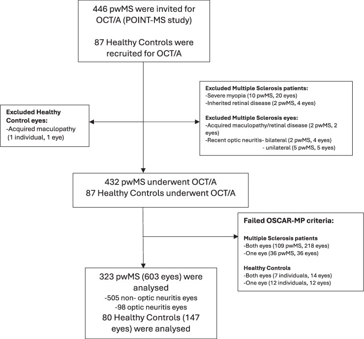 Figure 3
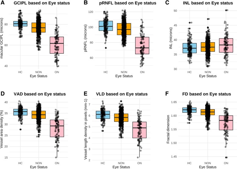 Figure 4
Peer Reviews
No public reviews on file for this paper yet. If you reviewed it on a platform where reviews are public (OpenReview, ICLR, NeurIPS, ICML), you can paste yours below so the community can read it here.
Videos
No videos yet. Explain this paper in a talk, walkthrough, or lecture? Add one.
Taxonomy
TopicsMultiple Sclerosis Research Studies · Ocular Diseases and Behçet’s Syndrome · Systemic Lupus Erythematosus Research