Applicability study of AI attribution methods for ophthalmic image classification
Ali Yavari, Tilman Schmoll, Rainer A. Leitgeb, Kim Lien Huber, Heiko Stino, Andreas Pollreisz, Wolfgang Drexler, Thomas Schlegl

TL;DR
This paper studies how well AI explanation methods work for interpreting deep learning models in ophthalmic imaging, focusing on diabetic retinopathy and fluid detection.
Contribution
The study evaluates and compares three attribution methods for their ability to highlight clinically relevant regions in ophthalmic OCT images.
Findings
AGI and AttEXplore show similar quantitative performance, but AttEXplore highlights clinically meaningful structures better in pathological cases.
High insertion or low deletion scores do not always correlate with clinically meaningful attributions.
All attribution methods require clinical expertise for proper interpretation in potential clinical use.
Abstract
Optical coherence tomography (OCT) enables early detection of vision-threatening diabetic retinopathy (DR) and retinal fluid accumulation, both major complications of diabetes. Despite high classification performance, deep learning models face limited clinical adoption due to poor interpretability. While attribution methods are effective for explaining predictions in the natural image domain, their applicability to medical imaging remains underexplored. To bridge this gap, our work explores how well these strong results transfer to the medical imaging domain. This study evaluates three cutting-edge attribution methods— DeepLIFT, AGI, and AttEXplore —for explaining predictions of a VGG16-based deep learning model in DR classification using widefield OCTA en face images and fluid detection in OCT B-scans. We assess attribution methods’ ability to highlight clinically relevant regions…
Genes, proteins, chemicals, diseases, species, mutations and cell lines named across the full text — each resolved to its canonical identifier and authoritative record.
Click any figure to enlarge with its caption.
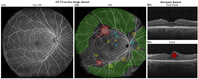 Figure 1
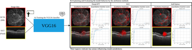 Figure 2
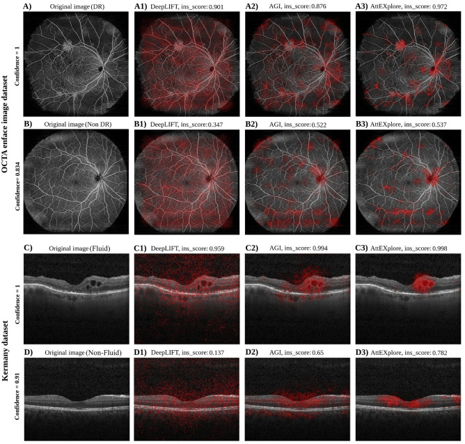 Figure 3
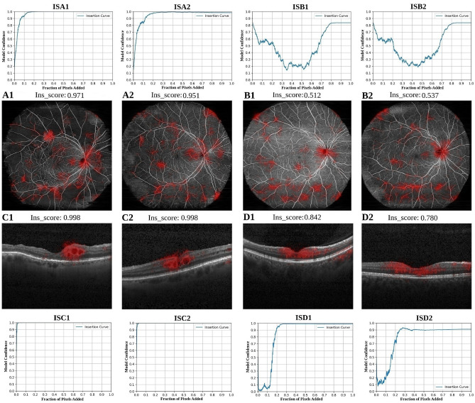 Figure 4
 Figure 5
Peer Reviews
No public reviews on file for this paper yet. If you reviewed it on a platform where reviews are public (OpenReview, ICLR, NeurIPS, ICML), you can paste yours below so the community can read it here.
Videos
No videos yet. Explain this paper in a talk, walkthrough, or lecture? Add one.
Taxonomy
TopicsRetinal Imaging and Analysis · Retinal Diseases and Treatments · Explainable Artificial Intelligence (XAI)