Intralabyrinthine MRI FLAIR as a predictive marker for hearing loss in vestibular schwannomas in Neurofibromatosis Type 2
Robert L. Walker, Maxwell T. Laws, H. Jeffrey Kim, Christopher Zalewski, Ashok Asthagiri, Sruthi Ranganathan, Christina Hayes, John D. Heiss, John A. Butman, Prashant Chittiboina

TL;DR
This study shows that a specific MRI signal in the inner ear can predict hearing loss in patients with a genetic disorder called Neurofibromatosis Type 2.
Contribution
The study introduces intralabyrinthine FLAIR MRI hyper-intensity as a novel predictive biomarker for hearing loss in NF2 patients with vestibular schwannomas.
Findings
FLAIR hyper-intensity in the inner ear was strongly associated with future hearing loss in NF2 patients.
Hearing loss occurred approximately 4 years after FLAIR signal changes in patients with normal hearing at baseline.
Surgery stabilized hearing but did not reverse FLAIR hyper-intensity.
Abstract
The onset of hearing loss due to vestibular schwannomas (VS) is inevitable but does not correlate with the size of the tumor. In patients with Neurofibromatosis Type 2 (NF2) and VS, we previously found an association between pre-contrast fluid-attenuated inversion recovery magnetic resonance imaging (FLAIR MRI) signal in the labyrinth and hearing loss. Here, we asked whether FLAIR hyper-intensity could serve as a predictive biomarker for hearing loss in NF2 patients with VS. A prospective longitudinal study (NCT00598351) of NF2 enrolled 168 subjects between 2008 and 2013. This study included 34 patients with small VS (total volume ≤ 500mm3). Middle fossa decompression surgery (n = 4 patients) was provided via a standard-of-care trial (NCT00060541). From 34 eligible subjects (mean age 26.8y) with NF2 and small VS, 53 ears met inclusion criteria. Abnormal hearing was recorded in 18 ears…
Genes, proteins, chemicals, diseases, species, mutations and cell lines named across the full text — each resolved to its canonical identifier and authoritative record.
Click any figure to enlarge with its caption.
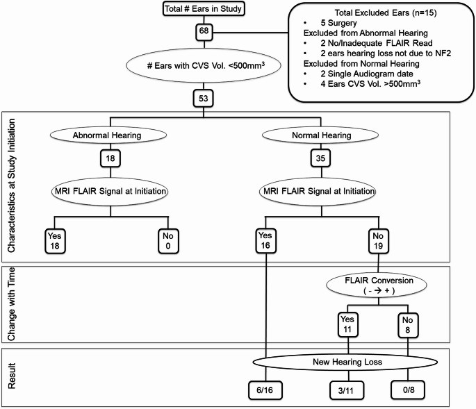 Figure 1
 Figure 2
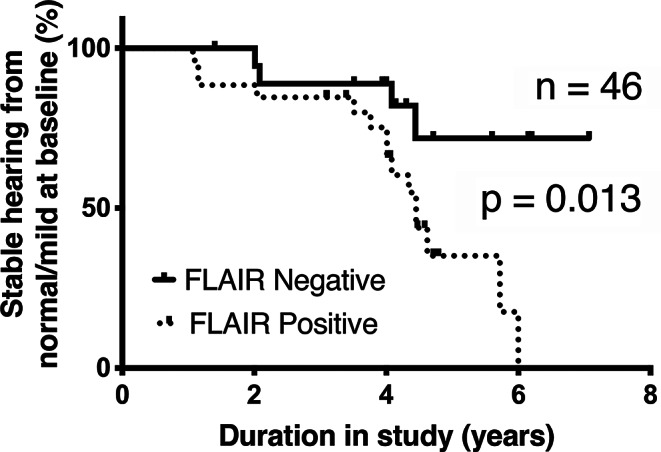 Figure 3
 Figure 4
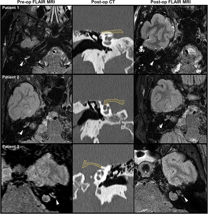 Figure 5
Peer Reviews
No public reviews on file for this paper yet. If you reviewed it on a platform where reviews are public (OpenReview, ICLR, NeurIPS, ICML), you can paste yours below so the community can read it here.
Videos
No videos yet. Explain this paper in a talk, walkthrough, or lecture? Add one.
Taxonomy
TopicsNeurofibromatosis and Schwannoma Cases · Meningioma and schwannoma management · Soft tissue tumor case studies