A BRN2:MYC transcriptional axis regulates interconversion between therapy-resistant and tumorigenic phenotypes in melanoma
Yuntian Zhang, Marcus A. Urquijo, Rebecca G. Zitnay, Kayla Marks, Rachel L. Belote, Maike M.K. Hansen, Montana Ferita, Hannah M. Neuendorf, Tong Liu, Eric A. Smith, Elnaz Mirzaei Mehrabad, Miroslav Hejna, Tarek E. Moustafa, Devin Lange, Min Hu, Fatemeh Vand-Rajabpour, Anne Done

TL;DR
The study identifies two key transcriptional states in melanoma cells that influence tumor growth and resistance to treatment, revealing potential new targets for therapy.
Contribution
The discovery of a BRN2:MYC transcriptional axis that regulates phenotypic switching in melanoma, distinguishing it from melanocytes.
Findings
Melanoma cells switch between an MYC-driven tumor-initiating state and a BRN2-high therapy-resistant state.
Transitions between these states involve MITF-high intermediate states and occur more frequently in melanoma than in melanocytes.
The BRN2-high state is present in both melanoma and melanocytes, but the MYC state is unique to melanoma.
Abstract
Metastatic spread and therapeutic resistance are the principal causes of cancer mortality. For melanoma, these processes rely on the capacity of cells to switch between transcriptional states. Although targeting transcriptional states pharmacologically is promising, the mechanisms by which melanoma cells switch between states—and how these processes differ from melanocytes—remain poorly understood. Here, we isolate distinct melanoma states with unique phenotypes: a MYC-driven state, essential for tumor initiation yet sensitive to BRAF inhibition, and a dedifferentiated, invasive BRN2-high state enriched in therapy-resistant cells but not directly tumorigenic. Transitions between phenotypes occur through intermediate, more differentiated states. Unexpectedly, the BRN2-high state is also present in melanocytes, whereas the MYC state is exclusive to melanoma. Melanoma cells also exhibit an…
Genes, proteins, chemicals, diseases, species, mutations and cell lines named across the full text — each resolved to its canonical identifier and authoritative record.
Click any figure to enlarge with its caption.
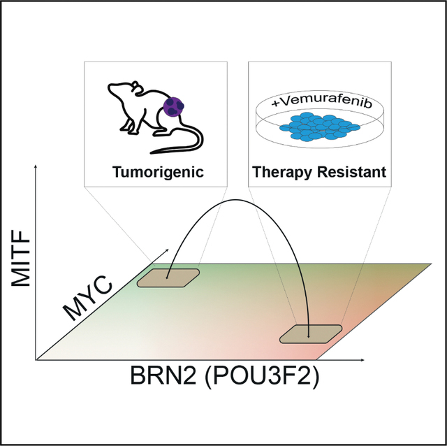 Figure 1
 Figure 2
 Figure 3
 Figure 4
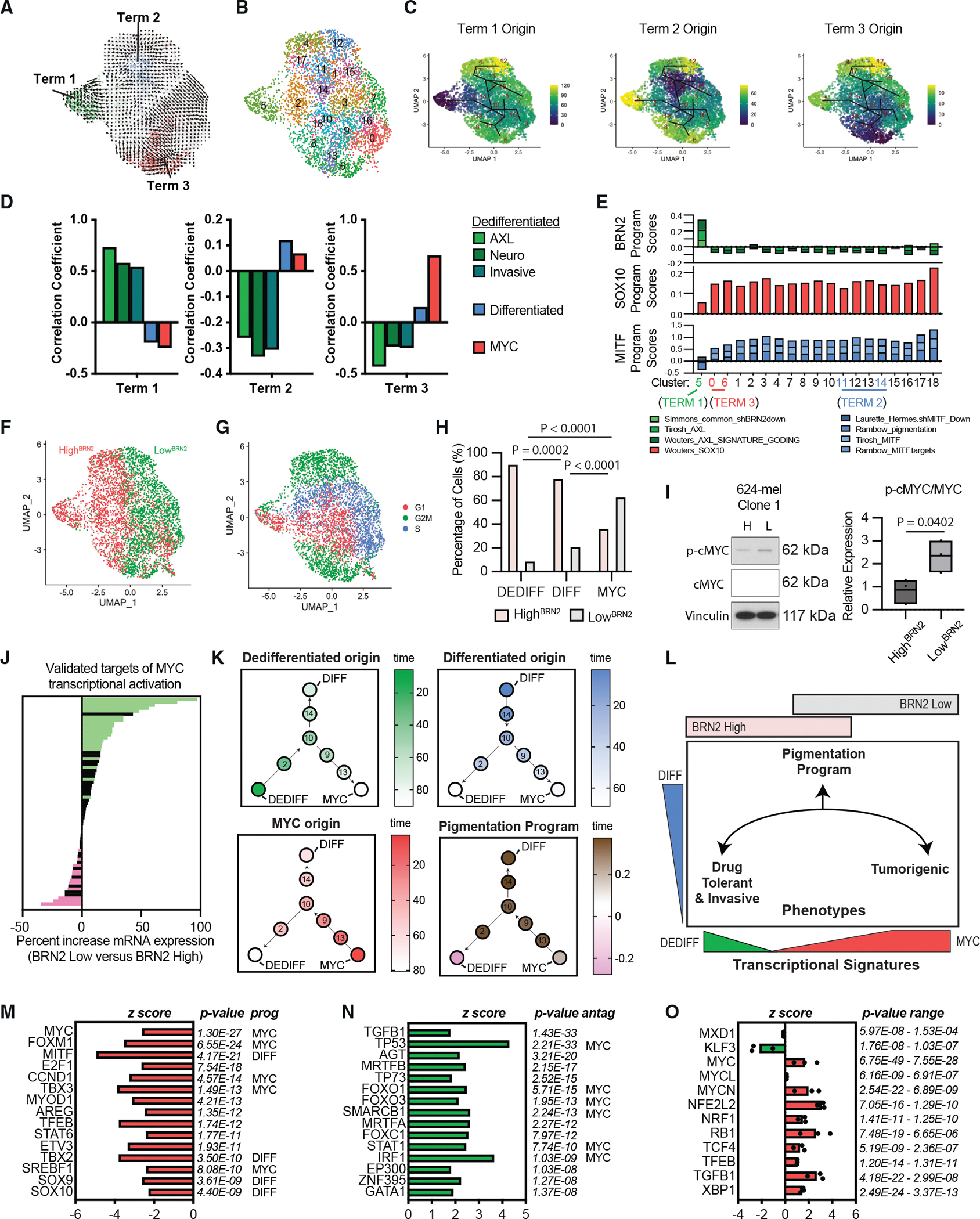 Figure 5
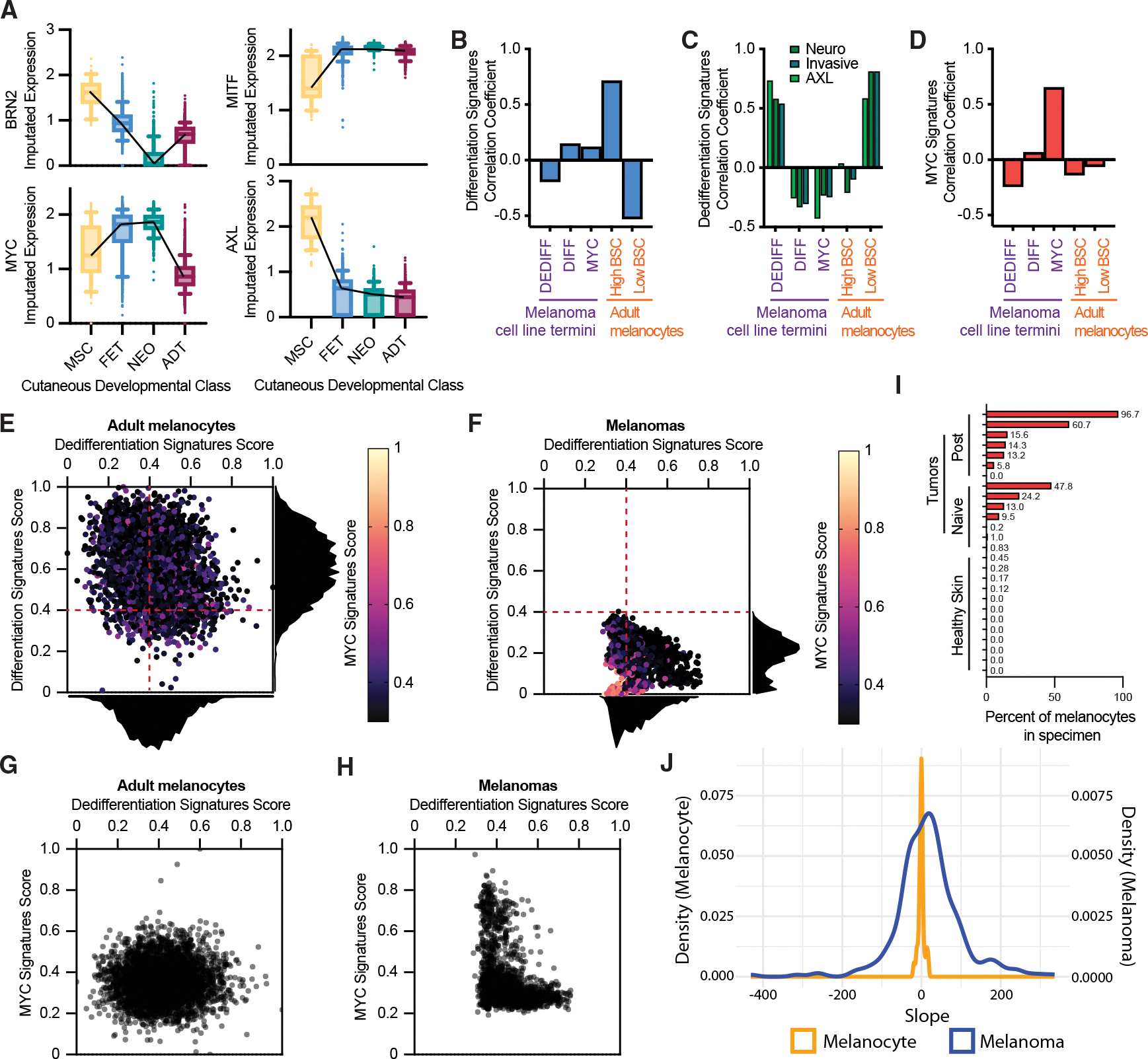 Figure 6
Peer Reviews
No public reviews on file for this paper yet. If you reviewed it on a platform where reviews are public (OpenReview, ICLR, NeurIPS, ICML), you can paste yours below so the community can read it here.
Videos
No videos yet. Explain this paper in a talk, walkthrough, or lecture? Add one.
Taxonomy
TopicsMelanoma and MAPK Pathways · Cutaneous Melanoma Detection and Management · melanin and skin pigmentation