Debonding characteristics of innovative 3D-printed lingual retainers following artificial aging and acid challenge
Noor Salam ALNUAIMY, Akram Faisal ALHUWAIZI, Akram Mohammed ALTAMIMI

TL;DR
This study evaluates the durability of 3D-printed lingual retainers after simulated aging and acid exposure, finding that semi-elliptical designs perform best.
Contribution
The study introduces and evaluates innovative 3D-printed retainers with different cross-sectional designs for orthodontic use.
Findings
3D-printed semi-elliptical retainers showed the highest debonding forces.
After acid challenge, semi-elliptical and oval retainers had no significant differences in debonding forces.
3D-printed retainers primarily exhibited cohesive fracture patterns.
Abstract
Traditional fixed retainers used after orthodontic therapy have many downsides. Innovative 3D-printed fixed retainers have been manufactured using nanoceramic hybrid resin-based material (SprintRay OnX®), investigating their durability to support long-term oral functions. One hundred acrylic blocks with mounted premolar pairs were allocated into five groups, including three groups of 3D-printed retainers with 1-mm round cross-section, one group with 1.5-mm oval cross-section, and one group with 1.5-mm semi-elliptical cross-section, and conventional multistranded stainless steel G&H® retainers and Respond® retainers. The models with bonded retainers were exposed to artificial aging or acid challenge. The debonding forces and the subsequent failure patterns were investigated. The 3D-printed semi-elliptical retainers exhibited the greatest debonding forces, while the 3D-printed oval,…
Genes, proteins, chemicals, diseases, species, mutations and cell lines named across the full text — each resolved to its canonical identifier and authoritative record.
Click any figure to enlarge with its caption.
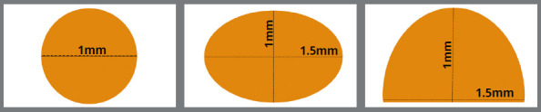 Figure 1
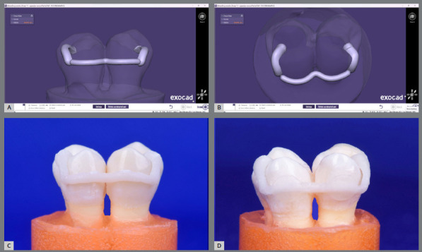 Figure 2
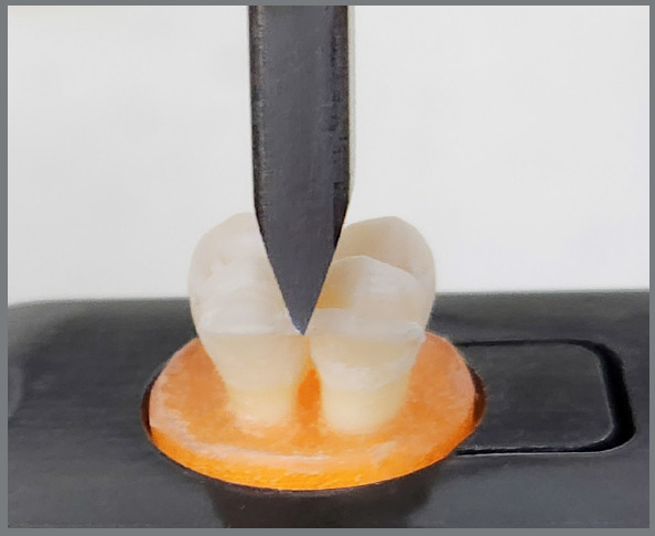 Figure 3
Peer Reviews
No public reviews on file for this paper yet. If you reviewed it on a platform where reviews are public (OpenReview, ICLR, NeurIPS, ICML), you can paste yours below so the community can read it here.
Videos
No videos yet. Explain this paper in a talk, walkthrough, or lecture? Add one.
Taxonomy
TopicsDental materials and restorations · Reconstructive Facial Surgery Techniques · Orthodontics and Dentofacial Orthopedics
INTRODUCTION
The traditional stainless steel multistranded wire (MSW) retainer is still largely considered the gold standard retainer.1^,^2 However, this retainer faces many challenges, such as a high rate of failure, negative impact on the maintenance of periodontal health, undesirable dental movements, and esthetic issues.3 This retainer is also not recommended for individuals with metal hypersensitivity and must be removed before a magnetic resonance imaging (MRI) scan.4 CAD/CAM customized fixed retainers using milling techniques have been introduced recently, promising greater success.2 However, these retainers do not appear to be clinically more effective than MSWs.5 They continue to have limitations, such as bonding difficulties, increased rate of failure, dependence on an external facility, and relatively high costs.2^,^6^-^8
The 3D printing is a cutting-edge technology in the manufacturing industry.9 The clinical orthodontic discipline has become more efficient due to the recent rapid advances in digital and 3D printing technologies offering cost-efficiency, lesser wasting materials, fabrication speed, superior quality and accuracy, and intricate object production.10^,^11 However, ordinary resin 3D-printed products still have some drawbacks related to their physical and mechanical properties. Incorporating nanoparticles or nanofillers into the raw material, utilizing their unique properties, including reactivity and high surface area, may push the boundaries of 3D printing technologies, facilitating the development of multifunctional materials with customized qualities.12^,^13
The present study sought to create innovative, 3D-printed personalized fixed retainers from nanoceramic hybrid resin-based material. This nanomaterial was used to fabricate prosthetic intraoral devices. It is a resin packed in nanoceramics to give it phenomenal strength and esthetic, a critical consideration when fabricating lingual retainer. The proposed benefits may include improved fitting accuracy, position, and interproximal adaptation, which may prevent direct occlusal contact with lower teeth, thereby reducing the chance of failure of the maxillary retainer. Passive fit is also essential, as the tension of the wire during bonding may result in unanticipated teeth movements. It is suitable for individuals with metal hypersensitivity or for MRI scans, due to its complete absence of metals. The objectives of this study were to measure the debonding forces and failure patterns after artificial aging and acid challenge protocols used to mimic the exposure of bonded retainers to intraoral challenges.
MATERIAL AND METHODS
PREPARATION OF SAMPLES
Ethical approval (Ref. number: 619) was obtained from the local Research Ethical Committee. Maxillary first premolars (245 teeth) were gathered from patients aged 15 to 30 years, which were extracted for orthodontic needs. The stereomicroscope with a magnification of 10x was used to inspect the teeth. The criteria for selecting the teeth included: intact lingual surfaces with no cracks, restorations or caries, and no previous bleaching or orthodontic treatments. Teeth of average size were selected, with a mesiodistal width of 7 to 7.5 mm, and a length of 22 to 23 mm. The chosen teeth were stored at ambient temperature in a weekly-changed 0.1% thymol solution. Each tooth crown was cut from a single proximal aspect by less than 1 mm, to imitate the close contact between the front teeth.
The analyzing rod of the dental surveyor was used to vertically mount each tooth pair in the acrylic into a cylindrical-shaped plastic mold. The lingual surfaces were aligned at the same level using a specially-made T-shaped instrument affixed to the surveyor. The mixed self-cure acrylic resin was poured 2 mm apically to the levels of cementoenamel junctions. These blocks remained at room temperature in distilled water until they were bonded.
The sample blocks were grouped into five sets of 20, in the following order (Fig. 1):
Figure 1:Cross-sections of the 3D-printed customized nanoceramic hybrid lingual retainers.
- » Group I (3D-printed round): 3D-printed customized round fixed retainers with a diameter of 1 mm.
- » Group II (3D-printed oval): 3D-printed customized oval fixed retainers with a thickness of 1 mm and a width of 1.5 mm
- » Group III (3D-printed semi-elliptical): 3D-printed customized semi-elliptical fixed retainers with a thickness of 1 mm and a width of 1.5 mm.
- » Group IV (G&H^®^ MSWs): G&H^®^ multistrand retainer wire; 0.0195-in 7-stranded stainless steel twisted retainers (G&H^®^ Orthodontics, USA).
- » Group V (Respond^®^ MSWs): Respond^®^ archwire; 0.0195-in 6-stranded stainless steel dead-soft coaxial retainers (Ormco Corp., USA).
Each group was further divided into 10 specimens subjected to debonding after either artificial aging or acid attack exposure.
The intraoral scanner (3Shape TRIOS 3; 3Shape, Denmark) was used to scan the affixed teeth of the 3D-printed groups, which produced STL files. The scanner had been calibrated earlier according to the manufacturer’s specifications. The retainers were designed using Exocad Dental CAD software. The retainer was conformed touching the middle third of the lingual surfaces of teeth, and adaptable to the interdental region. Two attached jigs, adapted to the occlusal and lingual surfaces, were designed distally to both bonding sites to facilitate accurate placement and handling (Fig. 2A, 2B). The Autodesk Meshmixer software was used to re-mesh the design’s STL format, enhancing the printing quality.
Figure 2:The 3D-printed customized lingual retainer: A, B) designing; C) before bonding; D) after bonding.
The retainers were printed following the manufacturer’s guidelines using the Pro 95 Printer (SprintRay, California, USA) as well as RayWare software (version 2.9.1). The SprintRay OnX nanoceramic hybrid resin-based material container (SprintRay, California, USA) was mixed well with an LC-3D Mixer before each printing process. The retainers were produced horizontally to the building base, to prevent failure due to the irregular fine wire structure, which prohibits vertical print. They were built with a 50-µm layer thickness and remained on the build base for 5-10 minutes. Next, they were cleansed by 91% isopropyl alcohol spraying, and dried using compressed air, to eliminate all residual uncured resin. Seven cleansing cycles were implemented. The retainers were then post-processed using the ProCure Post-Curing System (SprintRay, California, USA), with a 90-watt LED, 28.8 mW/cm2 light intensity and 365-405 nm wavelength, for 60 minutes at 60°C, to enhance strength, precision, and polymerization. The supports were cut using a diamond disc as close to the retainer as possible (Fig. 2C). To ensure sufficient print accuracy, the retainers were examined using an 10x magnification stereomicroscope and a digital caliper. Samples with flaws were discarded.
MSWs retainer of 15 mm length was manually gently curved, fitting passively over the middle thirds of the lingual teeth surfaces, close to the interdental region and parallel to the block bases.
BONDING METHOD
The lingual teeth surfaces were polished for 10 seconds with pumice free of fluoride, using rubber cups, rinsed with water spray for 10 seconds, and dried with compressed air for 10 seconds. Next, they were etched using Super-Etch etch gel with 37% phosphoric acid (SDI, USA) for 30 seconds, completely rinsed with water for 20 seconds, and dried out using compressed air for 20 seconds, until the area being etched turned chalky white. An even thin primer coating (Transbond^™^ XT; 3M Unitek, California, USA) was put on the lingual surfaces of teeth, and then delicately dispersed with a compressed air jet for 3 seconds.
A mini-mold wire bonder (4-mm diameter and 1.5-mm depth) was employed to standardize the bond pad’s form and dimensions, and confirm that the wire was accurately centered inside the adhesive bond. In line with the manufacturer’s directions, the adhesive (Transbond^™^ LR; 3M Unitek, California, USA) underwent light curing for 10 seconds using 3M^™^ Elipar^™^ DeepCure-L (3M ESPE, Germany), with a light intensity of 1470 mW/cm2 and wavelength of 430-480 nm (Fig. 2D). Next, the retainer jigs were meticulously eliminated with a diamond fine fissure bur.
DEBONDING FORCES
A. ARTIFICIAL AGING
Fifty bonded specimens were subjected to a two-year aging protocol, during which retainer failures were reported in the clinical studies.14 The thermocycling procedure was applied following ISO/TS.1140515 recommendation, to imitate the intraoral thermal fluctuation. Samples were placed inside a perforated stainless steel container of a custom-made thermocycler, and subjected to 20,000 cycles between 55±0.5°C and 5±0.5°C water baths, with 30 seconds dwelling time and 5 seconds transfer time each way.
The thermocycling was followed by a cyclic loading procedure using a custom-made cyclic loading machine, with an automatic cut-off calculator, when the specimen failed. A stainless steel chisel plunger (1 mm diameter) delivered an occluso-gingival vertical force to the center of the interdental region of the retainer at room temperature. To simulate masticatory forces, the specimens were subjected to 10^6^ cyclic loadings,16 a pre-set load of 20 N,17 and at 2 Hz,18 corresponding to the estimated oral chewing frequency. Cyclic loading was applied either until the retainer failed or until the limit of cycles was reached. Any failure during the mechanical fatigue was recorded.
B. ACID CHALLENGE
Fifty bonded specimens were immersed in a 2.5 pH acidic solution of diluted hydrochloric acid (HCl) for a 5-minute session, three times daily, and separated by 2-hour intervals of re-immersion in distilled water (pH 6) at 37°C. The samples were then stored in distilled water for the rest of the day at 37°C. Before and after each session, samples were rinsed under running water and air-dried. After each session, the acidic solution and the distilled water were replaced with new, freshly prepared solutions. This protocol was repeated for thirty days19, to mimic the impact of intraoral acidity fluctuation.
C. DEBONDING PROCEDURE
Following artificial aging and acid challenge, all blocks were debonded using a universal testing machine (Tinius Olsen; H50KT, England) equipped with a load cell of 50 kN at room temperature. The lower jaw of the machine was equipped with a sample-holding tool that was used to fix the sample individually. The upper moving jaw was supplied with a plunger with a diameter of 1 mm. The crosshead, imitating the force of bite, exerted a 1 mm/minute vertical load on the interdental midpoint. The applied load was raised gradually until failure happened on a minimum of one bonding site of the sample (Fig. 3).
Figure 3:The debonding test, with the block fixed into the sample-holding tool and the plunger exerting a vertical load on the interdental region.
FAILURE PATTERNS
Two operators investigated the enamel and wire surfaces in a stereomicroscope with 10x magnification, following the retainers’ failure. The examiner conducted assessments again after a month, and a different operator assessed the same specimens. The intra-examiner reliability and the inter-examiner reliability were verified using the Kappa test, which was evaluated according to the ratings of Landis and Koch.20 The results indicated a nearly perfect agreement (0.97 each). The failure patterns for the two bonding locations were recorded, and the data of the first to fail was analyzed. A smaller score was assigned in instances where the two bonding locations failed at the same time. The ranking system was altered from the one proposed by Foek et al.16, in the following manner:
- » Type 1: indicates complete debonding of the adhesive from the tooth surface.
- » Type 2: indicates partial debonding of the adhesive from the tooth surface.
- » Type 3: indicates that the adhesive was not debonded from the tooth surface; rather, the covering adhesive over the retainer has detached.
- » Type 4: indicates that the retainer was not debonded from the tooth surface; rather, it was fractured.
- » Type 5: indicates that the adhesive was not debonded from the tooth surface; rather, the retainer has dislodged.
STATISTICAL ANALYSES
The SPSS software program (version 25; IBM, USA) was used to conduct statistical analyses witha level of significance of p < 0.05. The reliability of failure mechanisms was evaluated using Kappa ratings. The debonding forces were subjected to the Shapiro-Wilk test and Levene’s test, which revealed that the data had a normal distribution, with identical variances. Thus, Tukey HSD post-hoc tests were implemented after the one-way ANOVA. The failure patterns were compared using the Kruskal-Wallis test, followed by the Pairwise comparison test.
RESULTS
The specimens survived thermocycling, cyclic loading, and acid challenge. The 3D-printed semi-elliptical retainers yielded the greatest debonding forces for the two debonding tests, with the 3D-printed oval, G&H^®^ MSWs, 3D-printed round, and Respond^®^ MSWs retainers following, in that order. Debonding forces showed statistically significant differences in all comparisons across groups, according to Tukey HSD post-hoc multiple comparisons. However, no significant disparities existed between 3D-printed semi-elliptical and oval retainers following the acid challenge (Table 1).
Table 1:Debonding forces (N) of 3D-printed customized retainers versus conventional retainers.TestsGroupsnMeanSDMinMaxANOVA** Post-hoc**significanceDebonding after aging3D-printed round1084.3102.25081.5088.30F = 3017.411 df = 49 p = 0.000All groups 3D-printed oval10112.2501.639109.70115.70All groups3D-printed semi-elliptical10114.3101.051112.80115.90All groupsG&H^®^ MSWs10107.3301.179105.50109.00All groupsRespond^®^ MSWs1050.7401.31048.5052.50All groupsDebonding after acid challenge3D-printed round1092.6101.76990.1095.20F = 1811.171 df = 49 p = 0.000All groups3D-printed oval10120.9102.409116.50124.503D-printed round* G&H^®^ MSWs* Respond^®^ MSWs3D-printed semi-elliptical10122.6802.426118.50125.603D-printed round Respond^®^ MSWs* G&H^®^ MSWsG&H^®^ MSWs10110.7302.609107.50114.40All groupsRespond^®^ MSWs1052.8501.20351.5055.00All groups** The difference is significant.
FAILURE PATTERNS
Most of the 3D-printed retainers presented type 4 failure. G&H^®^ MSWs experienced types 2, 3, and 5, whereas type 5 failures predominated in Respond^®^ MSWs during both debonding tests. The Kruskal-Wallis test showed that retainer type affected failure patterns after artificial aging, but not after acid challenge. Pairwise comparisons after aging demonstrated significant differences for G&H^®^ MSWs compared to 3D-printed round retainers and Respond^®^ MSWs. The significance values were adjusted by Bonferroni correction (Table 2).
Table 2:Failure patterns of 3D-printed customized retainers versus conventional retainers.TestsGroupsnFailure Patterns Kruskal Wallis Statistics Pairwise SignificanceType 1Type 2Type 3Type 4Type 5
Failure patterns after aging3D-printed round10000100H = 16.114 df = 4 p-value = 0.003G&H^®^ MSWs3D-printed oval1001090None3D-printed semi-elliptical1001090NoneG&H^®^ MSWs10072013D-printed round * Respond^®^ MSWs Respond^®^ MSWs1003007G&H^®^ MSWsFailure patterns after acid challenge3D-printed round1001090H = 4.617 df = 4 p-value = 0.329 3D-printed oval1002080 3D-printed semi-elliptical1002080 G&H^®^ MSWs1002305 Respond^®^ MSWs1003007
- The difference is significant.
DISCUSSION
Bonded lingual retainers are supposed to function in harsh oral environments, including long-term humidity, fluctuations in pH and temperature, and cyclic loading caused by mastication, occlusion, and intraoral parafunctional habits. Lifelong retention burdens the lingual retainers’ durability.2 However, the main problem associated with MSWs is the high rate of retainer failure, particularly in the maxilla. This failure is primarily due to bond failure, which may be caused by masticatory forces and occlusal interferences.1 On the other hand, a real-life orthodontic patient frequently consumes a highly acidic beverage. This study verified the long-term durability of the bonding and the new 3D-printed retainers under the influence of artificial in-vitro aging and acid challenge models.
All specimens survived thermocycling, cyclic loading, and acid attack, suggesting that the retainers complex could tolerate these aggressive stresses and, consequently, predict clinical durability. The 3D-printed semi-elliptical and oval retainers achieved the greatest debonding forces. Their digital designs and flat shapes resulted in a more even distribution of forces over the teeth surfaces, and improved durability when exposed to bending stress across their bigger dimensions. Additionally, the chemical robust bonds with the adhesive as well as the nanofiller particles were assumed to give superior bond and strength, respectively. However, 3D-printed retainers were fractured rather than debonded, following both aging and acid attacks. Water exposure (during thermocycling and acid challenge) might attack the ester bonds in methacrylate, the major constituent of the 3D-printed resin and composite adhesive. Water particle diffusion into the matrix could initiate two series of events in the composite material, including hygroscopic expansion and chemical degradation.21 It could act as a plasticizer of the polymer, thereby deteriorating the structural integrity of resins, decreasing their mechanical performance, and accelerating the process of failure.22 The hydrolytic degradation process of the 3D-printed resin matrix may be attributed to the polarity of the oxane bonds connecting the filler and silanes; thus, the polymer microstructure is significantly altered and irreversibly damaged. Thermocycling can potentially increase water sorption, particularly in warm temperatures.23 The flexural strength of 3D-printed resins deteriorated associated with thermocycling24^,^25 or related to water storage.26^,^27 On the other hand, TEGDMA, marked by hydrophilic ether linkages, exhibits the highest hydrophilicity, followed by bis-GMA; both are the ingredients of the Transbond LR adhesive,28 yet the water sorption property of 3D-printed resin is thought to be more prominent, due to its microstructure. Additive manufacturing generates multiple layers; therefore, water permeates in micro-spaces and displaces the layers from one another.22 Considering the possible incomplete polymerization at the interface between layers, microporosities, and residual monomers can also contribute to the increased hydrolysis potential. Moreover, 3D-printed materials have relatively low nanofiller content, as opposed to Transbond LR (high filler, 75-85%), and the relationship between surface degradation and filler content is inverse.29 Conversely, 3D-printed retainers may enhance thermal stability and mitigate mechanical fatigue formation in the retainer-adhesive-enamel complex, in comparison to MSWs. The adverse effect of thermal fluctuation and cyclic loading on materials with comparable coefficients of thermal expansion, thermal conductivity, and physical properties is more predictable and has an equivalent impact.16 However, a uniaxial compression of the 3D-printed sample resulted in the formation of numerous microcracks, potentially extending from the main cracks created between the layers. Also, the defective resin matrix caused by water diffusion could gradually undermine the reinforcing effects of the nanofillers, weakening the corroded subsurface layer. The direct cyclic loading renders it more susceptible to contact wear,30 a significant contributor to the dental materials’ deterioration. Multiple preceding factors could contribute to the fracturing of 3D-printed retainers. On the contrary, the physical properties differences between metal MSWs and adhesive are more remarkable than between enamel and adhesive. Consequently, the wire-adhesive bond would weaken with mechanical fatigue.
Notably, the 3D-printed oval and semi-elliptical retainers exhibited comparable debonding values for both debonding categories; although significant differences after aging signified different stress distribution patterns during cyclic loading. Additionally, the better adaptability of 3D-printed semi-elliptical retainers to the teeth’s lingual anatomy could potentially play a role. Nevertheless, these differences were not clinically noteworthy. This is indicative of their similar cross-sectional areas.
Bonding efficacy and the 3D-printed retainers’ strength could be compromised due to water sorption, erosive effects of the acidity, or both, resulting in degradation of the resin’s polymer network. Methacrylate degradation is known to be accelerated by pH variations.31 However, this process is impacted by additional intraorally variables, including the presence of enzymes and saliva’s buffering capacity, which mitigates the erosive effects of acids.
SprintRay OnX 3D-printed material was selected because, as stated by the manufacturer, it was designed to deliver superior flexural strength and modulus in its class, a critical consideration when utilizing a lingual retainer. Only one nanoceramic material was tested, which limits judgements about the overall efficiency of 3D-printed retainers. This research idea was not focused on a particular material; it was about the utilization of 3D printing for the fabrication of customized lingual retainers. In addition, the nanomaterials sector is always expanding and developing on a daily basis. Integrating nanotechnology and 3D printing can potentially customize the 3D printing material properties to meet certain clinical needs. This was shown by the material made with Nano-Fusion^™^ (OnX Tough), introduced by the same SprintRay company one year after the SprintRay OnX, with superior qualities. It guarantees denser polymer chains and more homogenous particle distribution. It might impact biomechanical behavior and, therefore, the longevity of 3D-printed retainers made on a manner similar to those employed in this study.
For the lingual comfort of the patient, the retainers’ lingual aspects were designed with rounded designs. In addition to round wire, semi-elliptical and oval cross-sectional wires were set up, so that their highest dimension were across the lingual surfaces of the teeth, to increase their strength and the surface area of retention, yet its thickness was held to a minimum, which would have allowed for a lower profile, avoiding bulky constructions and improving patient comfort. Nonetheless, any cross-sectional shape may be designed and produced with 3D printing. In the present study, only three cross-sectional designs were investigated; and a broader range of cross-sectional designs might reveal additional information about the best shapes for 3D-printed retainers.
The results of this study are inherently incapable of reproducing intricate clinical situations, as is the case with any in-vitro research. However, assessing debond forces following rigorous aging methods mitigated some of these variables. Clinical studies are required to evaluate the effect of the patient real mastication, saliva composition, physiologic teeth movement, tongue functional forces, mouth rinsing or brushing, and the presence of calculus and plaque, which may significantly impact the debonding process.
CONCLUSION
The 3D-printed nanoceramic hybrid semi-elliptical and oval retainers demonstrated desirable debonding forces, although the cohesive fracture pattern was dominant, following both artificial aging and acid challenge protocols. Nevertheless, long-term clinical studies are warranted.
The reference list from the paper itself. Each links out to its DOI / PubMed record.
- 11 Gelin E, Seidel L, Bruwier A, Albert A, Charavet C. Innovative customized CAD/CAM nickel-titanium lingual retainer versus standard stainless-steel lingual retainer: a randomized controlled trial. Korean J Orthod. 2020;50(6):373-82.10.4041/kjod.2020.50.6.373PMC 764223133144526 · doi ↗ · pubmed ↗
- 22 Roser CJ, Bauer C, Hodecker L, Zenthofer A, Lux CJ, Rues S. Comparison of six different CAD/CAM retainers vs. the stainless steel twistflex retainer: an in vitro investigation of survival rate and stability. J Orofac Orthop. 2023;86(2):119-28.10.1007/s 00056-023-00486-y PMC 1186124637378840 · doi ↗ · pubmed ↗
- 33 Kartal Y, Kaya B, Polat-Ozsoy O. Comparative evaluation of periodontal effects and survival rates of Memotain and five-stranded bonded retainers: a prospective short-term study. J Orofac Orthop. 2021;82(1):32-41.10.1007/s 00056-020-00243-532780168 · doi ↗ · pubmed ↗
- 44 Hasanin M, Kaplan SEF, Hohlen B, Lai C, Nagshabandi R, Zhu X, et al. Effects of orthodontic appliances on the diagnostic capability of magnetic resonance imaging in the head and neck region: a systematic review. Int Orthod. 2019;17(3):403-14.10.1016/j.ortho.2019.06.00131285157 · doi ↗ · pubmed ↗
- 55 Pullisaar H, Cattaneo PM, Gera A, Sankiewicz M, Bilinska M, Vandevska-Radunovic V, et al. Stability, survival, patient satisfaction, and cost-minimization of CAD/CAM versus conventional multistranded fixed retainers in orthodontic patients: a 2-year follow-up of a two-centre randomized controlled trial. Eur J Orthod. 2024;46(2):cjae 006.10.1093/ejo/cjae 006PMC 1088851838394353 · doi ↗ · pubmed ↗
- 66 Kadhum AS, Alhuwaizi AF. The efficacy of polyether-ether-ketone wire as a retainer following orthodontic treatment. Clin Exp Dent Res. 2021;7(3):302-12.10.1002/cre 2.377PMC 820402733314768 · doi ↗ · pubmed ↗
- 7Kadhum AS Alhuwaizi AF The effect of composite bonding spot size and location on the performance of poly-ether-ether-ketone (PEEK) retainer wires J Baghdad Coll Dent 202133219
- 8Ruwiaee RA Alhuwaizi AF Effect of artificial aging test on PEEK CAD/CAM fabricated orthodontic fixed lingual retainer J Baghdad Coll Dent 202234216