A rare pancreatic tumor unamenable to accurate diagnosis by EUS–guided tissue acquisition
Naho Kondo, Akihiko Kida, Jun Asai, Tatsuya Yamashita, Takeshi Urabe, Taro Yamashita

Abstract
Genes, proteins, chemicals, diseases, species, mutations and cell lines named across the full text — each resolved to its canonical identifier and authoritative record.
Click any figure to enlarge with its caption.
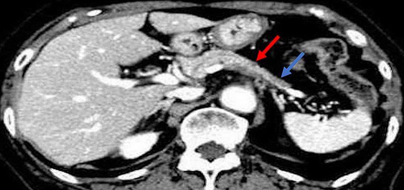 Figure 1
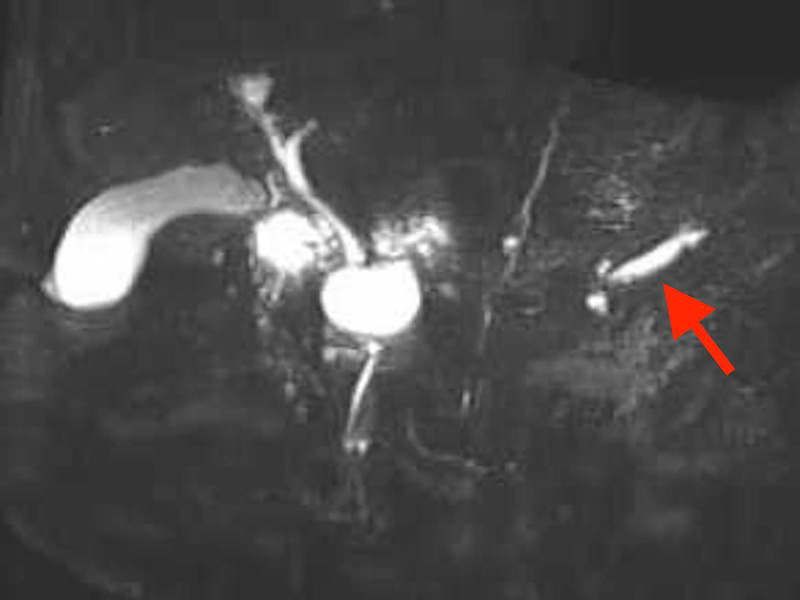 Figure 2
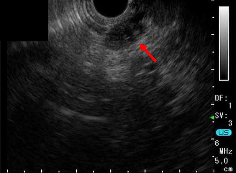 Figure 3
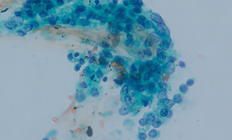 Figure 4
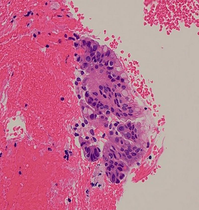 Figure 5
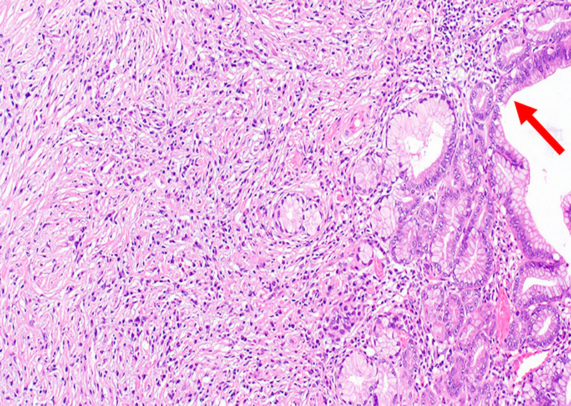 Figure 6
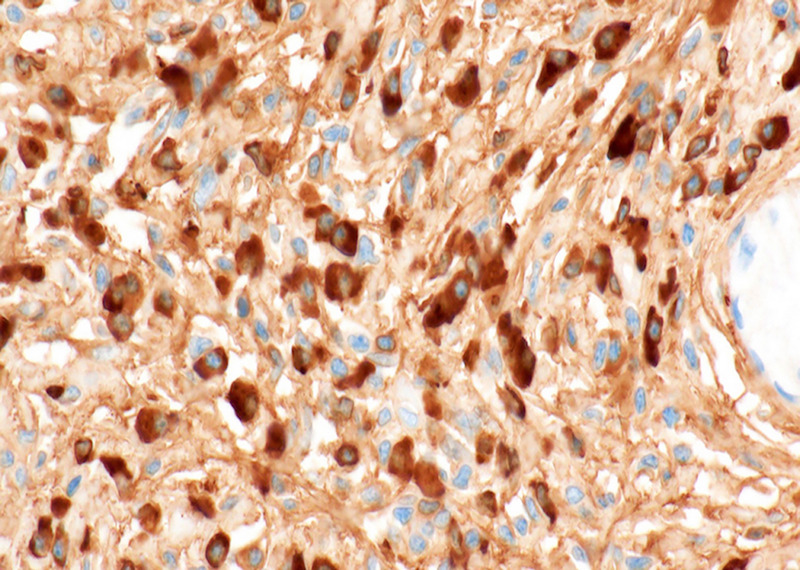 Figure 7
Peer Reviews
No public reviews on file for this paper yet. If you reviewed it on a platform where reviews are public (OpenReview, ICLR, NeurIPS, ICML), you can paste yours below so the community can read it here.
Videos
No videos yet. Explain this paper in a talk, walkthrough, or lecture? Add one.
Taxonomy
TopicsPancreatic and Hepatic Oncology Research · IgG4-Related and Inflammatory Diseases · Gastrointestinal disorders and treatments
An 85-year-old woman was diagnosed with an 11-mm nodular lesion in the pancreatic body, with mild dilation of the main pancreatic duct (MPD) in the pancreatic tail on computed tomography [Figure 1]. Blood tests showed normal CEA and CA19–9 levels. Magnetic resonance cholangiopancreatography revealed 3.5-mm MPD dilation in the pancreatic tail [Figure 2]. Fluorodeoxyglucose-positron emission tomography demonstrated mild accumulation at the nodular lesion. Because of the diagnostic challenge in distinguishing pancreatic cancer from other pancreatic diseases, EUS–guided tissue acquisition (EUS-TA) was planned. EUS revealed an 11-mm hypoechoic nodular lesion in the pancreatic body [Figure 3]. EUS-TA was performed using a 22-gauge Franseen needle, and pancreatic cancer was strongly suspected [Figures 4 and 5]. We performed distal pancreatectomy. Histological examination revealed infiltration of lymphocytes and plasma cells in the stroma surrounding low-grade pancreatic intraepithelial neoplasia (PanIN) in the pancreatic body [Figure 6]. Notably, no pancreatic cancer was observed. Immunohistochemistry revealed over 100 IgG4-positive plasma cells per high magnification field in the stroma surrounding PanIN [Figure 7]. In the tail region, no infiltration of lymphocytes or plasma cells was observed, and no PanIN was present. The postoperative diagnosis was coexistence of low-grade PanIN and focal type 1 autoimmune pancreatitis (AIP).
Although PanIN has been reported to increase with age and is found in 33% of men older than 60 years,^[1]^ it is unlikely that the coexistence of PanIN and AIP in our case was incidental. As PanIN lesions were observed only in areas of AIP, AIP may have contributed to the development of PanIN. Few studies have reported that AIP is associated with the development of PanIN, and whether AIP is a risk factor for the development of pancreatic cancer is controversial.^[2]^ Our case is valuable because it may suggest a process in which AIP is involved in the development of neoplastic lesions such as PanIN. Furthermore, although it is well known that EUS-TA has higher diagnostic yields for pancreatic solid tumors,^[3]^ it should be noted that unusual pancreatic solid tumor such as the coexistence of PanIN and AIP may be difficult to accurately diagnose even with EUS-TA.
Acknowledgments
None.
Source of Funding
None.
Ethical Approval
This case was conducted in accordance with the ethical standards described in the latest revision of the Declaration of Helsinki.
Informed Consent
Informed consent for patient participation and publication was received from the patient.
Conflicts of Interest
All the authors declare that no conflicts of interest exist.
Author Contributions
A. Kida did the concept and design. N. Kondo and A. Kida did the data acquisition. N. Kondo and A. Kida contributed to the analysis and interpretation of data. N. Kondo, A. Kida, J. Asai, T. Yamashita, T. Urabe, and T. Yamashita did the manuscript writing and review. T. Urabe and T. Yamashita supervised the study.
Data Availability Statements
All data relevant to the case are included in the article.
The reference list from the paper itself. Each links out to its DOI / PubMed record.
- 1Ito H Kawaguchi Y Kawashima Y, . A case of pancreatic intraepithelial neoplasia that was difficult to diagnose preoperatively. Case Rep Oncol 2015;8:30–36.25762925 10.1159/000371842 PMC 4342858 · doi ↗ · pubmed ↗
- 2Hart PA Kamisawa T Brugge WR, . Long-term outcomes of autoimmune pancreatitis: A multicentre, international analysis. Gut 2013;62:1771–1776.23232048 10.1136/gutjnl-2012-303617 PMC 3862979 · doi ↗ · pubmed ↗
- 3ASGE Standards of Practice Committee, Machicado JD Sheth SG Chalhoub JM, . American Society for Gastrointestinal Endoscopy guideline on the role of endoscopy in the diagnosis and management of solid pancreatic masses: Summary and recommendations. Gastrointest Endosc 2024;100:786–796.39387777 10.1016/j.gie.2024.06.002 · doi ↗ · pubmed ↗