Protective role of astaxanthin against bisphenol A induced biochemical and histopathological alterations in rat kidneys
Burcu Gültekin, Seda Çetinkaya Karabekir, İlknur Çınar Ayan, Hasan Basri Savaş, Serpil Kalkan

TL;DR
This study shows that astaxanthin can protect rat kidneys from damage caused by bisphenol A, a common toxin linked to kidney disease.
Contribution
The novel contribution is demonstrating astaxanthin's protective effect against BPA-induced kidney damage in a rat model.
Findings
Astaxanthin reversed BPA-induced inhibition of PON1 and ARE enzyme levels and reduced elevated urea levels.
BPA caused kidney tissue damage including vacuolization and collagen increase, which were improved by astaxanthin treatment.
Astaxanthin partially inhibited BPA-induced apoptosis in kidney tissue.
Abstract
This study investigates the ability of astaxanthin (ASTX), a powerful anti-oxidant, to protect kidney tissue from oxidative and cellular damage resulting from bisphenol A (BPA) toxicity, a widespread global toxin associated with chronic kidney disease. We used 32 male Wistar Albino rats, 16 weeks old, and weighing 250–300 g. The rats were randomly divided into four groups: Control, Sham, BPA, and BPA+ASTX. Following the experiment, serum samples were assessed for Paraoxonase 1 (PON1), Arylesterase (ARE), urea, and creatinine levels. Changes in kidney tissue induced by BPA were examined using histopathological methods. Also, the levels of apoptosis and collagen content were evaluated. ASTX treatment reversed the BPA-induced inhibition of PON1 and ARE levels, restoring them to control levels, and reduced the BPA-induced increase in urea levels. Creatinine levels showed no significant…
Genes, proteins, chemicals, diseases, species, mutations and cell lines named across the full text — each resolved to its canonical identifier and authoritative record.
Click any figure to enlarge with its caption.
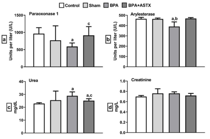 Figure 1
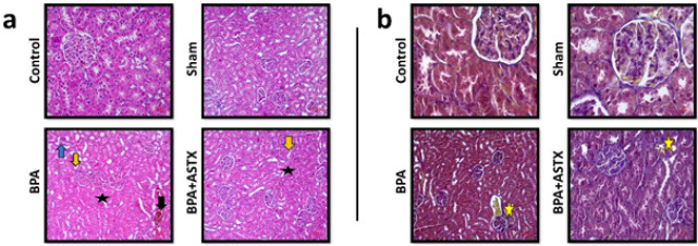 Figure 2
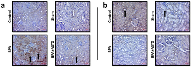 Figure 3
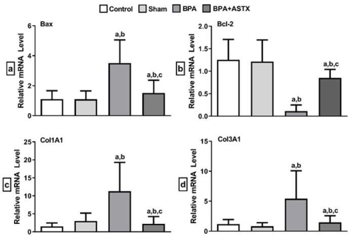 Figure 4
| Gene | Forward primer (5′- >3′) | Reverse primer (5′- >3′) |
|---|---|---|
|
| GCATTGCAGAGGATGGTAGAGAG | GCGGGAGAAGAAAGTCATGATTAG |
|
| GATGGCCTCCTCCTTTCCTACTTC | CTTCTTCCAGATGGTGAGTGAG |
|
| GGAGGATTGTGGCCTTCTTCTTT | GTCATCCACAGAGCGATGTT |
|
| CCAATGGTGCTCCTGGTATT | GTTCACCACTGTTGCCTTTG |
|
| GTGTGATGATGATGAGCCACTAGAC | TGACACAGGAGCAGGAGGTGTAGAA |
| Control | Sham | BPA | BPA+ASTX |
| |
|---|---|---|---|---|---|
| Last | 240.80±24.85 g | 231.10±24.65 g | 253.00±26.61 g | 237.13±21.32 g | 0.3136 |
| First | 221.14±16.94 g | 217.78±12.90 g | 238.13±18.88 g | 234.33±22.37 g | 0.3360 |
| Histopathological score | Bax score | Bcl-2 score | |
|---|---|---|---|
|
|
|
| |
| Control | 1.5 [1.16-2.0] | 0.50 [0.33-0.50] | 2.5 [2.25-2.66] |
| Sham | 1.58 [1.25-2.0] | 0.50 [0.37-0.66] | 2.5 [2.37-2.62] |
| BPA | 13.33 [13.0-13.46](a,b) | 2.83 [2.66-2.96](a,b) | 0.42 [0.33-0.50](a,b) |
| BPA+ASTX | 4.42 [3.37-4.50] | 1.50 [1.33-1.50]a | 1.92 [1.83-2.12] |
Peer Reviews
No public reviews on file for this paper yet. If you reviewed it on a platform where reviews are public (OpenReview, ICLR, NeurIPS, ICML), you can paste yours below so the community can read it here.
Videos
No videos yet. Explain this paper in a talk, walkthrough, or lecture? Add one.
Taxonomy
TopicsEffects and risks of endocrine disrupting chemicals · Skin Protection and Aging · Carcinogens and Genotoxicity Assessment
Introduction
Pollution involving hazardous chemicals and metals may stem from natural processes or human activities, with many of these pollutants inducing oxidative stress. For decades, oxidative stress has been implicated in the pathogenesis of numerous human diseases. Reactive oxygen species (ROS) are cytotoxic molecules capable of damaging nucleic acid bases, lipids, and proteins, ultimately resulting in cell death (1-3). Bisphenol A (BPA), along with octylphenols and nonylphenols, is an industrial pollutant classified as an endocrine-disrupting compound (EDC). BPA is commonly present in consumer plastic products, food packaging, resin-based materials, and some dental applications (4). As a result, human exposure to BPA is frequently unavoidable.
The kidney is a primary target organ for the accumulation of EDCs. In individuals with kidney disease, reduced kidney function and proteinuria are frequently observed, both of which adversely impact the recovery process (5). A study conducted in China demonstrated that reduced kidney function is associated with decreased urinary excretion of BPA. A correlation between urinary BPA excretion and albuminuria was demonstrated, revealing a potential link between BPA exposure and kidney damage (6). Renal dysfunction induced by BPA exposure impairs the elimination of BPA via the kidneys, thereby creating a vicious cycle that amplifies the toxic effects of continued BPA accumulation (7, 8). Histopathological assessments of rats exposed to BPA revealed infiltrative and dilatative alterations in kidney tissues, ultimately leading to renal failure (9). Furthermore, BPA is known to directly impact the mitochondria of kidney cells, inducing oxidative stress and mitochondrial dysfunction, ultimately causing damage to the entire organ (10). Indeed, exposure to BPA is a major source of oxidative damage in the body originating from the electron transport chain (ETC) located in the inner mitochondrial membrane (11). Reactive oxygen species (ROS) may be generated within the ETC, particularly during electron transfer from NADH to ubiquinone via the flavin mononucleotide and ubiquinone-reducing sites of complex I. Additionally, increased ROS production due to electron leakage has been reported at complex III. As a result, the increased levels of ROS impair mitochondrial energy metabolism and lead to tissue dysfunction (12).
Under pathophysiological conditions, endogenous antioxidant defenses become inadequate, necessitating external antioxidant supplementation. This helps neutralize ROS and prevents their detrimental effects on organs (13). Astaxanthin (3,3’-dihydroxy-b,b’-carotene-4,4’-dione, ASTX) is a natural carotenoid known for its potent antioxidant properties, which are attributed to its ability to neutralize free radicals. It is commonly found in marine organisms, including algae, crustaceans, salmon, shrimp, and crabs (14). Unlike other carotenoids, ASTX contains oxygenated groups in each of its ring structures, which imparts high polarity and substantially enhances its protective role against degenerative conditions. This property plays a key role in making ASTX a more potent antioxidant than β-carotene. Other studies comparing ASTX to antioxidant molecules like α-tocopherol and vitamin E have demonstrated that ASTX is more effective (15). Oxidative damage to mitochondria results in reduced oxidative phosphorylation efficiency, leading to increased mitochondrial permeability and the release of pro-apoptotic factors that initiate apoptosis (16). Research has demonstrated the protective effects of ASTX against oxidative damage in the liver, brain, and cardiovascular system (17). In addition, its therapeutic effects have been confirmed in in vitro epidermal-dermal interaction models of skin damage, primarily due to its anti-inflammatory properties (18). Moreover, ASTX has demonstrated a protective effect in an experimental model of ovarian damage induced by 3-nitropropionic acid (3-NPA) in rats (19). In another study, ASTX was found to exert protective effects against torsion/detorsion-induced injury through the modulation of autophagy (20). ASTX also demonstrates activity in anti-inflammatory and anti-apoptotic pathways, inhibiting mitochondrial dysfunction induced by oxidative stress, and providing protective effects to cells and organs in various pathological conditions (21). Studies have shown that ASTX exerts its antioxidant effects by increasing the activities of antioxidant enzymes such as SOD, GSH-Px, and CAT, while its anti-inflammatory effects are mediated through the modulation of the nuclear factor kappa-B (NF-κB) signaling pathway (22). Given the reasons above, the protective effects of ASTX on kidney cells may stem from its direct or indirect impact on mitochondria.
Paraoxonase-1 (PON1) and Arylesterase (ARE) are enzymes in the esterase group, encoded by the same gene, and are known to exhibit antioxidant properties (23). In the present study, oxidative damage induced by BPA exposure was evaluated by determining the levels of PON1 and ARE molecules.
This study aimed to investigate the biochemical changes in the blood serum and the histopathological alterations in the kidney tissue of rats exposed to BPA, as well as to reveal the therapeutic effects of ASTX on these changes.
Materials and Methods
Animals and experimental groups
Thirty-two male Wistar Albino rats (16 weeks old, weighing 200–250 g) were used in this study. The experimental protocol was conducted in accordance with the Declaration of Helsinki and was approved by the Local Ethics Committee of XX University on December 30, 2022 (Approval No. 2021-071). All animals were housed under standard laboratory conditions (temperature: 24 ± 1 °C, humidity: 45 ± 5%, light/dark cycle: 12 hr) and were fed standard laboratory chow and tap water ad libitum. The rats were randomly divided into four groups, with eight rats in each:
Group 1 – Control: No treatment was administered.
Group 2 – Sham (Olive Oil): 20 mg/kg of olive oil was administered via gavage once daily for two weeks.
Group 3 – BPA: 250 mg/kg of BPA, dissolved in olive oil, was administered via gavage once daily for two weeks (24).
Group 4** –** BPA + ASTX (20 mg/kg): 250 mg/kg BPA, dissolved in olive oil, was administered via gavage daily for two weeks. Two hours after each BPA administration, 20 mg/kg ASTX, also dissolved in olive oil, was administered via gavage (25).
In the study by Mashay Al-Anazi et al. (2022), BPA doses of 125, 250, and 500 mg/kg were administered based on previously reported oral LD50 values. They reported toxic effects at both 250 and 500 mg/kg doses. In our study, we selected the 250 mg/kg dose of BPA, as it demonstrated toxic effects while still being considered within a range that could be defined as safe (26). In the study by Toktay et al. (2023), ASTX was administered at doses of 10, 20, and 40 mg/kg, and it was found that the 20 mg/kg dose significantly inhibited markers of oxidative damage. Although they also reported that the 40 mg/kg dose achieved similar inhibition, the duration of administration was limited to 7 days. In our study, based on these literature findings, we selected the 20 mg/kg dose as a more reliable option and administered it for 2 weeks (27). Both BPA (≥99%, CAS No. 80-05-7, Sigma-Aldrich) and ASTX (pure 98%, CAS No. 472-61-7, Sigma-Aldrich) were freshly prepared before each administration. At the end of the 14 days, blood samples and kidney tissues were collected under anesthesia induced by intraperitoneal injection of 50 mg/kg Ketamine HCl and 10 mg/kg Xylazine HCl.
Biochemical analyses
All collected samples were stored under appropriate conditions until the day of the experiment. Paraoxonase 1 (PON1) and Arylesterase (ARE) were analyzed as oxidative stress parameters (28). To evaluate kidney function, urea and creatinine levels were measured in serum tissue (29). PON1 (Rel Assay Diagnostics Kits, Cat. no: RL0031, Mega Tıp San., Gaziantep, Turkey), ARE (Rel Assay Diagnostics Kits, Cat. no: RL0055, Mega Tıp San., Gaziantep, Turkey), Urea (Urea Assay Kit, Cat. no: OttoBC157, Otto Scientific, Ankara, Turkey), and Creatinine (Creatinine Assay Kit, Cat. no: OttoBC139, Otto Scientific, Ankara, Turkey) levels were determined according to the manufacturer’s procedures.
Histological examination
Following blood sample collection, the rats were euthanized, and the abdominal cavities were opened for macroscopic examination of the kidneys. After macroscopic evaluation, the excised kidney tissues were immersed in 10% neutral buffered formalin for 48 hr for fixation. Subsequently, tissues underwent routine histological processing, after which they were embedded in paraffin. Sections of 4 μm thickness were obtained from each paraffin block using a microtome (Leica RM2125RTS).
Hematoxylin and eosin (H&E) staining
Histological analysis of kidney tissue sections stained with hematoxylin and eosin (H&E) was performed based on the evaluation of several parameters, including congestion, vacuolization, tubular dilatation, desquamation, and mononuclear cell infiltration. Histopathological scoring was conducted according to the severity of the observed alterations, categorized as follows: no change [0], mild change [1], moderate change [2], and severe change [3]. Scores for each of the five parameters, each ranging from 0 to 3, were summed to yield a total histological score ranging from 0 to 15 (30). All histopathological evaluations were performed in a double-blinded manner using a Zeiss Lab.A1 light microscope and a Zeiss AxioCam ERc 5s camera imaging system.
Masson’s trichrome staining
Masson’s trichrome staining was performed on kidney tissue sections to evaluate the extent of fibrosis. In the stained sections, the amount of collagen fibers surrounding the Bowman’s capsule, blood vessels, and the basal lamina of renal tubules was visualized and assessed under a light microscope (31).
Immunohistochemistry (IHC) analysis
İn the immunohistochemical analysis, primary antibodies against Bax (Santa Cruz Biotechnology, catalog no: sc-23959) and Bcl-2 (Santa Cruz Biotechnology, catalog no: sc-7382) were employed. Four-micrometer-thick paraffin-embedded tissue sections were deparaffinized by immersion in xylene for 30 min. Following deparaffinization, sections were incubated in Super Block solution (ScyTek Laboratories, Logan, UT, USA) for 10 min, then rinsed with phosphate-buffered saline (PBS) for five minutes. Primary antibody incubation was performed overnight at 4 °C. After washing with PBS for five minutes, sections were incubated with the secondary antibody for 20 min, followed by another 5-min PBS wash. Subsequently, streptavidin-peroxidase was applied for 20 min and washed again with PBS. The chromogenic substrate AEC (Sigma Aldrich, AEC101-1KT) was then added and incubated for 15 min, followed by rinsing with distilled water for five minutes. Mayer’s hematoxylin was used as a counterstain once the immunoreactivity became microscopically evident. Slides were mounted using an aqueous mounting medium and examined under a Zeiss Lab.A1 light microscope, with images captured using the Zeiss AxioCam ERc 5s camera system. Immunoreactivity was semi-quantitatively scored based on staining intensity as follows: no staining [0], weak staining [1], moderate staining [2], and strong staining [3] (32). All histopathological evaluations were performed independently by two investigators blinded to the experimental groups.
RT-qPCR analysis
For molecular analyses, total RNA was isolated from one-third of the kidney tissue obtained from each experimental group, previously stored at –80 °C. RNA extraction was performed using RiboEX Total RNA Isolation Reagent (GeneAll, 301-001). To eliminate potential genomic DNA contamination, each sample was treated with DNase I enzyme (Thermo Fisher Scientific, #EN0521). Subsequently, cDNA was synthesized using the iScript™ cDNA Synthesis Kit (Bio-Rad, 170-8891), following the manufacturer’s instructions.
Primers for the target genes (Bax, Bcl-2, Col1A1, Col3A1) and the housekeeping gene (GAPDH) were designed using the IDT PrimerQuest Tool (https://www.idtdna.com/PrimerQuest/Home/Index). mRNA expression levels were quantified using 5x HOT FIREPol® EvaGreen® qPCR Master Mix Plus (ROX) (Solis BioDyne). Reactions were performed on a Bio-Rad CFX Connect real-time PCR system with the following thermal cycling conditions: initial activation at 95 °C for 12 min, followed by 40 cycles of denaturation at 95 °C for 15 sec, annealing at 60 °C for 20 sec, and extension at 72 °C for 20 sec. GAPDH was used as the reference gene for normalization (Table 1).
Statistical analysis
Normality of body weight and biochemical data was assessed using the Shapiro-Wilk normality test, and these data were found to be normally distributed. Statistical analysis for these data was performed using one-way analysis of variance (ANOVA) followed by Tukey’s post hoc test. Histopathological scores, as ordinal data, were analyzed using the Kruskal-Wallis test followed by Dunn’s post hoc test. Differences in mRNA expression levels between groups were analyzed using the 2((-ΔΔCT)) method. A p-value less than 0.05 was considered statistically significant. Data conforming to normal distribution are presented as mean ± standard deviation (SD), while non-normally distributed data are presented as median [interquartile range (IQR) – Q1, Q3]. All statistical analyses and calculations were performed using GraphPad Prism 8 and Microsoft Office 365 software.
Results
The evaluation of body weights across the experimental groups revealed no statistically significant differences, indicating that ASTX administration did not notably influence body weight in the context of BPA-induced toxicity (P>0.05; Table 2).
Effects of ASTX on oxidative stress and kidney damage biomarkers
PON 1 and ARE results were significantly lower in the BPA groups compared to the control group (P<0.05). This suggests that BPA exposure may lead to an increase in oxidative stress, thereby contributing to oxidative damage. However, ASTX administration resulted in a statistically significant increase in PON 1 and ARE levels compared to the BPA-induced group (P<0.05). These findings suggest that ASTX may exert its protective effects by enhancing the activity of antioxidant enzymes to combat oxidative stress. Urea levels were significantly higher in the BPA groups compared to the control group (P<0.05). ASTX treatment restored the urea levels to those observed in the control group. No statistically significant differences were observed in creatinine levels (P>0.05) (Figure 1). The lack of change in creatinine levels may support the hypothesis that the differences in creatinine levels observed in previous studies are dose-dependent, related to the BPA concentration.
Histopathologic staining
Hematoxylin-eosin staining
Light microscopic examination revealed that the glomeruli and renal tubules of the control group exhibited nearly normal structural organization in most kidneys. Kidney sections from the sham group also displayed typical histological renal architecture, similar to that observed in the control group. The BPA-treated group exhibited signs of vacuolization, congestion, tubular dilatation, desquamation, and infiltration. In contrast, the BPA+ASTX-treated group demonstrated a marked improvement in histological appearance compared to the BPA group. ASTX treatment was found to reduce the adverse effects induced by BPA on kidney tissue (Figure 2a, Table 3).
Masson’s trichrome staining
After Masson’s trichrome staining, no significant differences in fibrosis were observed in the kidney tissues of the control and sham groups. However, the BPA group exhibited an increase in fibrosis around the glomeruli and blood vessels. In the BPA+ASTX group, fibrosis was increased compared to the control and sham groups, but decreased when compared to the BPA group (Figure 2b).
Immunohistochemical staining for bax and Bcl-2
When compared to all other groups, the BPA group exhibited significantly higher expression levels of Bax, particularly in the renal tubules. ASTX treatment was found to reduce Bax expression around the tubules when compared to the BPA group (Figure 3a, Table 3). Conversely, immunohistochemical staining for Bcl-2 showed a decrease in Bcl-2 expression in the BPA group compared to all other groups (Figure 3b, Table 3). Our findings on Bax and Bcl-2 emphasize the increase in apoptotic activity in kidney tissue due to BPA exposure. As a consequence of the elevated apoptotic activity, the damage observed in renal tubules and the increase in fibrosis are consistent with our histopathological findings.
mRNA expression analysis of apoptosis and collagen-related genes
The effects of BPA and BPA+ASTX on the mRNA expression of apoptosis-related genes (Bax, Bcl-2) and collagen-related genes (Col1A1, Col3A1) were evaluated using RT-qPCR. Bax gene expression in the BPA group was significantly increased by 3.186-fold (P<0.05) compared to the control group and by 3.198-fold (P<0.05) compared to the sham group. In the BPA+ASTX group, Bax gene expression was reduced by 2.31-fold (P<0.05) compared to the BPA group (Figure 4a).
Bcl-2 gene expression in the BPA group was significantly reduced by 11.02-fold (P<0.05) compared to the control group and by 10.68-fold (P<0.05) compared to the sham group. When compared to the BPA group, Bcl-2 gene expression was significantly increased by 7.48-fold (P<0.05) in the BPA+ASTX group. No significant changes in Bcl-2 gene expression were observed when the BPA+ASTX group was compared separately to the control and sham groups (Figure 4b).
Col1A1 gene expression in the BPA group was significantly increased by 7.37-fold (P<0.05) compared to the control group and by 3.77-fold (P<0.05) compared to the sham group. In the BPA+ASTX group, Col1A1 mRNA expression was decreased by 5.09-fold (P<0.05) compared to the BPA group (Figure 4c). Col3A1 gene expression in the BPA-treated group was significantly increased by 4.58-fold (P<0.05) compared to the control group and by 6.63-fold (P<0.05) compared to the sham group. In contrast, Col3A1 mRNA expression was reduced by 3.7-fold (P= 0.05) in the BPA+ASTX group compared to the BPA group (Figure 4d).
Discussion
BPA is among the most extensively investigated EDCs, with its adverse effects well-documented in various laboratory animal models and sentinel human populations (33). Recent studies have demonstrated that BPA and structurally related environmental contaminants exacerbate oxidative stress within cells by compromising the endogenous antioxidant defense mechanisms, thereby promoting the accumulation of ROS and the generation of deleterious metabolic byproducts (34). While the hepatotoxic effects of BPA have been extensively characterized, data regarding its nephrotoxic potential remain comparatively scarce. The present study aims to evaluate the therapeutic efficacy of ASTX in mitigating BPA-induced oxidative damage in renal tissue, with a particular focus on histopathological alterations and functional impairments in kidney physiology.
Studies emphasize that ASTX exerts nephroprotective effects in various kidney pathologies through its antioxidant and anti-inflammatory properties. A key role in these effects is played by ASTX’s regulation of oxidative and inflammatory responses via the Nrf2/Keap1 or c-Src/Connexin 43 pathways. Additionally, ASTX’s nephroprotective effect is also demonstrated through a different mechanism by reducing mitochondrial ROS overproduction and inhibiting mitochondrial oxidative phosphorylation (35). Our study is based on the premise that BPA, a toxic substance, can cause oxidative damage, and consequently, oxidative injury may occur. It is designed around the hypothesis that ASTX may have therapeutic effects through the mechanisms described above.
Consistent with our findings, several researchers have reported that BPA exposure in rats and mice results in negligible differences in final body weight (36, 37). In contrast, other studies have demonstrated significant alterations in body weight, weight gain, and relative organ weights (38, 39). These inconsistencies in weight-related outcomes may be attributed to variations in dose, route of administration, exposure duration and timing, as well as differences in animal strains, sex, and dietary composition. Studies have indicated that elevated BPA levels are associated with a reduction in glomerular filtration rate, and increasing systemic BPA concentrations have even been proposed as potential biomarkers of renal injury (40). Therefore, investigations exploring the link between BPA exposure and kidney damage are crucial for the preservation of healthy renal function. PON1 and ARE are calcium-dependent antioxidant enzymes, localized in high-density lipoprotein (HDL), that function in esterase form and play a pivotal role in reducing oxidative stress in serum and tissues. These enzymes have been shown to hydrolyze hydrogen peroxide, the most commonly produced ROS, thereby preventing lipid peroxidation. Notably, their activities have been reported to decline in various pathological conditions (41). Ola-Davies and Olukole reported that oral administration of BPA at a dose of 10 mg/kg increased renal ROS levels while reducing the concentrations of endogenous antioxidants (42). Similarly, Kobayashi et al. observed that BPA exposure elevated the levels of plasma free radicals in rats (43). In line with these findings, Acaroz et al. demonstrated that BPA exposure exacerbated oxidative damage and decreased antioxidant enzyme levels (30). Consistent with the existing literature, our study also revealed that BPA exposure significantly reduced PON1 and ARE activities. However, supplementation with ASTX resulted in a marked increase in both PON1 and ARE capacities, a statistically significant finding.
Previous studies have reported that BPA exposure can lead to hyperglycemia, which may contribute to renal injury and dysfunction, ultimately resulting in significant increases in serum urea, creatinine, and uric acid levels (1). Suberg et al. and Saleh observed elevated serum urea and creatinine levels following oral administration of BPA, suggesting impaired renal filtration capacity (44, 45). In a separate study by Esplugas et al., mice exposed to a relatively low dose of BPA (25 mg/kg) exhibited signs of kidney damage, along with a significant increase in serum urea levels (46). However, no notable alterations were observed in creatinine concentrations or plasma clearance, indicating a dose- and biomarker-specific response to BPA-induced nephrotoxicity. Our findings are consistent with these studies, as BPA administration in our model also resulted in increased serum levels of renal function markers. Furthermore, ASTX supplementation at a dose of 20 mg/kg demonstrated a protective effect, which aligns with previous research indicating its efficacy in alleviating ROS-mediated oxidative stress and restoring the suppressed activity of endogenous antioxidant enzymes (47). In our findings, ASTX supplementation was shown to attenuate the BPA-induced elevation in serum urea levels, bringing them closer to those observed in the control group. This result was supported by statistical analysis. However, BPA exposure did not result in a statistically significant alteration in serum creatinine levels. This suggests that while urea may serve as a more sensitive early indicator of BPA-related renal impairment in this model, creatinine levels remained within a range that may not reflect subtle or early-stage nephrotoxicity.
Histopathological evidence from previous studies supports the nephrotoxic effects of BPA exposure. Dokmeci et al. (48) conducted H&E staining on renal sections of BPA-exposed rats and reported findings such as tubular epithelial desquamation, cytoplasmic vacuolization, and shrinkage. Similarly, Eid et al. (49) observed thickening of the glomerular basement membrane, increased cellular proliferation, and shedding of renal tubular microvilli in response to BPA exposure. Saleh et al. also reported a marked increase in collagen fiber deposition in renal tissues of BPA-treated animals compared to controls, suggesting progressive fibrotic changes (50). In contrast, studies investigating the protective effects of ASTX have reported its beneficial influence on renal histopathology under various pathological conditions. In a model of alloxan-induced diabetes, ASTX administration significantly improved histological damage such as glomerular and tubular degeneration, an effect attributed to its potent antioxidant properties (51). Erbaş et al. demonstrated that ASTX preserved normal renal histology in the context of lithium-induced toxicity, which was evidenced by reduced glomerular and tubular degeneration as well as diminished edema (22). These findings are in line with broader evidence suggesting that ASTX exerts protective effects against oxidative stress-induced cellular and tissue damage in both in vitro and in vivo models. Furthermore, ASTX has been reported to protect proximal tubular epithelial cells and diabetic nephropathy models from oxidative stress, inflammation, and apoptosis induced by high glucose levels (47).
In our study, ASTX administration was found to exert a protective effect on renal tissue by mitigating the histological alterations induced by BPA exposure. Based on our findings, it is believed that the observed renal damage may be primarily attributed to oxidative stress triggered by BPA, and that ASTX plays a significant role in attenuating this damage through its potent antioxidant activity. Consistent with our Masson’s trichrome staining results, the BPA+ASTX group exhibited reduced fibrotic tissue deposition compared to the BPA group, which demonstrated a markedly increased degree of fibrosis. This histological improvement in the ASTX-treated group suggests a potential antifibrotic role of ASTX in the context of BPA-induced renal injury. Furthermore, molecular analysis revealed a significant up-regulation of fibrosis-associated genes Col1A1 and Col3A1 in the BPA group, whereas their expression was notably down-regulated in the BPA+ASTX group. These findings support the hypothesis that ASTX not only modulates oxidative stress but also influences the transcriptional regulation of key fibrogenic pathways, thereby contributing to the preservation of renal structural integrity.
BPA exposure has been shown to down-regulate the expression of PI3K (phosphatidylinositol 3-kinase) and AKT (protein kinase B), while activating the Bcl-2/Bax–Caspase-9–Caspase-3 signaling cascade, ultimately leading to apoptosis and necrosis of renal cells (52). Tang et al. reported that the increase in apoptotic activity due to BPA exposure is accompanied by a disruption in mitochondrial membrane potential (53). Ma et al. reported that exposure to toxic substances activates the p53 pathway, leading to increased apoptotic activity in kidney tissue. It is thought that the increase in apoptotic activity highlighted by our study’s results may have occurred through similar mechanisms (54). Bosch-Panadero et al. also reported increased apoptotic activity in proximal tubular cells following BPA exposure (55). In line with these findings, Amin et al. demonstrated that oral administration of BPA at a dose of 50 mg/kg in male albino rats significantly up-regulated the expression of pro-apoptotic Bax protein and profibrotic TGF-β1, along with histopathological evidence of apoptosis and fibrosis (56). Supporting our results, previous studies have indicated that ASTX exerts cytoprotective effects on renal cells by preserving mitochondrial function and maintaining mitochondria in a more reduced state under oxidative stress conditions (22). Consistent with the study by Guo et al., which demonstrated that ASTX significantly reduced the number of apoptotic renal cells, an effect attributed to its inhibition of tubular apoptosis, our findings similarly showed that BPA exposure increased Bax expression and decreased Bcl-2 expression in renal tissue, as verified by both immunohistochemical staining and PCR analysis (47). Significantly, ASTX administration reversed these molecular alterations, resulting in down-regulation of Bax and up-regulation of Bcl-2 expression. These findings suggest that ASTX may reduce BPA-induced renal apoptosis by regulating the intrinsic apoptotic pathway, potentially through stabilization of mitochondrial integrity and inhibition of pro-apoptotic signaling. Consequently, the reduction of increased apoptotic activity caused by BPA exposure via ASTX treatment may have occurred through suppression of mitochondrial oxidative phosphorylation and mitochondrial oxidative overproduction, or inhibition of the p53 signaling pathway.
Studies have emphasized a strong association between BPA exposure and kidney pathologies in humans (57). Furthermore, individuals with chronic kidney disease (CKD) have been reported to be potentially at higher risk for adverse effects resulting from BPA exposure (58). The reported impacts of BPA exposure in humans align with our findings, thereby reinforcing the conclusion that BPA induces nephrotoxicity. Consequently, the development of protective, preventive, and therapeutic strategies against the detrimental effects of BPA exposure is of paramount importance in the current context.
BPA exposure emerges as a factor affecting kidney health in individuals. It is crucial to conduct qualified, long-term, and methodologically diverse studies for clinical practice and policy-making. Although ASTX shows promise in terms of safety and biological efficacy, particularly regarding kidney health, larger and more robust studies are needed to confirm its clinical benefits. In conclusion, while there is robust preclinical evidence supporting the protective effects of the BPA–ASTX interaction, confirming this effect through feasible, randomized clinical trials from a human health perspective will be a critical step in the future.
Parameters evaluated from blood serum using the enzyme-linked immunosorbent assay (ELISA) method
a. Pathological changes such as dilation (star), vascular congestion (black arrow), tubular epithelial desquamation (yellow arrow), and mononuclear cell infiltration (blue arrow) were observed in Wistar Albino rat kidney tissues stained with Hematoxylin and Eosin (H&E). b. Collagen fiber accumulation (yellow star) was observed in Masson's Trichrome-stained kidney sections
Effect of BPA exposure on apoptotic activity in Wistar Albino rat kidney tissue was evaluated through immunohistochemical expression of the pro-apoptotic protein Bax and the anti-apoptotic protein Bcl-2
mRNA expression analysis a. Bax, b. Bcl-2, c. Col1A1, d. Col3A1
Conclusion
This study was conducted on adult male rats and did not address potential differences related to age and sex. Moreover, it was carried out with a limited sample size. The dose–response relationships of both BPA exposure and ASTX supplementation need to be further explored in future studies. Additionally, the findings of this study are based solely on kidney tissue analysis. Investigations involving other tissues and different species may be considered in subsequent research. Nonetheless, our results suggest that ASTX supplementation may alleviate BPA-induced histopathological alterations and protect kidney tissue. These findings indicate that ASTX holds promise as a potential therapeutic or protective agent against BPA-induced nephrotoxicity. However, further studies are needed to elucidate the precise mechanisms underlying its protective effects.
The reference list from the paper itself. Each links out to its DOI / PubMed record.
- 1Morgan AM El-Ballal SS El-Bialy BE El-Borai NB Studies on the potential protective effect of cinnamon against bisphenol A- and octylphenol-induced oxidative stress in male Albino rats Toxicol Rep 20141921012896223010.1016/j.toxrep.2014.04.003PMC 5598475 · doi ↗ · pubmed ↗
- 2Akkoca ABüyükakıllıB BallıEGültekin BÖzbay E OruçDemirbağ H Protective effect of Mito TEMPO against cardiac dysfunction caused by ischemia-reperfusion: MCAO stroke model study Int J Neurosci 2024134158215933786200310.1080/00207454.2023.2273768 · doi ↗ · pubmed ↗
- 3Celen MC Akkoca A Tuncer S Dalkilic N Ilhan B Protective vs therapeutic effects of mitochondria-targeted antioxidant Mito TEMPO on rat sciatic nerve crush ınjury: A comprehensive electrophysiological analysis Biomedicines 202311330633183813752810.3390/biomedicines 11123306 PMC 10741406 · doi ↗ · pubmed ↗
- 4Koriem KMM Fertaric acid amends bisphenol A-induced toxicity, DNA breakdown, and histopathological changes in the liver, kidney, and testis World J Hepatol 2022145355503558229110.4254/wjh.v 14.i 3.535PMC 9055189 · doi ↗ · pubmed ↗
- 5Kitazawa T Sato T Nishiyama K Asai R Arima Y Uchijima Y Identification and developmental analysis of endothelin receptor type-A expressing cells in the mouse kidney Gene Expr Patterns 2011113713772156528410.1016/j.gep.2011.04.001 · doi ↗ · pubmed ↗
- 6Moreno-Gómez-Toledano R Arenas MIVélez-Vélez E Saura M Bosch RJ New evidence of renal and cardiovascular alterations promoted by bisphenol A Biomolecules 202111164916533482764710.3390/biom 11111649 PMC 8615835 · doi ↗ · pubmed ↗
- 7You L Zhu X Shrubsole MJ Fan H Chen J Dong J Renal function, bisphenol A, and alkylphenols: Results from the National Health and Nutrition Examination Survey (NHANES 2003-2006) Environ Health Perspect 201111952753310.1289/ehp.1002572 PMC 308093621147601 · doi ↗ · pubmed ↗
- 8Nicholson TM Nguyen JL Leverson GE Taylor JA Vom Saal FS Wood RW Endocrine disruptor bisphenol A is implicated in urinary voiding dysfunction in male mice Am J Physiol Ren Physiol 2018315 F 1208–F 121610.1152/ajprenal.00582.2017 PMC 629330230019933 · doi ↗ · pubmed ↗