Generation of an iPSC-derived alveolar rhabdomyosarcoma cell line during directed endothelial differentiation
Randolph K. Larsen, Madeline B. Searcy, Bradley T. Stevens, Katherine E. Gadek, Yang Zhang, Brian J. Abraham, Mark E. Hatley

TL;DR
This paper describes a method to create a cell model of alveolar rhabdomyosarcoma using iPSCs and an oncofusion protein during endothelial differentiation.
Contribution
A novel protocol for generating iPSC-derived alveolar rhabdomyosarcoma cells during endothelial differentiation using PAX3::FOXO1.
Findings
ARMS tumors can originate from non-myogenic cells like endothelial progenitors.
Enforced P3F expression during differentiation leads to aggressive myogenic tumor formation.
The iARMS model enables study of P3F's role in tumor transformation.
Abstract
Alveolar rhabdomyosarcoma (ARMS) is an aggressive soft tissue sarcoma typically driven by the oncofusion protein PAX3::FOXO1 (P3F). Despite ARMS tumor histology and transcriptome resembling skeletal muscle, these tumors arise in areas devoid of skeletal muscle, indicating that non-myogenic cells can give rise to ARMS. Our lab demonstrated that endothelial progenitors are a cell of origin for rhabdomyosarcoma. Here we provide a protocol for generating iPSC-derived alveolar rhabdomyosarcoma cells (iARMS) during endothelial directed differentiation through enforced expression of P3F. This model allows for dissection of how P3F mediates transformation of endothelial progenitors into aggressive myogenic tumors.
Genes, proteins, chemicals, diseases, species, mutations and cell lines named across the full text — each resolved to its canonical identifier and authoritative record.
Click any figure to enlarge with its caption.
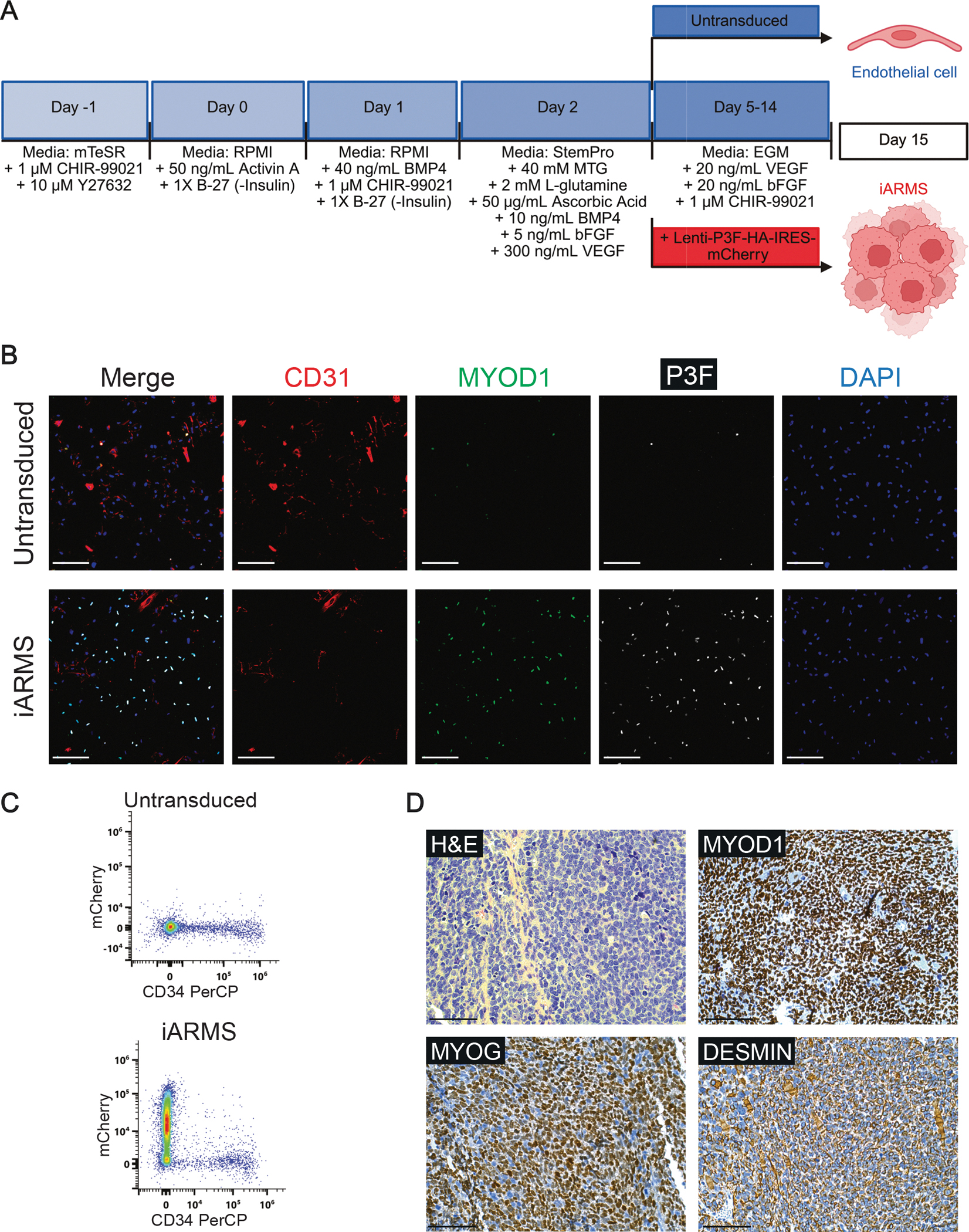 Figure 1
Peer Reviews
No public reviews on file for this paper yet. If you reviewed it on a platform where reviews are public (OpenReview, ICLR, NeurIPS, ICML), you can paste yours below so the community can read it here.
Videos
No videos yet. Explain this paper in a talk, walkthrough, or lecture? Add one.
Taxonomy
TopicsSarcoma Diagnosis and Treatment · FOXO transcription factor regulation · Angiogenesis and VEGF in Cancer
Resource utility
This protocol was developed to test the capacity of P3F to mediate transformation of endothelial progenitors into aggressive skeletal muscle tumor cells. This method allows for interrogation of the P3F-mediated processes that drive oncogenic transformation during development in a reproducible, scalable, and human system (see Table 1).
Resource details
Our laboratory has previously demonstrated that alveolar rhabdomyosarcoma (ARMS) driven by the PAX3::FOXO1 (P3F) oncofusion protein can arise when P3F is expressed in endothelial progenitors in mice (Searcy et al., 2023; Stevens and Hatley, 2025). This model allows for lineage tracing of endothelial progenitors that can be transformed into ARMS and comparison to murine models of ARMS arising from myogenic progenitors (Searcy et al., 2023; Keller et al., 2004). While useful, the murine models are costly, not fully penetrant, and tumors take over 100 days to develop, limiting their tractability for high-throughput screening or studying P3F structure/function. Other groups have developed cell line models of P3F-mediated transformation, but these either require the enforced expression of muscle fate-defining transcription factors or multiple oncogenic drivers to permit P3F-mediated transformation into ARMS (Naini, 2008; , Kalita et al., xxxx). These models provide insight into transformation of muscle progenitors into ARMS, but the artificial expression of myogenic and oncogenic factors may mask functions that P3F independently performs, which can only be revealed through transformation from non-myogenic cells. This protocol models ARMS transformation from a non-myogenic progenitor through the simple addition of P3F during differentiation. To accurately recapitulate the mutational landscape seen in human FP-RMS tumors, we generated TP53 knockout (TP53^KO^) human BJFF.6 iPSCs. Detailed methods on the CRISPR-Cas9 generated TP53^KO^ iPSCs were published previously (Searcy et al., 2023). Briefly, BJFF.6 iPSCs were nucleofected with precomplexed ribonuclear proteins (RNPs) consisting of chemically modified sgRNA, Cas9 protein, and pMaxGFP. GFP+ single cell clones were isolated by FACS and plated on 96-well plates. Knockout clones were identified, expanded, and sequenced confirmed by next generation sequencing analysis. In this protocol, TP53^KO^ iPSCs are differentiated sequentially to hemogenic mesoderm and then endothelial cells as previously described (Searcy et al., 2023; Palpant et al., 2017). When the hemogenic mesodermal cells are switched into endothelial growth media (EGM) for definitive endothelial differentiation, we transduce them with lentivirus expressing a P3F-HA-IRES-mCherry construct (Fig. 1A). The P3F-expressing cells transform into iPSC-derived alveolar rhabdomyosarcoma cells (iARMS), which lack expression of the endothelial markers CD31 and CD34 and gain expression of the myogenic marker MYOD1 (Fig. 1B–C) (Searcy et al., 2023). Furthermore, iARMS cells grafted into immunocompromised mice form tumors with 100 % penetrance that homogenously resemble human ARMS by immunohistochemistry (Fig. 1D) and gene expression (Searcy et al., 2023). This model provides a scalable human system to study how P3F cooperates with developmental cell state to drive transformation into a muscle tumor and allows for mechanistic dissection of cooperating genetic perturbations (see Table 2).
The reference list from the paper itself. Each links out to its DOI / PubMed record.
- 1Anders S, Pyl PT, Huber W, 2015. HT Seq–a Python framework to work with high-throughput sequencing data. Bioinformatics 31 (2), 166–169.25260700 10.1093/bioinformatics/btu 638PMC 4287950 · doi ↗ · pubmed ↗
- 2Chen Y-H, Pruett-Miller SM, 2018. Improving single-cell cloning workflow for gene editing in human pluripotent stem cells. Stem Cell Res. 31, 186–192.30099335 10.1016/j.scr.2018.08.003 · doi ↗ · pubmed ↗
- 3Kalita B, , PAX translocations remodel mitochondrial metabolism through altered leucine usage in rhabdomyosarcoma. Cell.10.1016/j.cell.2025.03.008PMC 1208529940185100 · doi ↗ · pubmed ↗
- 4Keller C, , 2004. Alveolar rhabdomyosarcomas in conditional Pax 3:Fkhr mice: cooperativity of Ink 4a/ARF and Trp 53 loss of function. Genes Dev. 18, 2614–2626.15489287 10.1101/gad.1244004 PMC 525542 · doi ↗ · pubmed ↗
- 5Kim D, Langmead B, Salzberg SL, 2015. HISAT: a fast spliced aligner with low memory requirements. Nat. Methods 12 (4), 357–360.25751142 10.1038/nmeth.3317 PMC 4655817 · doi ↗ · pubmed ↗
- 6Lee J, , 2015. Early induction of a prechondrogenic population allows efficient generation of stable chondrocytes from human induced pluripotent stem cells. FASEB J. 29 (8), 3399–3410.25911615 10.1096/fj.14-269720 PMC 4511207 · doi ↗ · pubmed ↗
- 7Love MI, Huber W, Anders S, 2014. Moderated estimation of fold change and dispersion for RNA-seq data with DE Seq 2. Genome Biol. 15 (550).10.1186/s 13059-014-0550-8PMC 430204925516281 · doi ↗ · pubmed ↗
- 8Naini S, , 2008. Defining the cooperative genetic changes that temporally drive alveolar rhabdomyosarcoma. Cancer Res. 68 (23), 9583–9588.19047133 10.1158/0008-5472.CAN-07-6178 PMC 2593800 · doi ↗ · pubmed ↗