The ER/Golgi Protein FNDC3B Facilitates Climbing Fibre to Purkinje Cell Synapse Elimination in the Developing Mouse Cerebellum
Céline Louise Mercier, Takaki Watanabe, Yuto Okuno, Kyoko Matsuyama, Kyoko Kushibe, Henry Denny, Taisuke Miyazaki, Miwako Yamasaki, Meiko Kawamura, Manabu Abe, Kenji Sakimura, Masahiko Watanabe, Naofumi Uesaka, Masanobu Kano

TL;DR
This study shows that the protein FNDC3B helps eliminate extra synapses on Purkinje cells in the developing mouse cerebellum.
Contribution
FNDC3B is newly identified as a facilitator of climbing fiber synapse elimination in the developing cerebellum.
Findings
FNDC3B is involved in synapse elimination from postnatal day 9 in mice.
FNDC3B deletion impairs synapse elimination and CF extension at P10 and P21, but effects are recovered by P40.
FNDC3B does not affect parallel fiber or inhibitory synaptic inputs to Purkinje cells.
Abstract
Synapse elimination during development is crucial for refining neural circuits by removing excess synapses formed around birth. In the neonatal cerebellum, Purkinje cells (PCs) are initially innervated by multiple climbing fibers (CFs) with similar synaptic strengths. During subsequent postnatal development, a single CF is strengthened and retained, while the other CFs are eliminated. Here, our PC‐specific RNAi knockdown (KD) screening revealed that fibronectin type III domain containing 3B (FNDC3B), an endoplasmic reticulum protein, was involved in CF synapse elimination from around postnatal day 9 (P9) in mice. We showed that FNDC3B mRNA was expressed in PCs during CF synapse elimination. In PC‐selective FNDC3B conditional knockout (FNDC3B‐cKO) mice, CF synapse elimination from P10 was impaired, and the extension of CFs along PC dendrites was reduced at P21. However, these phenotypes…
Genes, proteins, chemicals, diseases, species, mutations and cell lines named across the full text — each resolved to its canonical identifier and authoritative record.
Click any figure to enlarge with its caption.
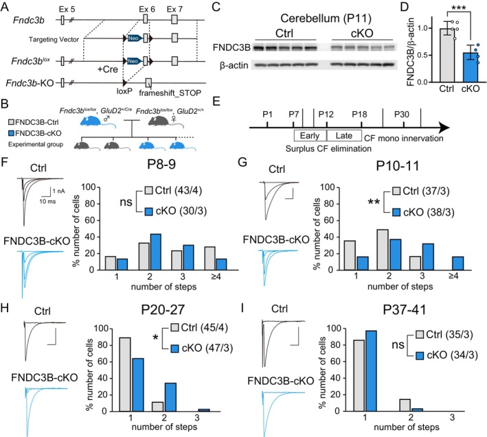 Figure 1
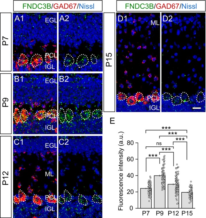 Figure 2
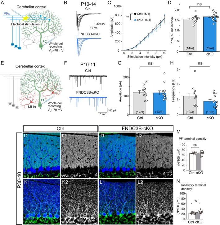 Figure 3
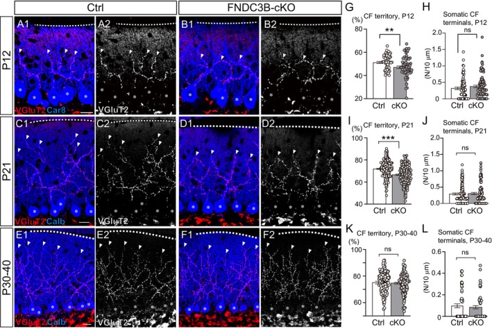 Figure 4
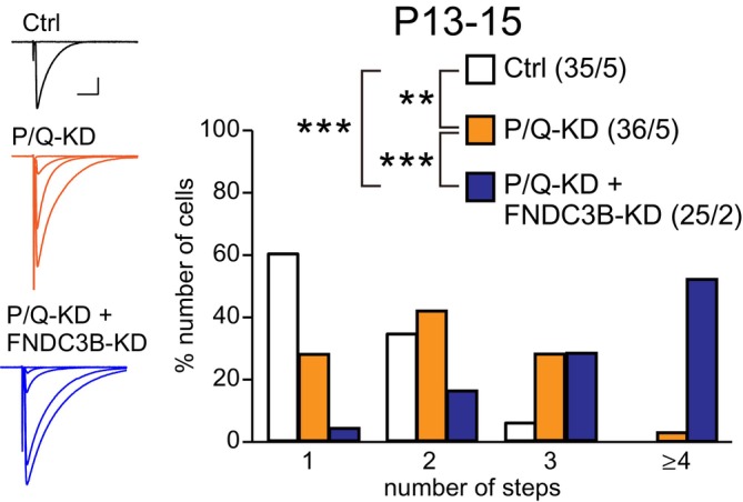 Figure 5
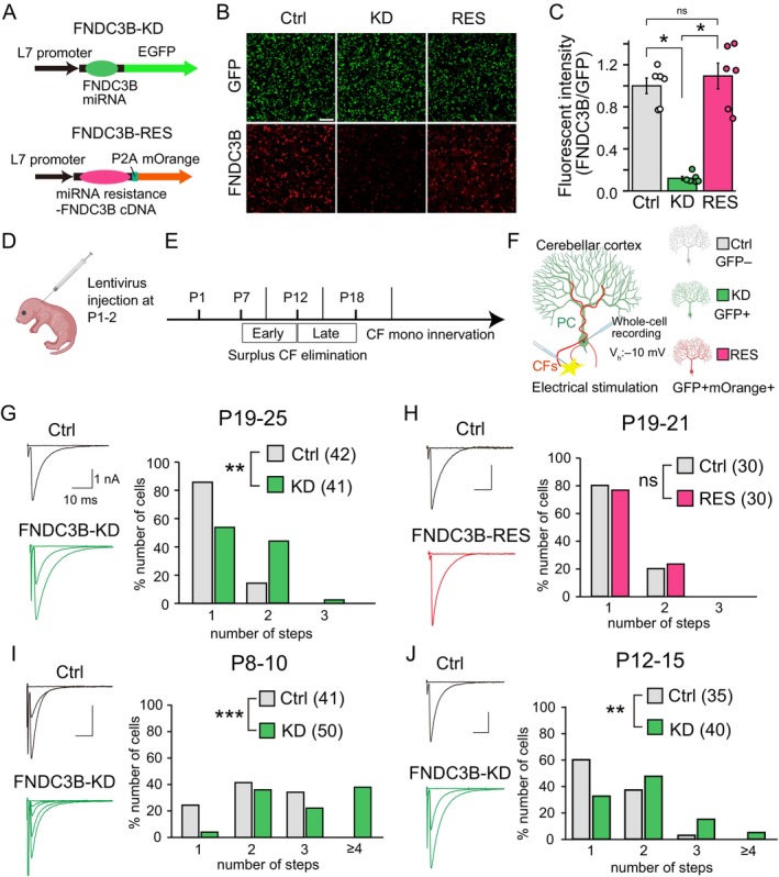 Figure 6
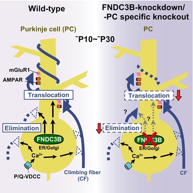 Figure 7
Peer Reviews
No public reviews on file for this paper yet. If you reviewed it on a platform where reviews are public (OpenReview, ICLR, NeurIPS, ICML), you can paste yours below so the community can read it here.
Videos
No videos yet. Explain this paper in a talk, walkthrough, or lecture? Add one.
Taxonomy
TopicsCellular transport and secretion · Endoplasmic Reticulum Stress and Disease · Microtubule and mitosis dynamics