After Traumatic Brain Injury, EPHA4 Enhances Endoplasmic Reticulum Stress to Promote M1 Microglial Polarization Through the MAPK Signaling Pathway
Yang Tan, Jing Xia, Mingwei Liu, Sangyang Deng, Haiying Wu, Chuanyun Qian

TL;DR
This study shows that EPHA4 promotes harmful microglial activity after brain injury by increasing stress signals, suggesting a new target for treatment.
Contribution
The novel finding is that EPHA4 drives M1 microglial polarization through endoplasmic reticulum stress and MAPK signaling in traumatic brain injury.
Findings
EPHA4 expression is upregulated in TBI rat brain tissue.
KYL peptide reduces M1 polarization and proinflammatory cytokines.
EPHA4 activates MAPK signaling to enhance endoplasmic reticulum stress and M1 polarization.
Abstract
Traumatic brain injury (TBI) is an important cause of disability and death worldwide. The development of neuroinflammation after TBI is related to the brain parenchyma. M1‐type microglia play important roles in this process, but the specific mechanism through which regulating microglia M1 polarization is still not fully understood. This study aimed to investigate the role of ephrin receptor A4 (EPHA4) in the M1 polarization of microglia after TBI. A TBI rat model was established by the controlled cortical impact (CCI) method, and M1 polarization of GMI‐R1 cells was induced by lipopolysaccharide (LPS) treatment. Target genes associated with the progression of TBI were screened by transcriptome sequencing; the expression of key genes and proteins was detected by real‐time quantitative PCR (RT‒qPCR), Western blot, ELISA, and immunofluorescence, and the damage of the rat brain tissue and…
Genes, proteins, chemicals, diseases, species, mutations and cell lines named across the full text — each resolved to its canonical identifier and authoritative record.
Click any figure to enlarge with its caption.
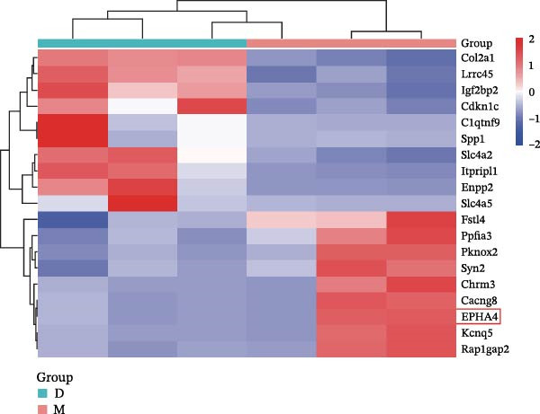 Figure 1
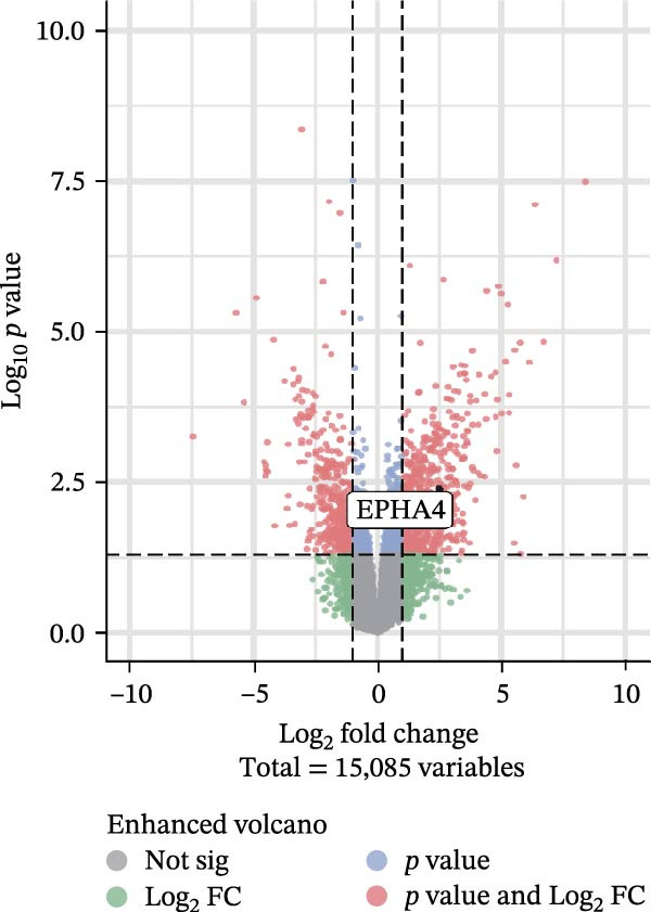 Figure 2
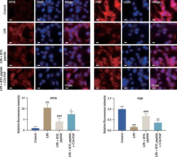 Figure 3
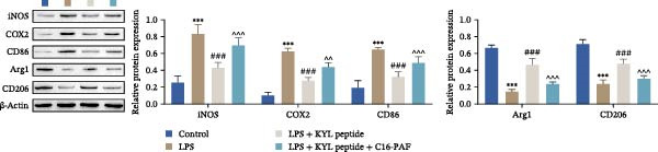 Figure 4
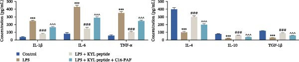 Figure 5
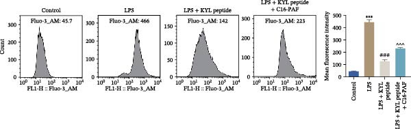 Figure 6
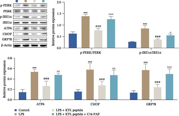 Figure 7
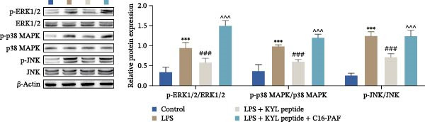 Figure 8
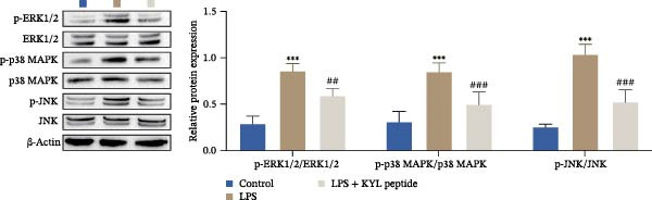 Figure 9
 Figure 10
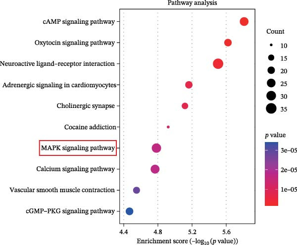 Figure 11
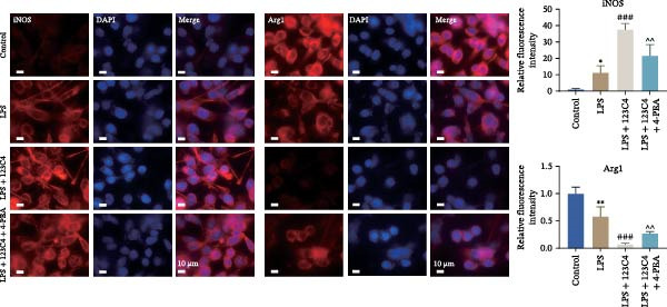 Figure 12
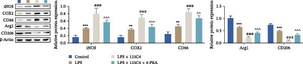 Figure 13
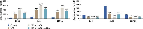 Figure 14
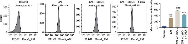 Figure 15
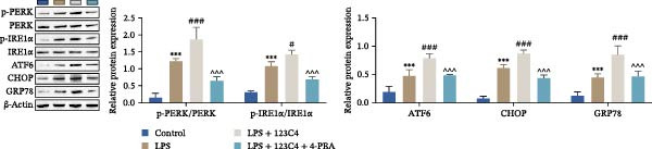 Figure 16
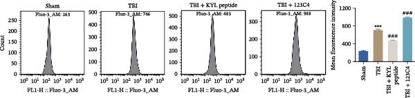 Figure 17
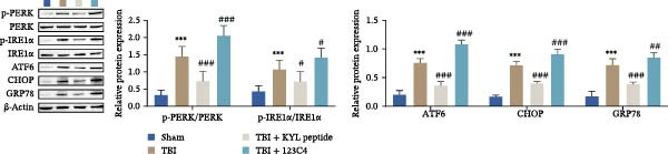 Figure 18
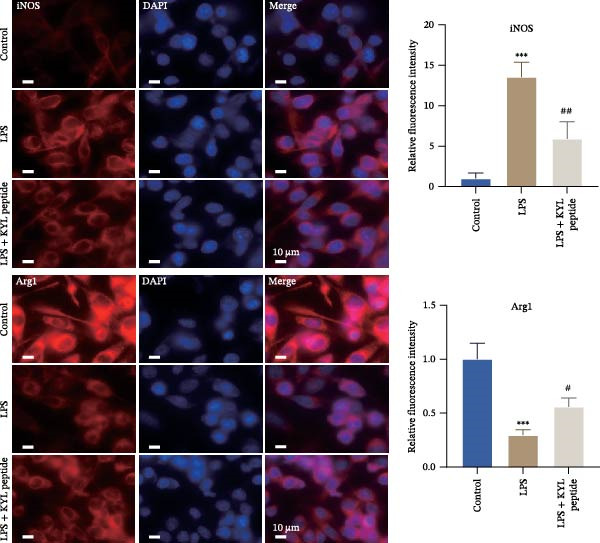 Figure 19
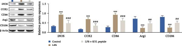 Figure 20
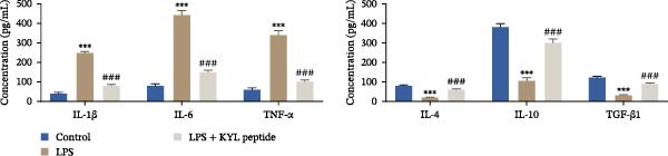 Figure 21
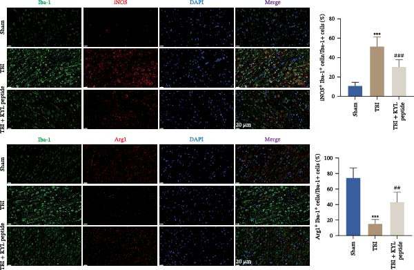 Figure 22
 Figure 23
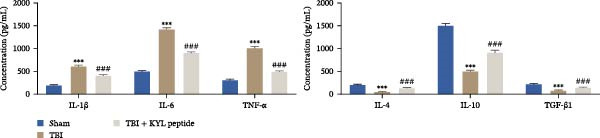 Figure 24
 Figure 25
 Figure 26
 Figure 27
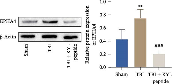 Figure 28
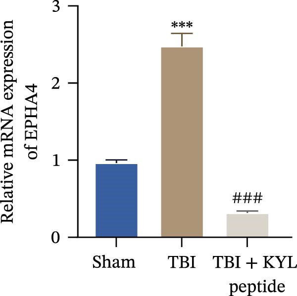 Figure 29
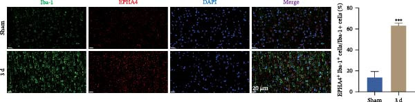 Figure 30
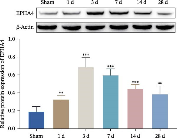 Figure 31
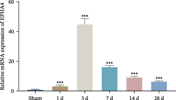 Figure 32
Peer Reviews
No public reviews on file for this paper yet. If you reviewed it on a platform where reviews are public (OpenReview, ICLR, NeurIPS, ICML), you can paste yours below so the community can read it here.
Videos
No videos yet. Explain this paper in a talk, walkthrough, or lecture? Add one.
Taxonomy
TopicsAxon Guidance and Neuronal Signaling · Neuroinflammation and Neurodegeneration Mechanisms · Barrier Structure and Function Studies